




 CRDOJ Home




Abstract
Background: Non-invasive body contouring procedures are primarily designed to reduce localized adiposity; however, objective assessment of their effects on the skin and dermal tissue remains limited.
Objective: To investigate dermal structural and biomechanical changes following a 1060-nm diode laser body contouring treatment using objective imaging and elasticity assessment methods.
Methods: A subject underwent a single abdominal treatment session using a 1060-nm laser system combined with electrical muscle stimulation. Four applicators were positioned according to a standardized abdominal protocol. Skin thickness, echogenicity, and elasticity were evaluated at baseline and 45 days after treatment using high-frequency ultrasound, image-processing analysis, and tensiometric measurements.
Results: Follow-up assessments demonstrated measurable changes in dermal architecture and biomechanical properties. Ultrasound and elastography evaluations revealed improvements consistent with dermal remodelling, increased tissue density, and enhanced skin firmness. Visual analysis of ultrasound images supported the quantitative findings. No adverse events were observed during the study period.
Conclusion: This report highlights the potential value of combining ultrasound imaging and elastography as objective, reproducible tools for monitoring skin remodelling after non-invasive body fat reduction treatments. Beyond adipose tissue targeting, the 1060-nm wavelength may contribute to dermal restructuring and skin tightening, supporting a more comprehensive approach to body contouring evaluation.
Keywords: 1060-nm Diode laser; Body contouring; Skin tightening; Ultrasound imaging
Introduction
To the Editor,
Conventional treatments targeting subcutaneous adipose tissue often carry the intrinsic risk of localized volume depletion, which can subsequently trigger or exacerbate secondary skin laxity. While Energy-Based Devices (EBDs) typically exert their therapeutic actions in a highly localized manner, an optimal body contouring approach should simultaneously induce dermal remodeling to effectively counteract and prevent this tissue laxity.
In our recent clinical research concerning non-invasive body contouring, we demonstrated the safety and efficacy of combining a 1060 nm laser with Electrical Muscle Stimulation (EMS) for localized adiposity reduction and muscle hypertrophy in both the male abdomen and female trochanteric/gluteal regions [1,2].
Moreover, we previously published a case study evaluating the positive impact of 2.45-GHz microwaves on facial skin, demonstrating a significant increase in dermal thickness and echogenicity quantified through a MATLAB-based image-processing algorithm [3]. We further extended this methodology in a split-face trial focused on periocular remodeling, establishing a strict framework that utilizes high-frequency ultrasound coupled with elastography probes to evaluate microstructural dermal modifications and viscoelastic properties [4].
For this reason, we designed our clinical protocols to meticulously investigate the biological responses of the dermal layer during non-invasive treatments primarily dedicated to fat reduction.
Following this exact integrated methodological framework, we have now reported a case report in which we performed a single treatment session on the abdomen using 4 applicators (PhysiQ360, Deka Mela, Italy), precisely following the abdominal protocol and setup previously described in our laser and electrostimulation trial [1].
Each applicator has a 60 × 40 mm laser area with a cooled window and electrodes for electrical muscle stimulation. The applicators are held in place by an elastic bandage, and transparent gel is applied to ensure good contact between the applicator's entire surface and the skin (as illustrated in (Figure 1).
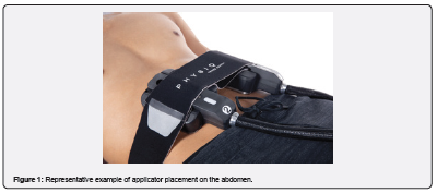
In this study the four applicators were positioned at four strategic points, using the navel as a reference point. A 4-cm-long line segment was drawn starting from the navel and extending vertically downward. At the end point of the line segment, the four points were identified on the horizontal line at 8 cm and 22 cm, in both directions.
To assess the outcomes and specifically monitor how a fat-reduction treatment affects skin tissue stability, we implemented the identical objective indicators established in our facial and periocular studies, namely ultrasound staging and tensiometric analysis.
Ultrasound images and skin elasticity measurements were acquired at the four points described above at baseline (T0) and 45 days after treatment (T1). For each time point, values from the four points were averaged and are reported as mean ± SD.
Skin elasticity was assessed using a dedicated device (DermaLab® Mini, Cortex Technology, Hadsund, Denmark), which applies negative pressure to elevate the skin and then records its retraction. The main parameter derived was the skin retraction time (R), defined as the time (in milliseconds) required for the skin to return from its maximum elevation to 33% of that value.
Skin thickness and echogenicity were selected as the most suitable parameters for evaluating dermal microstructural changes and were measured by ultrasound imaging (Clarius, Vancouver, Canada). Tissue echogenicity was assessed in MATLAB (MathWorks, Massachusetts, USA) via a custom script that binarized dermal ultrasound images (0 = black, 255 = white) through thresholding.
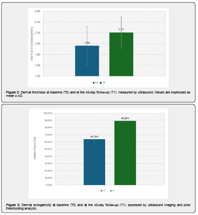
Our preliminary abdominal skin results fully mirror those structural and biomechanical trends. At the 45-days follow-up (T1), our quantitative assessments revealed an increase in mean dermal thickness of 13 % measured in via ultrasound, from 1,96±0,36 mm at baseline (T0) to 2,21±0,29 mm at T1(Figure 2). Dermal echogenicity, quantified by pixel thresholding analysis, increased by 26 %, consistent with collagen remodeling and neocollagenesis within the extracellular matrix (Figure 3). In addition, skin retraction time (R), measured by negative-pressure deformation, decreased by 6%, from 122,3± 9,8 ms at T0 to 114,7 ± 3,4 ms at T1, indicating a substantial restoration of dermoepidermal firmness and skin elasticity (Figure 4).
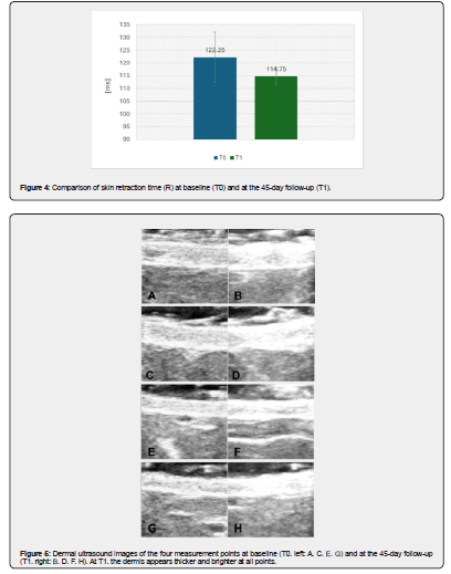
Changes in dermal thickness and echogenicity are directly visible on the ultrasound images. As shown in (Figure 5), all measurement points exhibited increased dermal thickness and brightness at 45 days after treatment (T1, right) compared with baseline (T0, left).
The safety and tolerability of the procedure were monitored throughout the study by clinical examination and patient self-reporting, including procedural pain. No adverse events were recorded.
These real-world outcomes confirm that tracking the exact indicators established throughout our consecutive trials provides an unparalleled, objective diagnostic tool, ensuring maximum reproducibility and precision in verifying non-invasive tissue-remodeling outcomes. Concurrently, our findings underscore the sophisticated mechanisms of the 1060 nm wavelength of the subject device; beyond inducing targeted adipocyte damage, it delivers a powerful stimulatory effect on the dermal district. This dual action is achieved thanks to a precise clinical balance between the mitigation of procedural pain, achieved by modulating nociceptor stimulation through integrated surface cooling, and the sustained, 30-minute thermal activation phase, which successfully triggers neocollagenesis without compromising patient compliance.
Author Contributions
Author Contributions: Conceptualization, AL, FC, TZ, PB; methodology, AL, FC, PB; software, AL, FC, JB, TZ, PB; validation, AL, FC, JB, IF, TZ, PB; formal analysis, AL, FC, JB, IF, TZ, PB ; investigation, AL, FC, JB, IF, TZ, PB; resources, AL, FC, TZ, PB; data curation, AL, FC, JB, IF, TZ, PB; writing—original draft preparation, AL, FC, JB, IF, TZ, PB; writing—review and editing, AL, FC, JB, IF, TZ, PB; visualization, AL, FC, JB, IF, TZ, PB; supervision, AL, FC, JB, IF, TZ, PB; project administration, AL, TZ, PB; funding acquisition, AL, TZ, PB. All authors have read and agreed to the published version of the manuscript.
Institutional Review Board Statement
Institutional Review Board Statement: The study was conducted in accordance with the principles of Declaration of Helsinki. No activity was carried out outside the scope of the device intended purpose or that no additional invasive or burdensome procedures were carried out compared to procedure performed under the normal condition of use of the device.
Conflicts of Interest
Conflicts of Interest: Authors Jorilda Biba, Irene Fusco and Tiziano Zingoni were employed by El.En. Group. The remaining authors declare that the research was conducted in the absence of any commercial or financial relationships that could be construed as a potential conflict of interest.
References
- Alessandro L, Jorilda B, Irene F, Tiziano Z (2026) Infrared Laser and Electrostimulation for the Reduction of Localized Fat in the Male Abdomen. Curre Res Diabetes & Obes J 18(5): 555997.
- Alessandro L, Jorilda B, Irene F, Tiziano Z (2026) Remodeling of the Female Trochanter and Gluteus Using Infrared 1060 nm Laser and Electrostimulation Techniques. An Ultrasound and Bioimpedance Evaluation. J Phy Fit Treatment & Sports11(5): 555830.
- Bonan P, Coli F, Biba J, Fusco I, Zingoni T, et al. (2026) Effects of 2.45 GHz Microwave Treatment on Facial Dermal Thickness and Echogenicity. A Case Study. JOJ Dermatol & Cosmet 7(3): 555712.
- Bonan P, Coli F, Biba J, Fusco I, Piccolo D et al. (2026) Periocular Skin Remodeling Induced by 2.45-GHz Selective Microwaves: Clinical, Ultrasonographic, and Elastographic Outcomes from a Split-Face Study. JOJ Dermatol & Cosmet 7(4): 555716.