




 JOJDC Home




Abstract
Background: Periocular aging is characterized by wrinkles, skin laxity, and structural deterioration of the dermal matrix, making periocular rejuvenation a major challenge in aesthetic medicine. Selective 2.45-GHz microwave technology has recently emerged as a non-invasive approach capable of inducing controlled dermal remodeling through collagen contraction and neocollagenesis.
Objective: To evaluate the clinical efficacy, structural effects, and safety of selective 2.45-GHz microwave treatment for periocular rejuvenation and to compare two delivery modalities, stamping and in-motion, in a split-face study design.
Methods: Twenty male subjects (mean age 51 ± 5 years; Fitzpatrick phototypes II-IV) presenting periocular wrinkles with a Lemperle Wrinkle Assessment Scale score ≥1 were enrolled. The right side of the face was treated using a stamping technique, while the contralateral side received in-motion treatment with the same 2.45-GHz microwave device. Four treatment sessions were performed at monthly intervals. Clinical assessments, standardized photography, skin elasticity measurements, and high-resolution ultrasound examinations were conducted at baseline (T0), before the third session (T4), and one month after the final session (T6).
Results: Both treatment modalities produced significant reductions in periocular wrinkle severity according to the Lemperle scale (p < 0.05). Skin retraction time decreased by 20% with stamping and 16% with in-motion treatment, indicating improved skin elasticity. Ultrasound evaluation demonstrated significant increases in dermal thickness, with gains of 18% and 12% for the stamping and in-motion techniques, respectively (p < 0.05). Increased tissue echogenicity suggested enhanced collagen remodeling and neocollagenesis. No burns, permanent pigmentary alterations, or severe adverse events were observed. Pain scores were lower with the in-motion technique (VAS: 0.4 ± 0.2) compared with stamping (VAS: 3.7 ± 1.2).
Conclusions: Selective 2.45-GHz microwave technology is a safe and effective non-invasive treatment for periocular rejuvenation. Both delivery modalities achieved significant clinical and structural improvements.
Keywords: Periocular rejuvenation; 2.45-GHz microwave technology; Split-face study; Standard Deviation; Post-Inflammatory Hyperpigmentation
Abbreviations: EBDs: Energy-Based Devices; HIFU: High-Intensity Focused Ultrasound; SMAS: Superficial Musculoaponeurotic System; PIH: Post-Inflammatory Hyperpigmentation; VAS: Visual Analog Scale; SD: Standard Deviation
Introduction
Periocular rejuvenation currently represents one of the most challenging and sought-after areas in aesthetic medicine and plastic surgery. The periocular region is intrinsically linked to facial expressiveness and the perception of biological age; however, it is also among the first areas to exhibit the signs of both chronological and environmentally induced skin aging. Periocular skin, characterized by its extremely thin structure, low density of sebaceous glands, and continuous mechanical stress exerted by facial mimetic muscles, undergoes early structural deterioration. This process is accompanied by progressive eyelid laxity, depletion of the subcutaneous fat compartments, and hyperpigmentation, which contribute to the appearance of wrinkles, under-eye bags, and dark circles [1]. In recent years, the therapeutic approach to this anatomical region has undergone a profound transformation, evolving from a purely surgical perspective toward a multimodal and minimally invasive strategy.
The current scientific objective is no longer limited to the excision of redundant tissue but rather focuses on three-dimensional volume restoration, improvement of skin texture, and tissue regeneration. Within the spectrum of non-invasive periocular rejuvenation treatments, energy-based devices (EBDs) have emerged as a cornerstone due to their ability to induce controlled tissue remodeling. Among these technologies, fractional CO₂ laser remains the gold standard for skin resurfacing. This technology creates microscopic vertical zones of thermal injury interspersed with unaffected tissue, thereby stimulating the immediate contraction of elastic fibers and extensive neocollagenesis over the subsequent months, resulting in a marked reduction in dynamic wrinkles and eyelid laxity [2]. Alongside laser technologies, fractional radiofrequency combined with microneedling has revolutionized the treatment of skin texture and laxity. By delivering thermal energy directly into the deep dermis through microneedles, this technique bypasses the epidermis, substantially reducing the risk of post-inflammatory hyperpigmentation, a crucial advantage for individuals with darker skin photos. Finally, High-Intensity Focused Ultrasound (HIFU) complements the non-surgical armamentarium by targeting deeper tissue layers, including the superficial musculoaponeurotic system (SMAS).
The thermal coagulation points generated by HIFU induce a biologically mediated lifting effect that elevates the brow region and enhances periocular tissue density, thereby redefining the eye contour without surgical intervention. Within this therapeutic landscape, microwave-based technology has emerged as a targeted, non-invasive approach for facial remodeling. The treatment relies on synergistic mechanisms whereby an integrated cooling system protects the epidermis while 2.45-GHz microwaves penetrate the deep dermis. The endogenous heat generated within the tissue induces immediate collagen fiber contraction followed by fibroblast activation, resulting in significant skin tightening and facial contour redefinition.
Collectively, the available evidence indicates that microwave technology promotes substantial structural remodeling, supporting its role as a safe, reproducible, and non-invasive modality for facial rejuvenation and skin tightening [3,4]. Preliminary findings from periocular treatments performed with 2.45-GHz microwave technology have demonstrated significant increases in dermal thickness and tissue echogenicity, indicating enhanced nucleogenesis and blastogenesis. These structural changes have been objectively detected and quantitatively assessed using high-resolution ultrasonography and advanced skin diagnostic imaging systems. The observed increase in dermal thickness and echogenicity provides indirect evidence of tissue remodeling, reflecting the deposition of newly synthesized collagen and elastin fibers within the extracellular matrix.
Such findings support the ability of 2.45-GHz microwave technology to induce clinically relevant dermal regeneration and structural rejuvenation of the periocular region [5]. These findings demonstrate the ability of 2.45-GHz microwave technology to induce substantial structural remodeling, further supporting its role as a safe, reproducible, and non-invasive treatment modality for facial rejuvenation and skin tightening. Based on this scientific background, the present study investigated the clinical and structural effects of selective microwave treatment in subjects with localized periocular skin aging. Emphasis was placed on evaluating improvements in skin texture and the restoration of dermoepidermal firmness in the periocular and zygomatic regions.
Materials and Methods
Cohort Selection and Eligibility Criteria
This clinical study enrolled male subjects presenting signs of skin aging characterized by a periocular wrinkle severity score of ≥1 according to the Lemperle Wrinkle Assessment Scale [6]. Baseline assessments were performed in the lower periocular and malar regions. The exclusion criteria were consistent with standard protocols commonly adopted for both ablative and non-ablative energy-based treatments. Specifically, subjects presenting active infections, skin lesions, or manifestations of atopic or seborrheic dermatitis within the treatment area were excluded. Patients with a history of keloid or hypertrophic scar formation, as well as those with a recent history of post-inflammatory hyperpigmentation (PIH), were also excluded. In addition, subjects who had received systemic retinoid therapy within the previous six months were considered ineligible. Finally, individuals who had undergone aesthetic procedures in the periocular region, including dermal fillers, botulinum toxin injections, or suspension thread treatments, within six months prior to enrollment were excluded from the study.
Study Design and Treatment Protocol
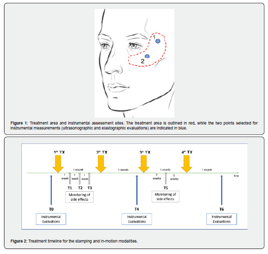
The study protocol involved targeted treatment of the lower periocular area and the malar region. Figure 1 illustrates the facial treatment area and the two anatomical points selected for instrumental assessments. To evaluate the efficacy and tolerability of different application modalities using the same facial handpiece of a 2.45-GHz selective microwave device (Onda Pro, DEKA M.E.L.A. Srl, Calenzano, Italy), a comparative split-face study design was adopted. The right side of the face was treated using a stamping technique, consisting of maintaining the handpiece in a fixed position for several seconds to deliver energy at predefined geometric points, with approximately 10% overlap between consecutive emissions relative to the handpiece dimensions. The contralateral side was treated using a continuous in-motion technique, characterized by smooth circular movements across the entire treatment area. The stamping technique enables the delivery of higher instantaneous energy than in-motion modality, resulting in a more rapid increase in skin temperature and shorter treatment times.
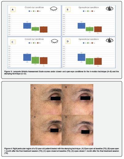
Conversely, the in-motion technique produces a gradual and homogeneous temperature increase across the entire treatment area, requiring longer treatment times but providing greater patient comfort. Both treatment modalities were performed until a predetermined total energy dose was delivered under continuous monitoring by the integrated contact cooling system maintained at 5°C. For the in-motion technique, a total energy dose of 12,000-15,000 J was delivered over an area of approximately 20-25 cm², according to patient tolerability. For the stamping technique, 2-3 passes were performed over a comparable treatment area (20-25 cm²), resulting in a total delivered energy of 3,000-4,500 J. The treatment protocol consisted of four sessions performed at monthly intervals for each patient, as shown in Figure 2. Clinical and instrumental evaluations were conducted at baseline (T0), before the third treatment session (T4), and one month after the final treatment session (T6). In addition to these time points, adverse events were monitored weekly during the first month following the initial treatment session (T1, T2, and T3) and two weeks after the third session (T5) (Figure 2).
Clinical and Instrumental Assessment
To evaluate treatment efficacy and the resulting dermal remodeling, standardized digital photographs (EOS 1100D, Canon Inc., Tokyo, Japan) and a dedicated device for assessing skin elasticity and dermal ultrastructure (DermaLab® Mini, Cortex Technology, Hadsund, Denmark) were employed at two predefined points within the periocular region (Figure 1). Specifically, the Ultrasound Probe provides high-resolution ultrasonographic images of the skin, enabling assessment of both dermal thickness and tissue echogenicity. When the emitted acoustic pulse encounters different skin structures, part of the signal is reflected while the remainder propagates deeper into the tissue. The reflected signal is detected by the ultrasound transducer, processed, and displayed as an image representing the intensity (amplitude) of the reflected echoes. Signal intensity is translated into a color scale, in which darker colors indicate areas of low reflectivity (i.e., homogeneous tissue composition), whereas brighter colors indicate areas of high reflectivity, corresponding to structural heterogeneity and significant variations in tissue density.
The Elasticity Probe employs a well-established method for evaluating the viscoelastic properties of the skin through controlled mechanical deformation. Skin elevation and retraction measurements provide information regarding the degree of skin aging as well as the collagen and elastin content of the tissue. Young skin typically elevates readily in response to negative pressure and rapidly returns to its original position, whereas aged skin may be easily elevated but demonstrates delayed recoil. Based on these two phases, a key parameter calculated by the instrument is the skin retraction time (R), defined as the time, expressed in milliseconds, required for the skin to retract from its point of maximum elevation to 33% of that elevation. Ultrasonographic images and skin elasticity measurements were acquired at the anatomical sites indicated in Figure 1 at baseline (T0), before the third treatment session (T4), and one month after the final treatment session (T6). Dermal thickness was measured from the ultrasound images at each assessment point, and the values were averaged across all subjects for each time point.
Similarly, skin retraction time values were collected and averaged for each evaluation time point. Wrinkle severity was assessed using the Lemperle Wrinkle Assessment Scale, a six-point ordinal scale that classifies facial wrinkles according to wrinkle depth and crease definition, where 0 indicates the absence of wrinkles and 5 indicates a very deep wrinkle. Periocular wrinkles were evaluated under two conditions, with eyes open and with eyes closed, by two independent evaluators at three different time points: baseline (T0), before the third treatment session (T4), and one month after the final treatment session (T6). Clinical improvement was defined as a reduction in the wrinkle score compared with baseline. In addition to efficacy outcomes, particular attention was paid to the clinical safety of the procedure. The occurrence of adverse events, including burns, erythema, and edema, as well as pain management and treatment tolerability, was carefully monitored. Patients were evaluated weekly during the first month following the initial treatment session (T1, T2, and T3), before the third treatment session (T4), two weeks after the third session (T5), and one month after the final session (T6). Treatment tolerability was assessed using a Visual Analog Scale (VAS) ranging from 0 to 10 (0 = no pain; 1-2 = mild pain; 3-6 = moderate pain; 7-8 = severe pain; 9-10 = intolerable pain).
Statistical Analysis
Data are presented as mean ± standard deviation (SD). Statistical analyses were performed using Student’s t-test to compare differences between pre-treatment and post-treatment values for each parameter evaluated. P-values were calculated to determine the statistical significance of the observed differences. A p-value < 0.05 was considered statistically significant.
Results
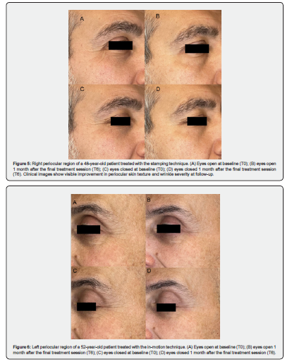
A total of 20 male subjects were enrolled, with a mean age of 51 ± 5 years and Fitzpatrick skin phototypes ranging from II to IV. At baseline, periocular wrinkle severity was assessed on both sides of the face using the Lemperle Wrinkle Assessment Scale under two conditions: eyes closed and eyes open. For the left side of the face, the mean score was 2.3 ± 0.7 with eyes closed and 2.2 ± 0.7 with eyes open. For the right side, the mean score was 2.4 ± 0.9 with eyes closed and 2.3 ± 0.9 with eyes open.
Wrinkle Severity Analysis and Clinical Assessment
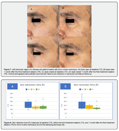
Clinical evaluation performed one month after the final treatment session demonstrated a statistically significant improvement in skin texture across the entire cohort. On the side treated with the stamping technique, the Lemperle analysis revealed a significant reduction in periocular wrinkle severity at the one-month follow-up after the final treatment session compared with baseline, decreasing from 2.4 ± 0.9 to 1.4 ± 0.9 with eyes closed and from 2.3 ± 0.9 to 1.2 ± 0.9 with eyes open (p < 0.05 for both comparisons). Similarly, on the contralateral side treated with the in-motion technique, a significant reduction in wrinkle severity was observed at the one-month follow-up after the final treatment session compared with baseline, decreasing from 2.3 ± 0.7 to 1.2 ± 0.7 with eyes closed and from 2.2 ± 0.7 to 1.2 ± 0.7 with eyes open (p < 0.05 for both comparisons). Both treatment approaches therefore demonstrated substantial clinical efficacy in reducing wrinkles of the lower periocular and malar regions compared with baseline values, as shown in Figure 3. Standardized photographic documentation further confirmed the restoration of skin firmness, with visible attenuation of periocular depressions on both sides of the face [Figures 4-7].
Instrumental Outcomes: Skin Elasticity and Dermal Thickness
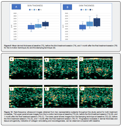
Tensiometric analysis demonstrated an increase in skin elasticity, as indicated by a reduction in skin retraction time (R), following both treatment modalities. For the area treated with the stamping technique, retraction time decreased from 153.5 ± 61.8 ms at T0 to 131.6 ± 21.2 ms at T4, corresponding to a 14% reduction, and further decreased to 122.3 ± 28.2 ms at T6, representing a 20% reduction compared with baseline. For the in-motion technique, retraction time decreased from 154.3 ± 42.4 ms at T0 to 126.4 ± 28.3 ms at T4, corresponding to an 18% reduction, and measured 130.2 ± 34.2 ms at T6, representing a 16% reduction compared with baseline [Figure 8]. In parallel, high-resolution ultrasound imaging revealed a significant increase in mean dermal thickness. For the stamping technique, dermal thickness increased from a baseline value of 1963.2 ± 208.9 μm to 2310.8 ± 307.1 μm at the one-month follow-up after the final treatment, corresponding to an 18% increase (p < 0.05). For the in-motion technique, dermal thickness increased from 1918.1 ± 191.5 μm at baseline to 2148.3 ± 180.5 μm at the one-month follow-up after the final treatment, corresponding to a 12% increase (p < 0.05) [Figure 9].
Safety Profile and Treatment Tolerability
No cases of skin burns, or permanent pigmentary alterations were observed with either treatment modality.
Post-procedural erythema and edema were generally mild to moderate in severity and resolved spontaneously within a few hours following treatment. Regarding pain tolerability, assessed using the Visual Analog Scale (VAS), patients reported a mean score of 3.7 ± 1.2 on the side treated with the stamping technique and 0.4 ± 0.2 on the side treated with the in-motion technique. These findings indicate excellent patient compliance with both approaches, largely attributable to the protective effect of the integrated contact cooling system.
Discussion and Conclusions
Selective 2.45-GHz microwave technology appears to be a highly effective and safe non-invasive option for periocular rejuvenation and remodeling of the midface region. Assessment using the Lemperle Wrinkle Assessment Scale demonstrated a significant reduction in wrinkle severity in both the lower periocular and malar regions with both treatment modalities when compared with baseline values. Standardized photographic documentation qualitatively confirmed these findings, showing improved skin firmness and a visible attenuation of periocular depressions on both sides of the face. The objective instrumental measurements further validated the clinical findings, demonstrating favorable changes in both the biomechanical and structural properties of the skin following treatment.
In addition, ultrasonographic examination revealed an increase in tissue echogenicity, an indirect histological marker of substantial neocollagenesis and increased elastic fiber density induced by the endogenous heat generated by microwave energy, as illustrated in Figure 10. Monitoring adverse events confirmed the excellent safety profile of 2.45-GHz microwave technology. Compared with surgical procedures and ablative laser treatments, this approach avoids the risks of infection, hypertrophic scarring, and prolonged recovery times while ensuring excellent treatment tolerability and the absence of severe complications such as burns. A key advantage of this technology lies in its biophysical selectivity. Unlike conventional radiofrequency, whose energy tends to disperse and propagate less predictably through tissue layers, the microwaves delivered by the microwave handpiece penetrate tissues in a targeted manner to a specific geometric depth.
This spatial confinement of thermal energy maximizes neocollagenesis and tissue tightening within the deep dermis while preserving the epidermis through the integrated cooling system, thereby providing a reproducible, safe, and minimally invasive treatment option. The results of this preliminary study demonstrate a marked improvement in skin texture, supported by objective increases in dermal thickness and skin elasticity as measured by ultrasound-based structural assessments, together with a significant reduction in wrinkle severity according to the Lemperle Wrinkle Assessment Scale. Both treatment modalities demonstrated comparable clinical efficacy, although the stamping technique showed a trend toward greater increases in dermal thickness. However, the in-motion technique was associated with better tolerability, with patients reporting virtually no pain during treatment.
Author Contributions:
Conceptualization, PB, FC, DP, TZ ; methodology, PB, FC, DP, TZ; software, PB, FC, JB, IF, DP, TZ; validation, PB, FC, JB, IF, DP, TZ ; formal analysis, PB, FC, JB, IF, DP, TZ ; investigation, PB, FC, JB, IF, DP, TZ; resources, PB, FC, JB, IF, DP, TZ; data curation, PB, FC, JB, IF, DP, TZ; writing-original draft preparation, PB, FC, JB, IF, DP, TZ; writing—review and editing PB, FC, JB, IF, DP, TZ,; visualization PB, FC, JB, IF, DP, TZ; supervision, PB, FC, JB, IF, DP, TZ; project administration, PB, TZ; funding acquisition PB, TZ,. All authors have read and agreed to the published version of the manuscript.
Institutional Review Board Statement:
The study was conducted in accordance with the principles of Declaration of Helsinki. No activity was carried out outside the scope of the device intended purpose or that no additional invasive or burdensome procedures were carried out compared to procedure performed under the normal condition of use of the device.
Informed Consent Statement
Informed consent was obtained from all subjects involved in the study.
Data Availability Statement
Data that support the study findings are available on request from the corresponding author (Irene Fusco).
Conflicts of Interest
Authors Jorilda Biba, Irene Fusco and Tiziano Zingoni were employed by El.En. Group. The remaining authors declare that the research was conducted in the absence of any commercial or financial relationships that could be construed as a potential conflict of interest.
- Tao BK, Butt FR, Dhivagaran T, Balas M, Nijhawan N, et al. (2025) Periocular Aging Across Populations and Esthetic Considerations: A Narrative Review. J Clin Med 14(2): 535.
- Bonan P, Campolmi P, Cannarozzo G, Bruscino N, Bassi A, et al. (2012) Eyelid skin tightening: a novel 'Niche' for fractional CO₂ rejuvenation. J Eur Acad Dermatol Venereol 26(2): 186-193.
- Zappia E, Bonan P, Coli F, Del Re C, Cassalia F, et al. (2025) An innovative microwave technology for the treatment of submental skin laxity. Lasers Med Sci 40(1): 28.
- Zappia E, Coli F, Amato S, Bennardo L, Nisticò SP, et al. (2024) An innovative microwave device for the treatment of facial skin appearance. Journal Of Plastic and Pathology Dermatology 20(3): 103-106.
- Paolo B, Federica C, Jorilda B, Irene F, Tiziano Z, et al. (2026) Effects of 2.45 GHz Microwave Treatment on Facial Dermal Thickness and Echogenicity. A Case Study. JOJ Dermatol & Cosmet 7(3): 555712.
- Lemperle G, Holmes RE, Cohen SR, Lemperle SM (2001) A classification of facial wrinkles. Plast Reconstr Surg 108(6): 1735-1750.