




 JPFMTS Home



Abstract
Objectives: this study evaluates the safety and efficacy of a 1060 nm laser device, utilizing electric stimulation in conjunction with laser for the treatment of trochanters adiposity in female patients.
Methods: A non-invasive system combining four independent 1060-nm laser applicators with integrated electrical stimulation was evaluated in 16 female patients. Applicators were symmetrically placed on the trochanteric region using elastic bandages and conductive gel. Patients underwent four 30-minute sessions at 2-week intervals; alternating treatment areas resulted in two sessions per site. Outcomes (BMI, photography, bioimpedance, circumference, and ultrasound) were assessed at baseline, during treatment, and at 1- and 3-month follow-ups, using standardized measurement protocols.
Results: All patients completed treatment and follow-up. BMI remained stable (22.6 ± 1.6 kg/m² at baseline to 23.2 ± 1.6 kg/m² at 3 months), with a transient increase at 1 month. Ultrasound showed a significant reduction (p<0.001) in adipose tissue of the trochanter and gluteus. Mean fat thickness decreased from 42.1 ± 3.5 at baseline to 32.7 ± 4.7 mm at 3-months follow-up in the trochanteric area and from 26.4± 2.7 at baseline to 17.4 ± 3.3 mm 3-months follow-up in the gluteal region. Bioimpedance showed improved phase angle (6.5° to 7.0°), reduced fat mass, and increased body cell mass. Mild transient hardening occurred in 3% of treatments.
Conclusion: The combination of 1060 nm laser and electrostimulation appear to provide a synergistic effect, promoting fat reduction alongside increased muscle tone and improved body composition.
Keywords: Infrared 1060 nm laser; Electrostimulation; Trochanter; Gluteus; Body Mass Index
Abbreviations: EMS: Electrical Stimulation; BMI: Body Mass Index; BIA: Bioelectrical Impedance Analysis; PhA: Phase Angle; BCM: Body Cell Mass
Keywords: Energy Drinks; Athletes; High School; Behavioral Science; Psychology; Sports; Dietitian
Introduction
The demand for non-invasive treatments for body contouring of the buttocks and trochanter area has seen significant growth, with an increasingly widespread demand among young women. This trend is driven by the need to safely sculpt the body, without resorting to the lengthy recovery times associated with cosmetic surgery. The risk-benefit ratio of non-invasive techniques can also benefit from the possibility of combining a targeted reduction of localized fat deposits with simultaneous muscle toning, resulting in increased gluteal muscle thickness and reductions in the adipose tissue some months after treatment, with an overall patient satisfaction rate exceeding 80% [1,2]. These procedures offer psychological benefits that add to the instrumental and postural improvements, significantly reducing anxiety related to perceived defects and alleviating stress, resulting in increased subjective well-being and an improved quality of life for patients [3].
The use of non-invasive energy-based devices is grounded in principles analogous to structured exercise regimens: aerobic activity and short targeted sessions primarily reduce fat, while resistance training is essential for maintaining muscle mass [4]. The 1060 nm laser wavelength has been shown to be both effective and safe in delivering energy to the fat under the skin, reducing fat in men and women, in different areas such as abdomen and trochanters [5]. Muscle stimulation has also been shown to be safe and effective in increasing muscle trophism [6] and, at the same time, a reduction of adipose tissue over the stimulated muscles [7]. In this context, an evaluation was conducted to ascertain the safety and efficacy of a 1060 nm laser device, utilizing electric stimulation in conjunction with laser in the same applicator for the treatment of trochanters adiposity in female patients.
Material and Methods
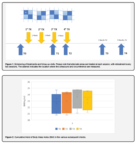
A novel non-invasive system featuring four independent 1060-nm laser applicators with integrated electrical stimulation (EMS) electrodes, sapphire cooling windows, and a 60 × 40 mm aperture (PHYSIQ 360, DEKA M.E.L.A. srl, Calenzano, Italy) was evaluated in this study. Symmetrically positioned on the trochanteric region of 16 female patients (mean age: 42.2 ± 9.1 years), the applicators were secured with elastic compression bandages over a transparent conductive gel. Patients underwent four 30-minute treatments in a supine position at 2-week intervals, during which they were required to maintain stable dietary and exercise regimens. While the full regime comprised four sessions, alternating anatomical zones between consecutive visits meant that each distinct region received only two treatments (Figure 1).
Laser energy profiles and EMS intensities were customized to the thickness of each patient's subcutaneous fat, targeting a mild-to-moderate subjective pain threshold and robust, tolerable muscle contractions. Safety and efficacy were monitored through systematic adverse event tracking and objective parameters assessed at baseline (T0), before sessions three (T1) and four (T2), and at 1- and 3-month post-treatment (T3 and T4). These outcome measures included Body Mass Index (BMI), standardized clinical photos, Bioelectrical impedance analysis (BIA), trochanteric circumference, and high-resolution ultrasonography, with circumference and ultrasound scans strictly replicated at fixed distances from specific anatomical landmarks to ensure maximum reproducibility.
Results
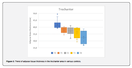
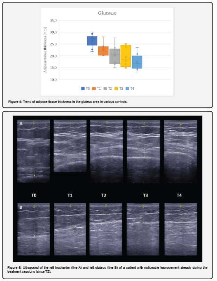
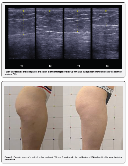
All patients completed treatment and follow-up visits. Patients' BMI remained unchanged, with a slight increase at T3 and subsequent decrease at T4, from a mean value of 22.6 ± 1.6 kg/m2 at baseline to 23.6 ± 1.7 kg/m2 at the 1-month follow-up (T3) and to 23.2 ± 1.6 kg/m2 at the 3-month follow-up (T4), as shown in Figure 2. Instead, ultrasound observations showed a significant reduction (p<0.001) in adipose tissue of trochanters and gluteus between baseline and the 3-month follow-up. Each measured point showed an improvement, as illustrated in Figures 3 and 4. The trochanter area decreased from a mean initial fat thickness (T0) of 42.1 ± 3.5 mm to a value of 32.7 ± 4.7 mm at 3-months follow-up (T4), with a mean reduction of 22% and a maximum reduction of 22.9 mm.
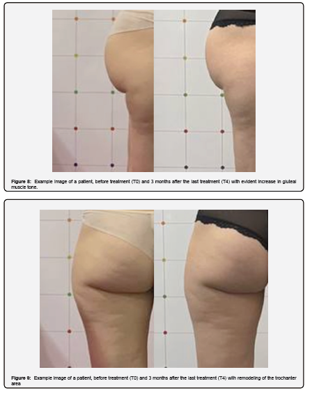
The gluteus area decreased from a mean initial fat thickness (T0) of 26.4 ± 2.7 mm to a value of 17.4 ± 3.3 mm at 3-months follow-up (T4), with a mean reduction of 34% and a maximum reduction of 12.8 mm. Figure 5 shows an ultrasound example of the left trochanter and gluteus of a patient with noticeable improvement already during the course of treatment. Figure 6 shows an ultrasound example of the left gluteus of a patient at different stages of follow-up with a late but significant improvement after the treatment sessions. While Figures 7, 8 and 9 show two photographic examples of the treated patients monitored at the third month after treatment. The culotte circumference decreased from an initial mean value (T0) of 101.4 ± 4.3 cm to a 3-month follow-up value (T4) of 100.1 ± 4.6 cm, with a maximum reduction of 2.0 cm, as shown in Figure 10.
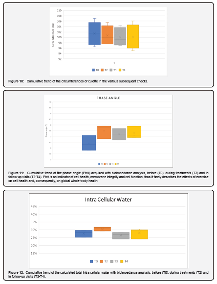
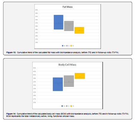
Some patients (3% of the treatments) reported mild hardening of the treated area for a few days after treatment which resolved spontaneously without any side effects. The bioimpedance data showed a progressive increase in the phase angle (PhA) from an initial mean value (T0) of 6.5±0.6° to a 3-month follow-up value (T4) of 7.0±0.6° as shown in Figure 11. The detail indicated a balance between the start and end of treatment for intracellular water, with a transient increase peak during treatments (T2), as shown in Figure 12. Finally, an estimated reduction in the cumulative trend of the calculated fat mass with bioimpedance analysis was observed before (T0) and in follow-up visits (T3-T4), as shown in Figure 13, and an opposite increase in the cumulative trend of the calculated body cell mass (BCM) with bioimpedance analysis, before (T0) and in follow-up visits (T3-T4), as indicated in Figure 14.
Discussion
The most noteworthy finding of this study was the significant reduction in adipose tissue demonstrated via ultrasonographic evaluation (Figures 3,4). In fact, a progressive reduction in fat thickness can be observed across the entire population with earlier or slower results but always evident at the 1- and 3-month follow-up (Figures 5 & 6). Ultrasound imaging represents the most effective methodology for quantifying these variations, offering superior spatial resolution and geometric precision compared to manual tape measurements and photographic documentation. Furthermore, it must be considered that neuromuscular stimulation enhances muscle trophism; this hypertrophic response induces an increase in localized volume that partially counterbalances the reduction in adipose mass. Consequently, while anthropometric measurements and photographic assessments remain valuable for postural analysis, they inherently underestimate the actual extent of lipid reduction [7].
Notably, this reduction was achieved with only two localized treatments per anatomical area, as the four total sessions were alternated across different regions. It is therefore reasonable to hypothesize that increasing the frequency of treatment sessions could yield even more pronounced outcomes. In addition to ultrasonographic evidence, the reduction in adipose tissue was corroborated by body composition analysis. This evaluation revealed a percentage decrease in fat mass concurrent with an increase in fat-free mass, which was directly attributable to the enhanced muscle trophism induced by electrical stimulation. Body composition analysis included the assessment of BCM through bioelectrical impedance measurements. Indeed, BCM constitutes the metabolically active component of fat-free mass, and it represents the most robust indicator of an individual’s nutritional status.
Measuring BCM is a well-established method for evaluating muscle mass and protein tissue states [8]. Photographic documentation further demonstrates an elevation of the gluteal region resulting from increased muscle tone (Figures 7, 14); this improvement was sustained throughout the 3-month follow-up period, with peak values also reflected in the Body Mass Index (BMI) and phase angle at one-month post-treatment. Finally, the efficacy of neuromuscular electrical stimulation is evidenced by a peak increase in intracellular water during the training phase (T2), signaling an immediate physiological response to exercise (Figure 12). Therefore, it can be concluded that improved cellular hydration (an increase in ICW) and higher PhA levels may lead to an increase in muscle glycogen stores. It should also be noted that improved intracellular hydration may contribute significantly to muscle hypertrophy by enhancing protein synthesis and inhibiting protein breakdown [9].
Ultimately, the direct action of the 1060 nm laser on adipocytes, combined synergistically with electrical muscle stimulation, drives a progressive lipolytic effect sustained by simultaneous muscular exertion.
Conclusion
This study demonstrates a significant reduction in adipose tissue in the trochanteric and gluteal regions, most accurately detected by ultrasound. Notably, improvements were achieved with only two treatments per area, suggesting that additional sessions could enhance outcomes. The combination of 1060 nm laser and electrostimulation appears to provide a synergistic effect, promoting fat reduction alongside increased muscle tone and improved body composition.
Author Contributions
Conceptualization AL, TZ; methodology, AL, TZ; software, AL, JB, TZ validation, AL, JB, IF, TZ; formal analysis, AL, TZ.; investigation, AL, TZ; resources, AL, TZ; data curation, AL, JB, TZ; writing—original draft preparation, AL, JB, IF, TZ; writing—review and editing, AL, JB, IF, TZ; visualization, AL, JB, IF, TZ; supervision, AL, JB, IF, TZ; project administration, AL, TZ; funding acquisition, AL, TZ. All authors have read and agreed to the published version of the manuscript.
Funding
: This research received no external funding.
Institutional Review Board Statement
The study was conducted in accordance with the principles of Declaration of Helsinki. No activity was carried out outside the scope of the device intended purpose or that no additional invasive or burdensome procedures were carried out compared to procedure performed under the normal condition of use of the device.
Informed Consent Statement
Informed consent was obtained from all subjects involved in the study.
Data Availability Statement
Data that support the study findings are available on request from the corresponding author (Irene Fusco).
Conflicts of Interest
Authors Jorilda Biba, Irene Fusco and Tiziano Zingoni were employed by El.En. Group. The remaining authors declare that the research was conducted in the absence of any commercial or financial relationships that could be construed as a potential conflict of interest.
References
- Fabi S, Dover JS, Tanzi E, Bowes LE, Tsai Fu F, et al. (2021) A 12-Week, Prospective, Non-Comparative, Non-Randomized Study of Magnetic Muscle Stimulation for Improvement of Body Satisfaction with the Abdomen and Buttocks. Lasers Surg Med 53(1): 79-88.
- Mazzoni D, Lin MJ, Dubin DP, Khorasani H (2019) Review of non-invasive body contouring devices for fat reduction, skin tightening and muscle definition. Australas J Dermatol 60(4): 278-283.
- Alzahrani KJ, Kattan AE, Ezzat LA, Alsaleh SA, Murad KA, et al. (2017) A Psychosocial Analysis of the Effect of Body-Contouring Surgery on Patients After Weight Loss. Eplasty 17: e12.
- Recchia F, Leung CK, Yu AP, Leung W, Yu DJ, et al. (2023) Dose-response effects of exercise and caloric restriction on visceral adiposity in overweight and obese adults: a systematic review and meta-analysis of randomised controlled trials. Br J Sports Med 57(16): 1035-1041.
- Bonan P, Fusco I, Madeddu F, Zingoni T, Ibrahim SMA (2025) Clinical Evaluation of a 1060-Nm Laser for Non-Invasive Fat Reduction of the Abdomen and Coulotte/Thighs in Men and Women. J Cosmet Dermatol 24(12): e70558.
- Negosanti F, Cannarozzo G, Zingoni T, Leone A, Fusco I (2022) Is It Possible to Reshape the Body and Tone It at the Same Time? Schwarzy: The New Technology for Body Sculpting. Bioengineering (Basel) 9(7): 284.
- Leone A, Piccolo D, Conforti C, Pieri L, Fusco I (2021) Evaluation of safety and efficacy of a new device for muscle toning and body shaping. J Cosmet Dermatol 20(12): 3863-3870.
- Talluri A, Liedtke R, Mohamed EI, Maiolo C, Martinoli R, et al. (2003) The application of body cell mass index for studying muscle mass changes in health and disease conditions. Acta Diabetol 40 Suppl 1: S286-S289.
- Annunziata G, Paoli A, Frias-Toral E, Marra S, Campa F, et al. (2024) Use of phase angle as an indicator of overtraining in sport and physical training. J Transl Med 22(1):1084.

























