




 NFSIJ Home



Summary
Introduction: Microcytic anaemia is a drop in Hb levels associated with a MCV of less than 80 fl. It constitutes a major public health problem in the world and is frequently encountered in our daily practice.
Material and method: We carried out a retrospective, descriptive and analytical study of records of hospitalized patients aged 15 years and above with HB levels less than 12 g/dl in women and 13 g/dl in men associated with a MCV of less than 80 fl at the internal medicine department of the mame Abou Aziz Sy hospital in Tivaouane over a period of 2 years. (October 2021 to October 2023).
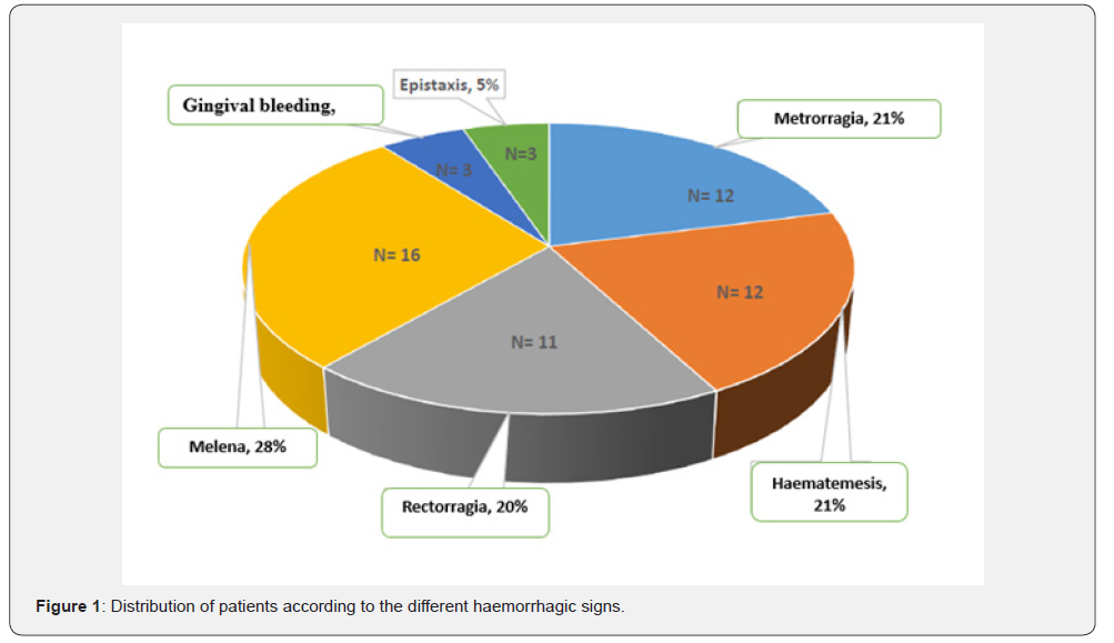
Results: During the study period, microcytic anaemia was found in two hundred and twenty (220) patients, i.e. a prevalence of 25%. The mean age was 44 years [15-92 years]. The 15-39 age group was more represented (47%). Females predominated, with a sex ratio of 0.83. Hypertension (14.55%) and diabetes (11.36%) were the main comorbidities. Symptoms were dominated by asthenia (83%), dyspnoea (28%) and headache (18%). The main physical signs were pale conjunctiva (70%), tachycardia (56%), fever (36%), pulmonary condensation syndrome (31%) and external bleeding (25%). The latter consisted of: melena (28%), metrorrhagia (21%), haematemesis (21%) and rectal (20%). The mean Hb level was 7.66 g/dl [1.8-12 g/dl]. The leading causes of anaemia were anaemia of inflammation (66.8% of cases), followed by iron deficiency anaemia (18.20%). Their aetiologies were mainly infectious (45.91%), tumoral (12.27%), non-tumoral digestive (11.81%) and undetermined (10.45%). 29 cases of death (13.18%) were recorded, of which four (04) were related to anaemia (1.81%).
Conclusion: Microcytic anaemia is common in internal medicine, with an estimated prevalence of 25%. Iron deficiency assessment markers play an important role in determining the type of anaemia. infectious causes dominated the list of aetiologies.
Keywords:Anaemia; Microcytic; Internal medicine; Ferritin
Abreviations:REV: Rupture of Esophageal Varices; PHP: Portal Hypertension; MCHC: Mean Corpuscular Hemoglobin Concentration; CST: Transferrin Saturation Coefficient; BAAR: Acid-Fast Bacillus
CKD: Chronic Kidneys Diseases; HG: Haemoglobin
Introductıon
Microcytic anemia is defined as anemia with an MCV of less than 80 fl. It is the most common cause of anemia. It is a very heterogeneous group of diseases that can be either acquired or hereditary. This anemia is defined by age and sex [1,2]. Microcytic hypochromic anemia may result from defects in globin (hemoglobinopathies or thalassemias) or from heme synthesis or iron availability, or from its acquisition by erythroid precursors [3]. The diagnosis of microcytic anemia is very important in order to establish an appropriate treatment plan. We carried out a study whose objective was to determine the prevalence of microcytic anemias, epidemiological, clinical and biological characterizations and identify etiologies.
Material and Method
We carried out a retrospective study in the internal medicine department of the Abdou Aziz Sy Hospital in Tivaouane over the period from October 1, 2021 to October 1, 2023. It concerned all patients hospitalized in the department and who presented microcytic anaemia during their exploration. We included all inpatients aged 15 years and older in the AASH Internal Medicine Department during the study period. Data were collected from hospitalized patient records and then completed a survey form for each patient. This sheet contained the following information: sociodemographic data, medical, surgical and gynaecologicalobstetric history, habits and lifestyle, reason for hospitalization or consultation, clinical signs, biological data (complete blood count, serum iron, transferrin, ferritinemia, CST, inflammatory assessment, liver test, HB electrophoresis, Genexpert/sputum BAAR), serologies (HBV, HCV, HIV)). It also contained the fibroscoscopy report (FOGD, Colonoscopy), the results of medical imaging, the etiologies of anaemia, the therapeutic modalities and the evolution.
Results
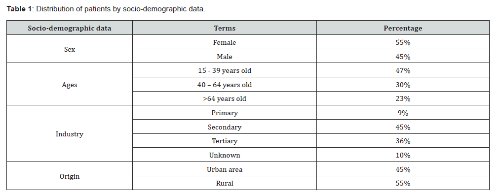
Sociodemographic data: We included 220 patients, i.e. a prevalence of 25%. The average age of our patients was 44 years with extremes of 15 years and 92 years. Female sex was predominant with a sex ratio of 0.83. We have listed the various socio-demographic data in Table 1.
Clinical data: Clinical signs are polymorphic and depend on etiology for the most part. The main antecedents found were arterial hypertension (14.55%), diabetes (11.36%), sickle cell anemia (5.45%). They are followed by tuberculosis (3.18%), connective tissue disease (3.18%), abortions (3.18%), ulcerative epigastric disease (2.73%). Tobacco was used by two of our patients and herbal medicine by two patients.
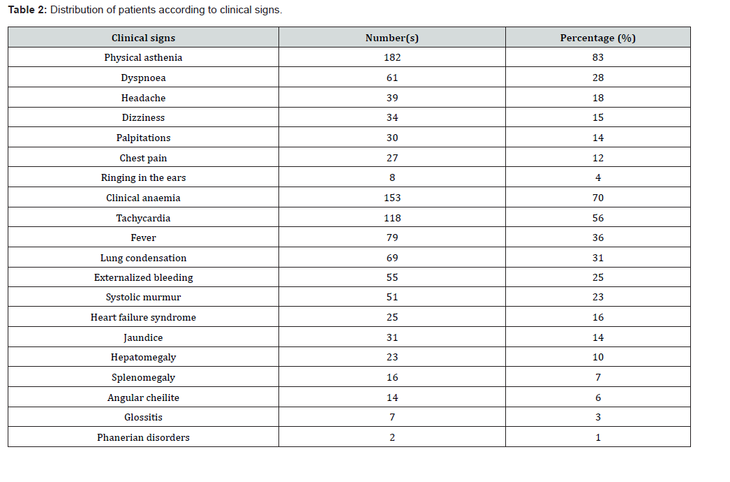
The main clinical signs were: asthenia (83%), clinical anemia (70%), tachycardia (56%), fever (36%), dyspnoea (28%), externalized bleeding (25%). These signs are listed in Table 2.
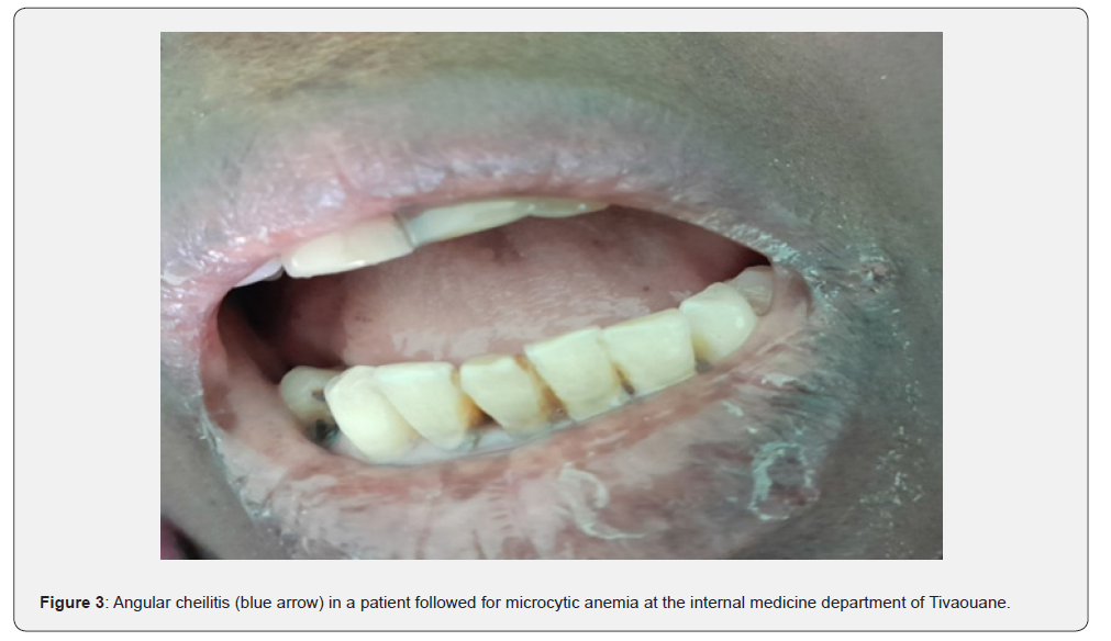
The different types of bleeding are listed in Figure 1. Figures 2 & 3 are illustrative of clinical anemia and angular cheilitis in 2 of our patients followed for microcytic anemias.
Biological data: The mean haemoglobin level was 7.66 g/ dl with extremes of 1.8g/dl and 12g/dl. Anaemia was severe in 89 patients (40.5%), mild in 77 patients (35%) and moderate in 54 patients (24.5%). Anemia was predominantly hypochromic at 65% according to MCHC and 94% according to MCHT. The C reactive protein was increased in 169 patients (85.8%). We found hyperleukocytosis in 118 patients (53.9%), leukopenia in 6 patients (2.7%). Thrombocytopenia was found in 25 patients (11.4%) and thrombocytosis in 88 patients (40%).

Serum iron was measured in 29 patients and lowered in 19 patients. Ferritin was measured in 31 patients; it was low in 6 patients and increased in 9 patients. Transferrin was normal in 4 patients and low in 3 patients. The transferrin saturation coefficient was measured in 2 patients and it was lowered. The total iron binding capacity of transferrin was measured in 2 patients and was lowered. In (Figure 4), we have represented the profile of patients according to the abnormalities of the martial assessment.
Haemoglobin electrophoresis was performed in 9 patients and was abnormal in 7 patients.

During these explorations, we found inflammatory anemia in 66.80% of patients, followed by anemia due to iron deficiency 18.20%. It was indeterminate in 6.40% of cases and mixed in 8.60% of cases.
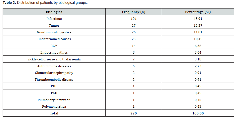
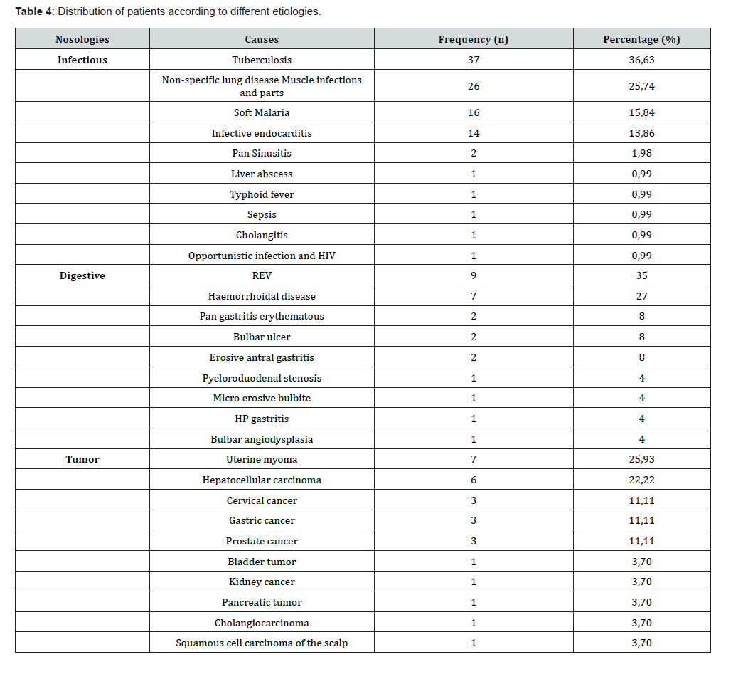
Etiologies: The main etiologies found were infectious (45.91%), tumor (12.27%), non-tumor digestive (11.81%) and undetermined (10.45%) causes. The different etiologies and their distribution are described in (Table 3 & Table 4).
Table 4 shows the distribution of patients according to etiologies. The frequencies are calculated according to the number of patients in each nosological setting.
Treatment: Blood transfusion was performed in 98 patients (45%). Martial therapy was prescribed in 51 patients (23%), antibiotic therapy in 135 patients (61%), corticosteroid therapy (6%), immunosuppressive treatment (3%).
Evolution:
The evolution was favorable in 87% of cases.
In multivariate analysis:
• Anaemia was more severe in females, although the
difference was not significant with P value = 0.8781.
• Anaemia was more severe for the 15 to 39 year and 40
to 64 year age groups with a non-significant difference, p value =
0.910.
• Anaemia was more severe in patients with a non-tumoral
digestive etiology, which is partly explained by the hemorrhagic
syndrome, pvalue = 0.0075 significant; It was also severe in
patients with a tumor etiology with a non-significant difference,
pvalue = 0.1490.
Discussion
During our study period, the prevalence of microcytic anemia was 25%. Tarek A Guseibat et al [4], in their study on the prevalence of microcytic anemia in the city of Benghazi in Libya, found a prevalence of 59.19% [4]. In the work of Naz et al [5] on microcytic anemia in 2021 at Balochistan Hospital in Pakistan, there was a prevalence of 32.95% [5]. This variation in prevalence could be explained by the fact that their cohort was much smaller and the study period was short.
The average age of our patients was 44 years with extremes of 15 and 92 years. The most representative age group is the under 40s, i.e. 47%. She was predominantly female for the age groups between 30-44 and 45-60 years. The sex ratio (M/F) was 0.83. Our results corroborate with data from the literature. In the study by Naz et al [5], the mean age of patients was 33.8 years with a female predominance [5]. The study by Tarek A Guseibat et al [4] revealed a female predominance and the most affected age group was women (21-40 years) [4]. In the Prabhat Kumar et al [6] study, there was a predominance of women with 61% women and 39% men. The 61-70 age group (26%) predominated, followed by the 21-30 age group (22%) [6]. The work published by Zanetti in 2021 most often revealed the juvenile age of patients [7]. This could be explained by gynaecological pathologies, heavy menstruation, pregnancy, poverty, difficulties in accessing care, and the predominance of inflammatory pathologies among women.
In our study, the mean hemoglobin level was 7.66 g/dl with extremes of 1.8g/dl and 12g/dl and a standard deviation of 2.68.
Mild anaemia predominated for all age groups, followed by severe anaemia for the 15-39 and 40-64 age groups. Anaemia was severe in 89 patients (40.5%), followed by mild anaemia in 77 patients (35%) and moderate anaemia in 54 patients (24.5%). These data on the severity of anemia are different from those found in the literature.
In the work of Prabhat Kumar et al [6], moderate anaemia predominated in 62% and followed by severe anaemia in 34% of patients [6]. In the study by Naz et al [5], there was mild anemia at 21.4%, moderate anaemia at 42.77% and severe anaemia at 35.83% [5]. Indeed, the ferritin assay was performed in only 31 patients (14.09%), serum iron in 29 patients (13.18%), transferrin in 08 patients (3.63%), CST in 2 patients (1%) and CFT in 2 patients (1%).
However, the low dosage of martial balance markers in our series may underestimate the true prevalence of iron deficiency in our population.
In our study, inflammatory anemias were the main causes with 66.8% of cases. Etiologies were dominated by infectious (45.91%), tumor (12.27%), non-tumor digestive (11.81%) and undetermined (10.45%) causes.
This same trend was observed in the studies of Joosten E et al [8] on iron deficiency anemia and chronic disease anaemia in 2015, revealing 70% of patients with inflammatory anemia. These authors found: acute infection (71%), malignancy (12%), chronic infection 16% and CKD in 22 patients (16%) [8].
In the 2021 study by Prabhat Kumar et al [6] in India on the etiological assessment of microcytic anemia, inflammatory anemias were observed in 28% of patients. The main causes were infections (39.2%), followed by autoimmune diseases (17.8%) and chronic kidney disease (10.7%) [6].
In our work, tuberculosis infection was retained in 36.63% of patients (27 pulmonary TB cases, 7 multifocal TB cases and 3 peritoneal TB cases), the other causes in 63% of infections (pleuropneumonia 25.74%, muscle and soft tissue infections 15.84%). The association with malaria found in our study is supported by Pradhan’s studies, which had demonstrated the presence of anaemia during malaria. This can be microcytic, normocytic or macrocytic due to the effect of NO on erythropoiesis [9].
After these, we find tumor causes 12.27%, CKD (5.45%), autoimmune diseases.
In the literature, the most frequent etiologies are infections and tumors. This high prevalence could be explained by the endemicity in our regions of certain infectious diseases such as tuberculosis (in 2022, 10.6 million cases of tuberculosis worldwide), HIV infections (a prevalence of 0.32% according to CNLS in 2021) and the increase in the prevalence of gynecological cancers (more than 11,000 new cervical cancer cases in Senegal in 2020), digestive systems, etc. [10-11].
Chronic anemia and inflammation most often complicate a specific underlying condition. It is triggered by cellular immune mechanisms and pro-inflammatory cytokines (tumor necrosis factor, interleukin 1 and interferons). The production of the latter would increase the synthesis of hepcidin [12].
Knowledge of the pathophysiology of cytokines during inflammation could lead to the development of a more specific treatment for this syndrome [13-14].
Iron deficiency anemia is the leading cause of deficiency anemia found in most studies. In 2019, the WHO estimated that iron deficiency was one of the main causes of disability due to anaemia. Globally, it involved 40% of children aged 6 to 59 months, 37% of pregnant women and 30% of women aged 15 to 49 years [15]. Iron deficiency is the most common nutritional deficiency. In Africa, it is one of the leading causes of anaemia [16].
In our series, anemia due to iron deficiency was found in 18.20% of patients.
In Morocco, Ali Zenibi et al [17] in a retrospective study on the etiological profile of anaemia in an internal medicine department in 150 patients, iron deficiency anaemia clearly predominated with 60% of cases [17].
The study conducted by Ida M Ngongang on iron deficiency anemia in the internal medicine department in Mali in 2020 over a period of 11 years revealed a prevalence of 0.40% [18].
These results are lower than ours. This could be explained by the fact that their cohort was smaller and their duration of study longer than our study.
The prevalence of iron deficiency varies with age. The most affected are premature or growing children, the elderly, pregnant women and women of childbearing age [15]. In our series, non-tumoral digestive causes (55%) predominated, followed by gynecological causes (30%). In the context of the search for foci of gastrointestinal bleeding, twenty-six (26) FOGDs (11.8%) were performed showing HP gastritis, esophageal varices, microerosive bulbitis, pyeloroduodenal stenosis, bulbar angiodysplasia and two (2) total colonoscopies (1%) highlighting hemorrhoidal thrombosis and colonic ulcerations in nail cutting. For gynecological causes, explorations were carried out in conjunction with the gynaecological patients. The main causes found were: uterine fibroids (7 cases), cervical cancer (3 cases) and polymenorrhea due to hormonal dysfunction.
In Ben Ahmed’s study of 40 cases in Tunisia, iron deficiency predominated (60%). The latter was most often related to a lack of intake (83%), a gynaecological cause (8.4%) and digestive causes (8.4%) [19].
The work of Kafle S et al [20] in 2016 revealed that iron deficiency anaemia was the most common etiology (49% of cases) and the main cause was uterine bleeding (20.8%) [20].
Contrary to Ben Salem et al [21], the main etiologies found were gynaecological (21.4%), digestive (12.8%) and intake deficiencies (15%) [21].
The observation noted in our series with a low rate of martial deprivation would probably be linked to a deficit of martial explorations because of its high cost but also poverty, malnutrition, lack of contraception, inaccessibility to care, close pregnancies, gynecological bleeding.
Indeed, the low dosage of markers of the martial assessment could underestimate the real prevalence of martial deficiency
In the study by Prabhat Kumar et al [6] on microcytic anemias, 11% of patients had hemoglobinopathy [6].
In our series, there were 07 cases of hemoglobinopathies (3.2%), including 5 cases of SS sickle cell disease and 2 cases combining sickle cell anemia AS-alpha thalassemia.
In our series, tumor causes occupied the second position with 12.27% of cases. They affect both sexes with a female predominance (F/M ratio 1.45). In our study, digestive tumors predominated at 41%, followed by gynecological tumors at 37%, prostate tumors at 11% and other rarer causes at 11% (bladder, kidney, scalp).
In our work, twenty-three (23) patients (10.45%) had no etiologies found. These data are close to those found in the study by Cappellini MD et al [22], where in a third of patients, anemia could not be explained by a fundamental disease or by a specific pathological process, and for this reason it is defined as “unexplained anemia”. It could be due to the progressive resistance of erythroid progenitors in the bone marrow to erythropoietin and to a chronic sub-clinical pro-inflammatory state [22].
This high rate in our cohort could be explained by the fact that the markers of the martial assessment or the other explorations necessary to search for a possible cause of anemia were incomplete.
Etiological research for microcytic anemias is often limited in our contexts. This is due to the fact that the explorations are most often the responsibility of the patient; And in the majority of cases, patients do not have health care.
CKD is the fourth etiological group. In our series, 14 patients (6.36%) had CKD and 9 patients were terminally ill. It most often affects the elderly.
Iron deficiency anemia is a common complication during CKD and this iron deficiency is both absolute and functional. The unavailability of iron could be due to increased hepcidin synthesis [23].
Mechanisms responsible for this phenomenon in CKD include decreased GFR and erythropoietin levels and increased inflammatory cytokines [24-25].
Half of our patients who had CKD had a background of high blood pressure (14.45%), diabetes and hypertension (8.3%). CKD remains one of the major and serious complications of these longterm pathologies.
Conclusion
Microcytic anemia is common in internal medicine consultations. There are many causes, with infectious causes being the most prevalent. Treatment must be tailored to the patient and based on a rigorous diagnostic approach.
References
- McLean E, Cogswell M, et al. (2009) Worldwide prevalence of anemia,Vitamin and Mineral Nutrition Infromation System, 1993-2005. Public Health Nutr 12(4): 444-454.
- WHO (2011) Haemoglobin concentrations for the diagnosis of anaemia and assessment of severity. Vitamin and Mineral Nutrition Information System.
- Leporrier M, Seguin A (2004) Anaemia due to iron deficiency. Rev Prat 54(2): 217-223.
- Tarek AG, Mona S (2020) A comparative study on the prevalence of microcytic anemia and associated age of anemic patients from Benghazi city. JOPAS 19(1): 27-32.
- Shabana N, Hamid I, Javeria A, et al. (2023) Outcome of hypochromic microcytic anemia in patients attending tertiary care hospital of balochistan. World J Pharm Res 12(16): 43-50.
- Prabhat K, Arun KS, Anju B, et al. (2023) Etiological Evaluation of Microcytic Hypochromic Anemia at a Tertiary Care Hospital in the Eastern Part of India. Int J Res Pharm Sci 14(1): 26-34.
- Zanetti R, Feldman B, Porea T (2021) Microcytic Anemia. Pediatr Rev 42(1): 41-43.
- Joosten E, Lioen P (2015) Iron deficiency anemia and anemia of chronic disease in geriatric hospitalized patients: How frequent are comorbidities as an additional explanation for the anemia? Geriatr Gerontol Int 15(8): 931-935.
- Pradhan P (2009) Malarial anaemia and nitric oxide induced megaloblastic anaemia: a review on the causes of malarial anaemia. J Vector Terminal Dis 46(2): 100-108.
- (2025) National Council for the Fight against AIDS 2021 Annual Report.
- World Health Organization (2025) Cervical Cancer Country Profiles 2021.
- Wiciński M, Liczner G, Cadelski K, Kołnierzak T, Nowaczewska M, et al. (2020) Anemia of Chronic Diseases: Wider Diagnostics-Better Treatment? Nutrients 12(6): 1784.
- Ganz T, Nemeth E (2011) Hepcidin and disorders of iron metabolism. Annu Rev Med 62: 347-360.
- Means RT (1995) Pathogenesis of the anemia of chronic disease: a cytokine mediated anemia. Stem Cells 13(1): 32-37.
- Global Health Measures (2019) Level 1 deficiency anemia. Lancet 393.
- Leporrier M, Seguin A (2004) Anemia due to iron deficiency. Rev Prat 54(2): 217- 223.
- Zinebi A, Eddou H, Moudden KM, et al. (2017) Etiological profile of anaemia in an internal medicine department. Pan Afr Med J 26:10.
- Ida MN (2021) Epidemio-clinical, paraclinical, etiological and therapeutic aspects of iron deficiency anemia in the internal medicine department of the CHU du point-G, 2020. Medical thesis.
- Ben AI, Ben DF, Ben AA, et al. (2011) Diagnosis of anemia in the Tunisian Cap Bon region: about 40 cases. Diabetes and Metabolism 37(1-1): A56.
- Kafle S, Lakhey M (2016) Etiological study of microcytic hypochromic anemia. J Pathol Nep 6(12) : 994-997.
- Ben ST, Laanani A, El OA, et al. (2014) Etiological profile of iron deficiency anemias in an internal medicine department : about 187 cases. Revmed 35(1): A93.
- Cappellini MD, Motta I (2015) Anemia in Clinical Practice-Definition and Classification: Does Hemoglobin Change with Aging? Semin Hematol 52(4): 261-269.
- Gafter-Gvili A, Schechter A, Rozen-Zvi B (2019) Iron Deficiency Anemia in Chronic Kidney Disease. Acta Haematol 142(1): 44-50.
- Babitt JL, Lin HY (2010) Molecular mechanisms of hepcidin regulation: implications for the anemia of CKD. Am J Kidney Dis 55(4): 726- 741.
- Van der WNC, Grooteman MP, Bots ML, et al. (2012) Hepcidin-25 in chronic hemodialysis patients is related to residual kidney function and not to treatment with erythropoiesis stimulating agents. PLoS One 7(7): E39783.

























