Sigmoid Colon Perforation Caused by Bone: A Rare Case Report
Mayank Mahajan*
Mahajan Hospital, India
Submission: August 28, 2017; Published: September 26, 2017
*Corresponding author: Mayank Mahajan (Consulting Surgeon), Mahajan Hospital, Magajpura Road, Dhar (M.P.) -454001, India, Email: drmayankmahajan@gmail.com
How to cite this article: Mayank Mahajan. Sigmoid Colon Perforation Caused by Bone: A Rare Case Report. Adv Biotech & Micro. 2017; 6(3): 555686. DOI: 10.19080/AIBM.2017.06.555686
Abstract
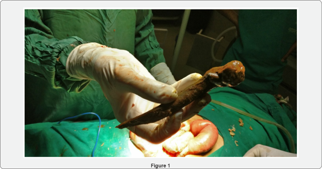
The accidental ingestion of a foreign body is common, and the majority of ingested foreign bodies pass through the gastrointestinal tract without complication. Perforation is one of the rarest complications and commonly occurs in the terminal ileum and recto-sigmoid junction. The sigmoid colon is an extremely rare site of perforation because of its anatomical features of a thick wall, large diameter, and non-angulation. Here, we present a case of sigmoid colon perforation caused by bone insertion per rectally. A 60-year-old male patient presented with generalized abdominal pain for 4 days n not passing stool and flatus for 4 days. He had history of penile erectile dysfunction for which he went to some quack and for this, that quack inserted a piece of bone into his rectum 1 month back in crematorium and he has no surgical history. His abdomen was distended and the whole abdomen was tender, with signs of generalized peritoneal irritation. X-Ray abdomen standing revealed a linear radio-dense foreign body in abdomen with gas under diaphragm. An emergency exploratory laparotomy was performed. During the operation, a 15-cm-long bone was found protruding from the sigmoid colon. Intraoperative bowel wash, primary resection, and anastomosis were performed. The postoperative course was uneventful, and the patient was discharged on the eleventh postoperative day. The case represents an unusual case of sigmoid colon perforation caused by bone inserted per rectally. Because colon perforation by bone is extremely rare, and its preoperative diagnosis is difficult, meticulous history taking is crucial for the correct diagnosis and prompt management in the emergency setting.
Keywords: Bone; Foreign body; Colon; Perforation; History taking
Introduction
Ingestion of a foreign body is not uncommon, but rarely results in perforation of the gastrointestinal tract. The majority of ingested foreign bodies pass through the gastrointestinal tract without complication. Only less than 1% may cause bowel perforation, depending on the size and the shape of the foreign body [1]. Common sites of perforation are the narrow parts of the bowel. Perforation of the colon other than at recto-sigmoid junction is rare [2]. Here, I am present a case of sigmoid colon perforation caused by a bone inserted per rectally.
Case Report
A 60-year-old male patient was referred to the emergency department after presenting with whole abdominal pain. The pain had started acutely approximately 4 days prior to presentation and had intensified gradually over time. There were no aggravating or relieving factors for the pain. He had history of penile erectile dysfunction for which he went to some quack and for this, that quack inserted a piece of bone into his rectum 1 month back in crematorium and he has no surgical history. The vital signs at admission were body temperature, 38.0 °C; blood pressure, 150/90mmHg; heart rate, 116 beats/ minute; and respiratory rate, 26 breaths/minute. The abdomen was distended and the whole abdomen was tender, with signs of generalized peritoneal irritation. Laboratory results were within the normal range, except for a white blood cell count of 18,880/|iL. There were gas under diaphragm with some bone in abdominal cavity in abdominal radiography. A clinical diagnosis of perforation peritonitis was made based on the results of physical examination, simple abdomen radiography, and laboratory examination.
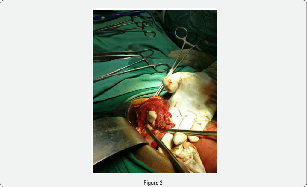
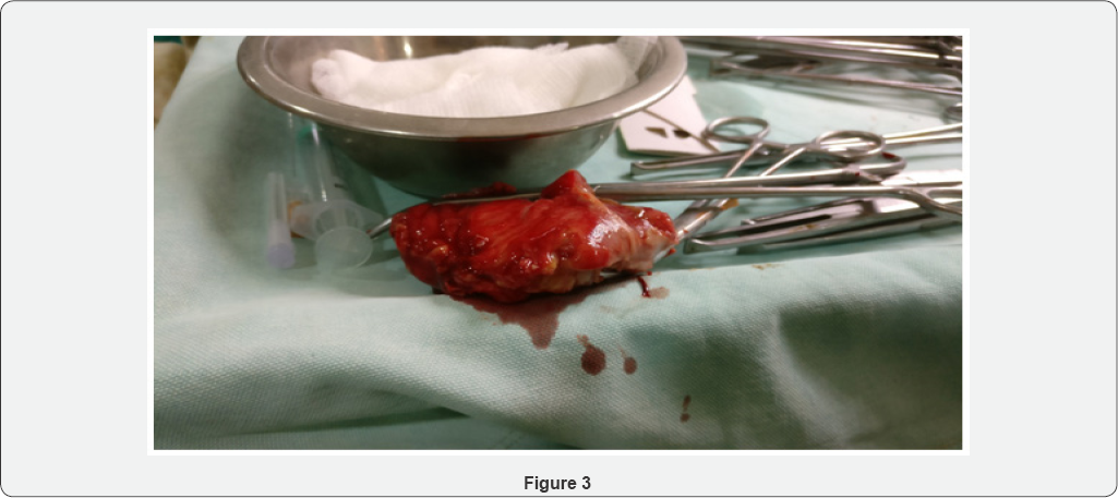
With a diagnosis of perforation of the bowel, an emergency operation was performed. During the operation, an approximately 15-cm-long bone was found protruding from the sigmoid colon with severe inflammation. After removing the bone, primary repair of the colon was not feasible owing to inflammation and fragility of the bowel. As the degree of intraperitoneal soiling was not severe, intra operative bowel wash and primary resection of the sigmoid colon, including the affected segment, with anastomosis were performed. Postoperative period was uneventful, patient rapidly recovered (Figure 1-3).


Discussion
Anorectal foreign objects are rare cases in emergency services. They mostly appear to involve 30-40-years-old patients, with two-thirds being males [3,4]. Anorectal foreign objects are generally things made from plastic, aluminium or glass bottles, eggplant, carrot or wood. These objects may be used erotically or for diagnosis and treatment purposes (Table 1) [5]. Foreign objects in the rectum can be of different sizes, and the larger ones may cause more complications. Therefore, these cases must be handled as complicated cases and must be considered in terms of a systemic treatment approach and not as cases needing local treatment [6,7]. Treatment of psychiatric and forensic reviews should not be neglected after application of the patients [5,6,8]. Systemic antibiotic therapy should be applied with tetanus prophylaxis, while preventing the possible complications, and if there are complications, they should be treated.
Because of the shame of the situation, patients usually refrain from consulting a doctor. Abdominal pain, rectal pain and bleeding are common symptoms [8]. Some of the patients consult doctors for perforation, sepsis or bleeding resulting from trying to remove the object themselves [8]. For the classification of rectal organ injury, the use of a system for penetrating and blunt injuries created by the Association of American Trauma Surgery (AAST) is helpful for evaluation of rectal foreign objects (Table 2) [9].
However, a rectal examination is a basic requirement for diagnosis, and it should be performed after abdominal and pelvic radiography. The water-soluble contrast graphics are helpful for diagnosis and also provide information both on the localisation of object and if there is any perforation. Objects placed distally can be defined and removed easily if the process will not cause additional trauma. However, foreign objects placed over peritoneal reflection cannot be detected by rectal touch and thus does not allow guessing of an injury level. It is also important to determine if there is any sphincter damage. Upon physical exam, the abdomen must be well evaluated, and severe pelvic pain, abdominal pain, tachycardia and fever could be a warning for organ perforation. If there is a stable perforation suspected during the vital findings, thickness increases in the rectal wall, along with air and liquid collections, they should be treated as full-thickness wall injury until proven otherwise by evaluation with computerised tomography. If foreign object was removed, evaluation of rectal injury should be made by endoscopy [5,7,8].
For patients who do not discuss a colorectal foreign object and do not present any rectal pathology, the diagnosis can be made by perirectal pain, reduction in sphincter tone and directly imaging the object. If the patient informs about a foreign object, the properties of the object and sphincter functions must be evaluated by clinical examination. To prevent wrong decisions when managing the case, the findings should be evaluated carefully. As a result of assessment if there is a possibility of complications related to obstruction or directly to the foreign object, physicians should be ready for operation. For the cases unlikely to have complications, the exam and extraction can be made under sedation or general anaesthesia [6].
Techniques for extraction are determined according to the size, placement height and structure of object. For objects placed on the rectosigmoid junction, the possibility of being passed into the rectum and transanal extraction should be assessed. Foreign objects under the rectosigmoid junction must be assessed according to the breakage risk and determined if they have sharpened surfaces. The vacuum effect could lead to damage after manipulation. Possibly, sharpen objects can cause injury to full-thickness walls. Therefore, the colorectal zone should be assessed for injury by endoscope. After operation, sphincter tone should be assessed, and after recording, the patient should be contacted for follow-up [5] and control of faecal incontinence [8,10].
Keeping in mind that the most dangerous complication is perforation, the stabilisation of the patient, place of perforation and faecal leakage should be assessed. The four D rules must be remembered for rectal injuries: diversion, debridement, distal wash and drain. Using a trauma surgery approach, the applied primary repair with/without diverting stoma for patients consulting in an earlier phase can result in minimal pollution and damage. However, for later consulting patients who are not stabilised, and who have additional comorbidities, diversion is the most advised method. In our reported case, there was full sphincter function. Therefore, a resection and anastomosis was done for the patient. The patient was observed, and antibiotic treatment was administered via the intravenous route.
Conclusion
According to Danielson & Holmes [6], 8% of adolescent youths suffer sexual abuse. Therefore, they stated that personality disorders observed in youths include anxiety disorders, post- traumatic stress disorders, suicide, substance abuse, self-harm and a damaging environment [6]. After the elapsed time the caused damage to the sigmoid colon and tissue around was less than we anticipated. Complications such as perforation septicemia, septic shock, faecal fistula, Severe pelvic sepsis were expected complications in this case. As in this case, a surgical approach may eliminate dissection planes, increasing morbidity and mortality related to the injuring of surrounding bodies during object extraction. In cases assessed for anorectal foreign objects, patients have the possibility of sepsis leading to death from minimal mucosal bleeding after late consultation for object extraction.
References
- Mapelli P, Head LH, Conner WE, Ferrante WE, Ray JE (1980) Perforation of colon by ingested chicken bone diagnosed by colonoscope. Gastrointestinal Endosc 26(1): 20-21.
- Singh RP, Gardner JA (1981) Perforation of the sigmoid colon by swallowed chicken bone: case reports and review of literature. International Surgery 66(2): 181-183.
- Clarke DL, Buccimazza I, Anderson FA, Thomson SR (2005) Colorectal foreign bodies. Colorectal Dis 7(1): 98-103.
- Lake JP, Essani R, Petrone P, Kaiser AM, Asensio J, Beart RW (2004) Management of retained colorectal foreign bodies: predictors of operative intervention. Dis Colon Rectum 47(10): 1694-1698
- Atila K, Sokmen S, Astarcioglu H, Canda E (2004) Rectal foreign bodies: a report of four cases. Ulus Travma Acil Cerrahi Derg 10 (4): 253-256
- Danielson CK, Holmes MM (2004) Adolescent sexual assault: an update of the literature. Curr Opin Obstet Gynecol, 16(5): 383-388.
- Goldberg JE, Steele SR (2010) Rectal foreign bodies. Surg Clin North Am 90(1): 173-184
- Yanar H, Yanar F, Agcaoglu O, Mentej B, Bulut MT, et al. (2011) Anorectal Foreign Bodies, Selim Diseases of the Anorectal Region. Benign Anorectal Diseases, pp. S329-S339.
- Moore EE, Cogbill TH, Malangoni MA, Jurkovich GJ, Champion HR, et al. (1990) Organ injury scaling II: pancreas, duodenum, small bowel, colon, and rectum. J Trauma 30(11): 1427- 1429.
- Kurer MA, Davey C, Khan S, Chintapatla S (2010) Colorectal foreign bodies: a systematic review. Colorectal Dis 12(9): 851-861.


