Evaluation of the Killing Virulence of Pigmented and Non-Pigmented Clinical Isolates of Pseudomonas Aeruginosa in Mice
Pambuk CA1*, Husein Al-Jubury SA2 and Kamal MA2
1College of Dentistry, Tikrit University, Iraq
2Biology Department, Tikrit University, Iraq
Submission: May 30, 2017; Published: July 26, 2017
*Corresponding author: Chateen I Ali Pambuk, College of Dentistry, Tikrit University, Iraq, Tel: 009647701808805; Email: dr.chatin2@yahoo.com
How to cite this article: Pambuk CA, Husein Al, Kamal MA. Evaluation of the Killing Virulence of Pigmented and Non-Pigmented Clinical Isolates of Pseudomonas Aeruginosa in Mice. Development of General Gompertz Models and Their Simplified Two-Parameter Forms Based on Specific Microbial Growth Rate for Microbial Growth, Bio-Products and Substrate Consumption. Adv Biotech & Micro. 2017; 4(4): 555641. DOI: 10.19080/AIBM.2017.04.555641
Abstract
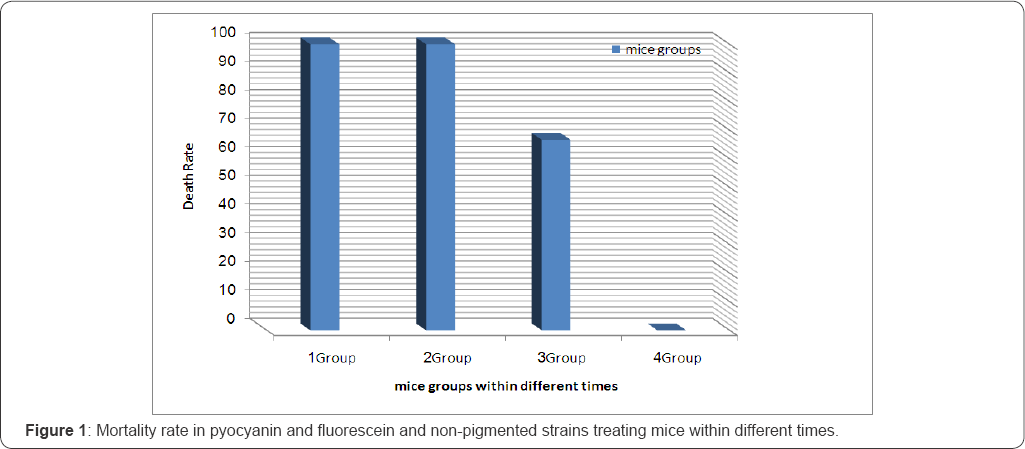
Pseudomonas aeruginosa employ a large virulence armamentarium to overcome host defenses, including the production and dispersal of Pyocyanin exotoxin and other phinazine molecules that are toxic to their hosts. The aim of the present study is to evaluate the mice killing capacity of different clinical isolates of pigmented and non-pigmented Pseudomonas aeruginosa. Three reference isolates isolated previously from otitis media and otitis external (pyocyanin highly producer, fluorescein highly producer, non-pigmented strain) where chosen to be inoculated intra peritoneally in mice. The results of the present study showed that the Mortality occurred within 24h in group one (pyocyanin producer) by 100% of mortality rate and within 48h in group two (fluorescin producer strains) by 100% of mortality rate whereas mortality occurred in group three (non-pigmented strains) at the end of 96h post infection by 66.6% of mice death when all compared with control group (Intra peritoneally saline injection). Our study concludes the highly significant mice killing capacity of highly pyocyanin P. aeruginosa producer when compared to other pigmented and non-pigmented and these different isolates retain the capability to develop otitis media.
Keywords: Pseudomonas aeruginosa, Pyocyanin , Fluorescin, Killing Virulence, pigmented and non-pigmented, , mice
Introduction
Pseudomonas aeruginosa is an opportunistic pathogen that causes extensive morbidity and mortality in individuals who are immune compromised or have underlying medical conditions such as, urinary tract, respiratory tract and skin infections and primarily causes of nosocomial infections [1]. It's a non sporulating, gram negative, oxidase positive motile bacterium with a polar flagellum [2]. P aeruginosa is a common nosocomial pathogen because it is capable of thriving in a wide variety of environmental niches [3]. It is a leading cause of hospital associated infections in the seriously ill, and the primary agent of chronic lung infections in cystic fibrosis patients [4]. They exist in very large numbers in the human environment and animal gut, they are capable of inhabiting/contaminating water, moist surface and sewage, hospital environment usually have resident P. aeruginosa [5].
Despite the apparent ubiquity of P. aeruginosa in the natural environment and the vast array of potential virulence factors, the incidence of community-acquired infections in healthy subjects is relatively low. However, in the hospital environment, particularly in immune suppressed, debilitated and burns patients, the incidence of P. aeruginosa infection is high [6].
It produces many numbers of extracellular toxins, which include phytotoxic factor, pigments, hydrocyanic acid, proteolytic enzymes, phospholipase enterotoxin, exotoxin and slim [1]. P aeruginosa grows well on media and most strains elaborate the blue phenazine pigment pyocyanin and fluorescein (yellow), which together impart the characteristic blue-green coloration to agar cultures [5]. Pyocyanin is a blue redox-active secondary metabolite [7], which induces rapid apoptosis of human neutrophils, with a10 fold acceleration of constitutive neutrophil apoptosis in vitro but no apoptosis of epithelial cell or macrophages [8]. The redox active exotoxin pyocyanin is produced in the concentration up to 100mol/l during the infection of CF patients and other bronchiectatic airways. The contributions of pyocyanin during infection of bronchiectatic airways are not appreciated [9]. Notably pyocyanin mediated ROS inhibit catalase activity, deplete cellular antioxidant reduced glutathione and increased the oxidized reduced glutathione in the bronchiolar epithelial cell [10,11]. Excessive and continuous production of ROS and inhibit of antioxidant mechanisms overwhelm the antioxidant capacity, leading to tissue damage, also pyocyanin inhibit ciliary beating of the airway epithelial cell [12]. Pyocyanin also increases apoptosis and inactivates 1-protease inhibitor [13]. Reducing agents such as GSH and NADPH can reduce pyocyanin to pyocyaninradical, which then mono-or divalently reduce O2 to form superoxide anion O2 or H2O2 [14]. Pyoverdin per contra is the main siderophore in iron gathering capacity its function as a powerful iron chelator, solubilizing and transporting iron through the bacterial membrane via specific receptor proteins at the level of outer membranes. Pyoverdin is important because it has a high affinity for iron, with an affinity constant of 10(32) [15]. Moreover, has been shown to remove iron from transferring in serum, probably assisting growth within, and ultimate colonization of the human host by P. aeruginosa [16]. Moreover experiments studying the burned models of P. aeruginosa infections have shown that ferric-pyoverdine is reuired infection and /or colonization, underlining the importance of ferric-pyoverdin to virulence of P. aeruginosa [15]. P. aeruginosa it is highly resist to antibiotics this resistance can be conferred by the outer membrane which provides an effective intrinsic barrier in the cell wall (or) cytoplasmic membrane (or) within the cytoplasm and modifications in outer membrane permeability via alternations in porin protein channel represent a component of many resistance mechanisms. In addition in activating enzymes released from the inner membrane can function more efficiently within the confines of the periplasmic space, the mechanisms by which intracellular concentrations of drugs are limited include decreased permeability through the outer membrane and active efflux back out across the cytoplasmic membrane [17].
The production of β-lactamase is the most prevalent mechanisms of resistance to (β-lactam antibiotics, the (β-lactamase have been reported to hydrolyze all anti-pseudomonal agents. Moreover, P. aeruginosa cell particularly in patients with chronic infections can develop a bio-film, in which bacterial cells are enmeshed into amucoidexopolysaccharide becoming more resistant to beta-lactams as well as decrease the outer membrane permeability that enable bacteria to gain resistance development [17,18].
Materials and Methods
Bacterial isolates
Three reference isolates isolated previously from otitis media and otitis externa (pyocyanin highly producer, fluorescein highly producer, non-pigmented strain) where chosen to be inoculated in mice. All strains passed in mice to retain their virulence. Stock cultures were maintained at 70 °C in brain heart infusion broth containing 5% glycerol.
Laboratory animals
Swiss albino male mice were purchase from (institute of biological and pharmaceutical research laboratory, Baghdad) aged 4-8 week and weigh 22-30gm were bred at animal breeding house at the College of Science, Tikrit University, all mice were kept at 22-25 °C in plastic cage and fed pellet and water every day.
Experimental infection
Swiss albino mice treated with multiple previously referenced isolates of P. aeruginosa (highly pyocyanin producer isolates, fluorescein producer andnon-pigmented isolates). Bacterial culture adjusted to 0.5 Mcfarland and each mice (5 in each group) challenged in traperitoneally with 1ml of bacterial suspension and mortality rate calculated for 5 days and in compared with control (injected only with normal saline).
Result and Discussion
Effect of pigmented P. aeruginosa on the laboratory animals
The results of the present study showed that the Mortality occurred within 24h in group one (pyocyanin producer) by 100% of mortality rate and within 48h in group two (fluorescin producer strains) by 100% of mortality rate whereas mortality occurred in group three (non-pigmented strains) at the end of 96h post infection by 66.6%) of mortality rate when all compared with control group (Intraperitoneally saline injection). The present results are in agreement with Al-shamaa et al. [19] that elucidate pyocyan in is the important virulence factor among many virulence factors of P. aeruginosa which caused the death of injured rat within 24h. Where aspyoverdin treated rat death within 4h, pyocyanin also alter specific immune defenses and potentiates and per pretauates harmful inflammatory reactions in the infected cystic fibrosis. O'Malley et al. [20] also recorded that pyocyanin exhibits paradoxical pro-oxidant property. Azwitter ion that can easily penetrate biological membranes, pyocyanin can directly accept electrons from reducing agent such as NADPH and reduced glutathione, then transfer the electrons to oxygen to generate ROS such as peroxide and single oxygen, also in harmony with Finlayson et al. [21] who elucidate pigmented strains of P. aeruginosa were highly virulence than non pigmented strains. Furthermore, virulence factor is produced in large ratio than non pigmented strain in which pigmented strains produce significant more (P<0.05) DNase, elastase, protease and siderophore. Pyocyanin is the highest virulence factor which altered the host immune response in several ways to aid evasion of immune system and establish chronic infection, evidence suggest that pyocyanin could prevent the development of an-effective T-cell response against P. aeruginosa and prevent activation of monocyte and macrophage [22], also pyocyanin in neutrophils induce a sustained increase in ROS and subsequent decrease in intracellular Camp, which triggers the time and concentration dependent acceleration of apoptosis [8]. As confirm in studies using wild type and isogenic pyocyanin deficient mutant P. aeruginosa, pigment dependent acceleration of neutrophil apoptosis and admonished release of chemokine might represent an immune suppression mechanism of the pathogen [23]. The fundamental ability of pyocyanin to alter the redox cycle and increase oxidative stress appear central to its divers detrimental effect on host cell, for example pyocyan in disrupt Ca+2 homeostasis in human airway epithelial cells by oxidant-dependent increases in inosited triphosphate and abnormal releases of Ca+2 from intracellular stores, because Ca+2 is important for regulating ion transport, secretion and ciliary beat. These alterations probably have important ramification for P. aeruginosa lung infection [24].

Also pyocyanin function as inhibitor of ATPase and this explains the pyocyanin toxicity including ciliary dysmotility, disruption of calcium homeostasis and dimished apical membrane localization of the cystic fibrosis trans-membrane conductance regulator (CFTR) [25]. Other potential toxic effects of pyocyanin include preturbance of cellular respiration, epidermal growth inhibition, prostacyclin release from lung endothelial cell and alter balance of protease-antiprotease activity in the cystic fibrosis lung [10,11]. The pro-oxidant effect of pyocyan in can thus augment such innate immune response circuits, for example, pyocyanin increases the release of the neutrophil chemokine (IL-8) from lung epithelial cells and up regulates the expression of the neutrophil receptor intracellular adhesion molecule (ICAM-1) [26,27]. In spite of all above toxic effects of pyocyanin, pyocyanin producer strains show highly virulence because pyocyanin act as a signaling molecule for quorum sensing regulation, which is regulated virulence factor expression [10], in spite of also pyoverdin (PVD) importance virulence factor which is function as a powerful iron chelators solubilizing and transporting iron through the bacterial membrane via specific receptor process before it reaches its targets Oberhardt [29]. Elucidate that PVD is essential element in vivo iron gathering and virulence expression in P. aeruginosa who found that PVD deficient mutants demonstrated no virulence when injected into burned mice [27-32] (Figure 1).
References
- Pollack M (2000) Pseudomonas aeruginosa. In principle and practice of infection Diseases. In: Mandell GL, Bennett JE Dolin R (Eds.), Philadelphia Churchill Livingstone, USA, pp. 2310-2335.
- Akanji BO, Ajele A, Onasanya Oyelakin O (2011) Genetic Fingerprinting of Pseudomonas aeruginosa Involved in Nosocomial Infection as Revealed by RAPD-PCR Markers. Biotech 10(1): 70-77.
- Romling U, Wingender J, Muller H, Tummler B (1994) A major Pseudomonas aeruginosa clone common to patients and aquatic habitats. Appl Environ Microbiol 60(6): 1734-1738.
- Brooks GF, Butel JS, Morse SA (2001) Jawetz, Melnik and Adelberg's. Medical Microbiology. Medical East edn, Appleton Large, USA, pp. 229-231.
- Anderlini P, Przepiorka D, Champlin R, Korbling M (1996) Biologic and clinical effects of granulocyte colony-stimulating factor in normal individuals. Blood 88(8): 2819-2825.
- Holder IA (1977) Epidemiology of Pseudomonas aeruginosa in a burn hospital. In Pseudomonas aeruginosa: Ecological Aspects and Patient Colonization. In: Young VM (Ed.), Raven Press, New York, USA, pp.77- 95.
- Xu H, Lin W, Xia H, Xu S, Li Y, et al. (2005) Influence of ptsP gene on pyocyanin production in Pseudomonas aeruginosa. FEMS Microbiol 253(1): 103-109.
- Usher LR (2002) Induction of neutrophil apoptosis by the Pseudomonas aeruginosa exotoxin pyocyanin: a potential mechanism of persistent infection. J Immunol 168(4): 1861-1868.
- Zychlinsky A, Sansonetti P (1997) Perspectives series: host/pathogen interactions: apoptosis in bacterial pathogenesis. J Clin Invest 100(3): 493-495.
- Lau GW (2004) The role of pyocyanin in Pseudomonas aeruginosa infection. Trends Mol Med 10(12): 599-606.
- Lau GW, Hassett DJ, Britigan BE (2005) Modulation of lung epithelial functions by Pseudomonas aeruginosa. Trends Microbiol 13(8): 389397.
- Wilson RD, Sykes A, Watson D, Rutman A, Taylor GW, et al. (1988). Measurement of Pseudomonas aeruginosa phenazine pigments insputum and assessment of their contribution to sputum sol toxicity for respiratory epithelium. Infect Immun 56(9): 2515-2517.
- Shellito J, Nelson S, Sorensen RU (1992) Effect of pyocyanine, a pigment of Pseudomonas aeruginosa, on production of reactive nitrogen intermediates by murine alveolar macrophages. Infect Immun 60(9): 3913-3915.
- Cheluvappa R, Jamieson HA, Hilmer SN, Muller M, LeCouteur DG (2007). The effect of Pseudomonas aeruginosa virulence factor,pyocyanin, on the liver sinusoidal endothelial cell. J Gastroenterol Hepatol 22(8): 1350-1351.
- Meyer JM, Neely A, Stintzi A, Georges C, Holder IA (1990) Pyoverdin is essential for virulence of Pseudomonas aeruginosa. Infect Immun 64(2): 518-523.
- Cox CD (1985) Iron transport and serum resistance in Pseudomonas aeruginosa. Antibiot Chemother 36: 1-12.
- Giamarellou H, Antoniadou A (2001) Antipseudomonal Antibiotics. Medical Clinics of North America 85(1).
- Henwood CJ, Livermore DM, James D, Warner M (2001) Antimicrobial Chemotherapy Disc Susceptibility Test. J-Antimicrob-chemother 47(6): 789-799.
- Bonfiglio G, Carciotto V, Russo G, Stefani S, Schito G, et al. (1998) Antibiotic Resistance in P aeruginosa: an Italian Survey. J AntimicrobChemother 41(2): 307-310.
- Al-Shamaa SD, Bahjat SA, Nasir NS (2011) Production of extracellular pigments as a virulence factor of Pseudomonas aeruginosa. colle of edu resear J 11: 689-697.
- O'Malley YQ, Reszka KJ, Spitz DR, Denning GM, Britigan BE (2004) Pseudomonas aeruginosa pyocyanin directly oxidizes glutathione and decreases its levels in airway epithelial cells. Am J Physiol Lung Cell Mol Physiol 287(1): 94-103.
- Finlayson EA, Brown PD (2011) Comparison of Antibiotic Resistance and Virulence Factors in Pigmented and Non-pigmented Pseudomonas aeruginosa. West Indian Med J 60(1): 24-31.
- Winstanley C, Fothergill JL (2008) The role of quorum sensing in chronic cystic fibrosis Pseudomonas aeruginosa infections. FEMS Microbiol Lett 290(1): 1-9.
- Allen L (2005) Pyocyanin production by Pseudomonas aeruginosa induces neutrophil apoptosis and impairs neutrophilmediated host defenses in vivo. J Immunol 174(6): 3643-3649.
- Denning GM (1998) Pseudomonas pyocyanin increases interleukin-8 expression by human airway epithelial cells. Infectlmmun 66(12): 5777-5784.
- Kong F (2006) Pseudomonas aeruginosa pyocyanin inactivates lung epithelial vacuolar ATPase-dependent cystic fibrosis transmembrane conductance regulator expression and localization. Cell Microbiol 8(7): 1121-1133.
- Look DC (2005) Pyocyanin and its precursor phenazine-1-carboxylic acid increase IL-8 and intercellular adhesion molecule-1expression in human airway epithelial cells by oxidant-dependent mechanisms. J Immunol 175(6): 4017-4023.
- Xie L, Bourne PE (2008) Detecting evolutionary relationships across existing foldspace, using sequence order-independent profile-profile alignments. Proc Natl Acad Sci USA 105(14): 5441-5446.
- Merriman TR, Merriman ME, Lamont IL (1995) Nucleotide sequence of pvdD, a pyoverdine biosynthetic gene from Pseudomonas aernos Pav: d D has similariw to peptide synthetases. J Bacteriol 177: 252-258.
- Oberhardt MA, Puchalka J, Fryer KE, Martins dos Santos VA, Papin JA (2008) Genomescale metabolic network analysis of the opportunistic pathogen Pseudomonas aeruginosa PAO1. J Bacteriol 190(8): 27902803.
- Stover CK, Pham XQ, Erwin AL, Mizogushi SD, Warrener P, et al. (2000) Complete genome sequence of Pseudomonas aeruginosaPAO1, an opportunistic pathogen. Nature 406: 959-964.
- Voggu L, Schlag S, Biswas R, Rosenstein R, Rausch C, et al. (2006) Microevolution of cytochrome bd oxidase in Staphylococci and its application in resistance to respiratory toxins released by Pseudomonas. J Bactriol 188(23): 8079-8086.


