Green Synthesis, Characterization of Metal Antibacterial Nanoparticles and Removal of Fluoride from Waste Water
Shabnam S Shaikh1, Meenu S Saraf1* and Yadavendra K Agrawal2
1Depatment of Microbiology and Biotechnology, Gujarat University, Gujarat
2Institute of Research and Development, Gujarat Forensic Science University, Gujarat
Submission: February 06, 2017; Published: March 06, 2017
*Corresponding author: Meenu Saraf, Department of Microbiology and Biotechnology, School of sciences, Gujarat University Ahmedabad, Gujarat, Tel: +91-79-26303225; Fax: +91-79-26851704; Email: sarafmeenu@gmail.com
How to cite this article: Shabnam S S, Meenu S S, Yadavendra K A. Green Synthesis, Characterization of Metal Antibacterial Nanoparticles and Removal of Fluoride from Waste Water. 006 Adv Biotech & Micro. 2017; 2(3): 555590. DOI:10.19080/AIBM.2017.02.555590
Abstract
In the present study stable ZnO nanoparticles were biologically synthesized by using microbial strains, the average particle size and charge potential varies in all the three preparation, which was 328.3 d.nm with -3.09 charge potential in solution synthesized by A. terrus, followed by A. niger with particle size 633 d.nm with -3.97 charge potential and particle size 870 d.nm with +2.59 charge potential in solution synthesized by P. aeruginosa. The particle size of ZnO nanoparticle from A. niger after sonication ranges from 68-100 d.nm. The structural morphology and fluorescence properties were characterized by zeta sizer, UV-Vis spectrophotometer, spectrofluorometer and scanning electron microscopy (SEM).
Nanoparticles were evaluated for their antibacterial properties and decontamination of fluoride from waste water. Nanoparticles shows toxicity against various Gram positive and Gram negative bacterial cultures with strong antibacterial activity against B. subtilis establishing 14 mm zone of inhibition. Interactions of ZnO nanoparticles with Standard sodium fluoride and fluoride containing waste water were estimated using spectrofluorescence. Results revealed that a fluoride complex with ZnO nanoparticle and thus the adsorption of fluoride is achieved up to 65%from waste water concentration. Thus biologically synthesized zinc oxide nanoparticle can be effectively used as an antibacterial compound and in decontamination of fluoride from waste water.
Keywords: Nanoparticles; Zinc oxide nanoparticle; Antibacterial activity
Introduction
Green chemistry can be recognized as suitable environmentally friendly procedures, which widely reports intrinsic use of non-hazardous, inexpensive and stable materials with their multiple applications [1]. The material which is natural or manufactured possessing an external diameter in the range of 1-100 nm is considered as nanoparticles. In recent years nanotechnology is associated approaching field contributing the simple procedures with minimizing the chemical use with distinctive properties [2]. Green synthesis of nanoparticles is proved to be valuable alternatives as compared to conventional chemical methods using expensive and harmful reducing agents [3].
Among inorganic materials metal oxide nanoparticles are significantly important due to their distinct properties such as catalytic, high mobility, wide electrochemical, optical, magnetic and electrical properties [4]. Zinc oxide belonging to a group of metal oxides is characterized by photo catalytic and photo oxidizing ability against various living cells [5]. Zinc oxide nanoparticles when synthesized by biological methods are inexpensive [6]. ZnO nanoparticles have wide range of applications including, sensors piezoelectric, transparent conductors, surface wave devices, and in biomedical as well as chemical industry' [7]. Exploiting the nanoparticles in various field have attracted much attention, with their unique properties including antimicrobial properties for acceptable applications [8]. Nanoparticles are reported to possess antimicrobial properties against several microorganisms including bacteria, fungi and certain extremophiles [9]. The properties of ZnO nanoparticle is strongly influenced by the particle size and mechanism of cell inhibition. The properties includes disruption of cell membrane, altering the permeability, electrostatically binding to the cell surface and accumulation of nanoparticle in cytoplasm [10].
Nanoparticles of metal oxide plays efficient role in waste water industry as a photocatalyst and can degrade both chemical and biological contamination [11]. ZnO nanoparticle shows photo catalytic activity with large number of active sites, where higher area is available and more absorption of the target molecule is achieved. ZnO nanoparticle is an attractive material for waste water treatment as it can absorb visible light and is capable of binding with many ions. It has effective properties than other metal oxides like TiO2 in water purification and other environmental remediation process. Considerable scientific interest is recorded for the removal of harmful chemical contaminant with the use of ZnO nanoparticles [12].
Materials and Methods
All the media and chemicals were obtained from Himedia Laboratories and Sigma Aldrich USA of analytical grade. ZnO nanoparticles were synthesized by fungal strain of Aspegillus terrus using biological procedure. The characterization of the nanoparticle was studied by U.V-Visible spectrophotometer, zeta sizer, and scanning electron microscopy (SEM) Antibacterial study of nanoparticles were carried out against Gram positive and Gram negative bacteria [8]. Florescence intensity of ZnO Nanoparticle was recorded in Spectrofluorometer (JASCO, Japan) and adsorption of fluoride with the assistance of nanoparticles was achieved from standard fluoride solution (sodium fluoride) and fluoride containing waste water sample.
Green synthesis of ZnO nanoparticles
The microbial strains were isolated from rhizospheric soil of Ahmedabad at 23.0793° N latitude and 72.4957° E longitude. The isolates were identified on the basis of 16s r RNA and 18s R RNA sequence as Pseudomonas aeruginosa, Aspergillus terrus and Aspergillus niger deposited in gene bank under the accession number: KF984467, KU962937 and KU962935 respectively. These microbial species were screened for green synthesis of nanoparticles by the published method [13]. The Microbial cultures were activated in their basal medium and then inoculated into Pikovskaya's broth.
They were allowed to grow for six days on rotatory shaker (200 rpm at 30 °C) and later treated with 1.0% NaCl for inoculum preparation. Twenty-five ml of this inoculum was diluted with 75ml of modified Pikovskaya's broth. It was amended with 0.1% ZnO and allowed to grow for ten days on rotatory shaker (200 rpm). These samples were centrifuged for 20 min at 10,000 rpm and the resulting supernatant was probe sonicated and analyzed for nanoparticle size and charge [6]. Using zeta seizer (Malvern, U.K). The synthesized nanoparticles from all the three cultures were probe sonicated at 30 amplitudes for 30 sec, 30 amplitude for 1min and 50 amplitude for 1min to select the optimum combination where the nanoparticle size is 100d.nm.
Among the nanoparticles synthesized, A. Terrus was most efficient and stable for 10 days. They were selected for the further optimization with respect to the different concentration of ZnO (100 mg and 200 mg) added in growth medium and Days of incubation (8 and 10) for the nanoparticle synthesis.
Characterization of ZnO nanoparticles
U. V spectrophotometer analysis: The optical absorbance spectra of ZnO nanoparticles were recorded in double beam Shimadzu UV-1800 at room temperature in the wavelength from 200 to 800 nm.
FT-IR analysis: The spectrum of ZnO nanoparticles were characterized by Fourier Transform - Infrared Spectrometer (Perkin Elmer Spectrum GX Range)The FT-IR spectrum was analyzed between the wave number of 400 and 4000 cm-1.
Antibacterial properties of nanoparticles: Antibacterial properties of nanoparticle and complex with Sodium fluoride solution was evaluated against Gram positive (Bacillus subtilis, Bacillus cereus, Bacillus megaterium, Micrococcus luteus and Staphylococcus aureus) and Gram negative (E. coli and P. aeruginosa) bacterial cultures. Standard agar diffusion method was used to examine antibacterial activity, 100 |il of Nanoparticle containing solutions was poured into well and incubated at 37 °C for 24 hrs. The antibacterial activity was determined by zone of inhibition in mm [8].
Spectrofluorescence study of ZnO nanoparticles: Florescence intensity for ZnO nanoparticles were recorded in JASCO spectrophotometer with single emission, initially prescan was performed at wavelength of 400-700nm to determine excitation and emission. Further all analysis was carried out at excitation 400 nm and emission 450 nm with data pitch of 1nm and band width of 3 nm.
Spectrofluorescence of ZnO nanoparticles with NaF solution: The negatively charged ZnO nanoparticles were allowed to react with different concentration of 0.1% Sodium fluoride (NaF) standard solution to determine the florescence intensity of ZnF complex. The aliquots for nanoparticles were kept constant (40ml) and different concentration of NaF solution (from 1 ml to 10 ml) was added and further florescence spectral intensity of each mixture were recorded in spectrofluorometer. Further the effect of incubation time on reaction of ZnO nanoparticle with NaF solution was carried out where spectrofluorescence intensity was studied with respect to the incubation time after preparation of the mixture (ZnO and NaF solution) to determine the effect of incubation time in ZnF complex formation. The experimental sets were prepared and incubated for different time, 0 min, 15 min, 30 min, 45 min and 60 min respectively.
Adsorption of fluoride from waste water
The waste water sample was obtained from chemical industry effluent from Gujarat Industrial Development Corporation Ahmedabad having 100-200 mg concentration of fluoride. The adsorption of fluoride from waste water was carried out with treatment of ZnO nanoparticles. Different aliquots of waste water (1 ml -10 ml) were treated with constant volume of nanoparticles (40 ml) and fluorescence intensity was recorded for each sample. Waste water without addition of nanoparticles was kept as control. Analysis of the residual fluoride concentration was carried out on a UV-vis spectrophotometer according to the procedures outlined in Standard methods of APHA [14].
Result and Discussion
Green synthesis of nanoparticles
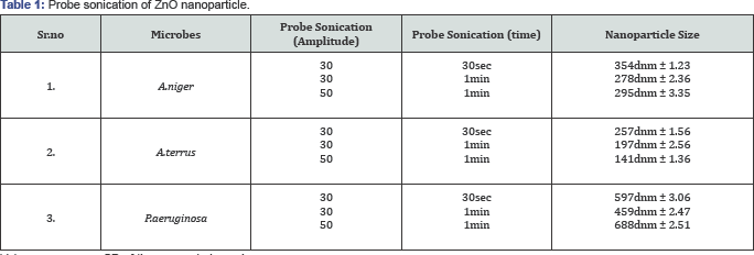
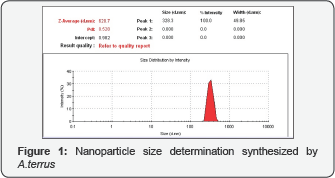
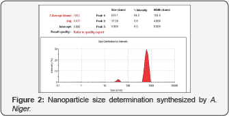
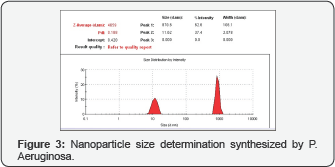
Three microbial samples were screened for green synthesis of nanoparticles including one bacterium and two fungi, Pseudomonas aeruginosa, Asperigillus terrus and Aspergilus niger respectively. After synthesis, their particle size and charge potential were carried out by zeta seizer. The average particle size and charge potential varies in all the three preparation, which was 328.3 dnm with -3.09 charge potential in solution synthesized by A. terrus, followed by A. niger with particle size 633 dnm with -3.97 charge potential and particle size 870 dnm with +2.59 charge potential in solution synthesized by P aeruginosa (Figure 1-3) There are reports on green synthesis of nanoparticles, where soluble starch was used for the green synthesis of nanoparticles [15]. Shamsuzzan et al. [6] also reported the green synthesis of nanoparticle using Candida albicans as eco-friendly reducing agent [6]. To study the spectrofluorescence of ZnO nanoparticle, the particle size was further decreased with the assistance of probe sonication with different amplitude at different incubation time (Table 1). The nanoparticle size 141dnm was obtained by A. terrus with probe sonication of 50 amplitude for 1min.
Values are mean ± SD of three sample in each group.
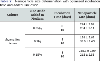
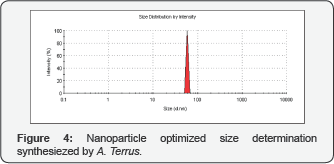
Optimization of nanoparticles synthesis by A. Terrus: After the synthesis of nanoparticles, further optimization of the synthesis process was carried out where the condition for the synthesis process was kept constant. The conditions altered was incubation time and zinc oxide concentration, which was altered as 8 and 10 days and 0.05%, 0.1, 0.15%, respectively (Table 2). The nanoparticle size less than 100 d nm was obtained (68 d nm) (Figure 4&5), with the condition where A. terrus was used, with the incubation time 10 days, 0.1% ZnO and probe sonication constant at 50 amplitude for 1min
Values are mean ± SD of three sample in each group.
Characterization of ZnO nanoparticles
UV-Vis spectrophotometer analysis: The UV-Vis absorption spectrum demonstrates the unique method for the determination of ZnO nanoparticles in basal medium and the reference uninoculated. The sharp typical absorption peak was obtained at 381 nm corresponding to the characteristics of ZnO nanoparticles. ZnO nanoparticle shows a sharp absorption band at 362nm which confirms the synthesis of nanoparticles [8].
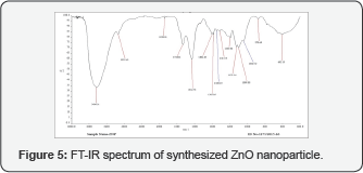
The FT-IR absorption spectra of the synthesized ZnO nanoparticle: The spectrum of biologically synthesized nano material obtained shows the peak at 603.18 cm-1 which is the characteristic absorption of Zn-O bond and the broad absorption peak at 3,463.21 and 2,935.62 cm-1 indicate the presence of -OH and C=O residues, indicating presence of atmospheric moisture and CO2 respectively. The literature shows the Zn-O absorption band near 430 cm-1 and the peaks at 3,450 and 2,350 cm-1 showing the presence of -OH and C=O residues. Hong et al., 2008 also reported the Zn-O spectrum peak at 472.54 cm-1 which is the characteristic absorption of Zn-O bond and the broad absorption peak at 3438.26 cm-1 can be attributed due to absorption of hydroxyls [16].
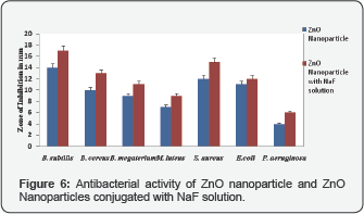
Antibacterial properties of nanoparticles:Antibacterial properties of nanoparticles were studied against Gram positive (B. subtilis, B. cereus, B. megaterium, M. Luteus and S. aureus) and Gram negative (E.coli and P. aeruginosa) bacterial cultures. Antibacterial activity was studied against both Gram positive and Gram negative culture using as the zone of inhibition (Figure 6). Maximum zone of inhibition was observed against B. subtlis 14 mm and minimum inhibition was observed against P. aeruginosa. Antibacterial properties of ZnO nanoparticle of E. coli, P.aeruginosa and S. aureus was reported by Premanthan et al. [8]. Xia et al. [2] also observed antibacterial properties of nanoparticle, where zone of inhibition increases with increase in concentration of nanoparticles. In both Gram positive and Gram negative bacteria, cell wall and cell membrane are crucial in maintaining the osmotic balance and integrity of cell. Damage of bacterial cell integrity could be the reason in present study [17].
Spectrofluorescence study of Nanoparticles: Florescence spectra were observed in spectrofluorometer from prescan the exitation at 400 nm and emission 450 nm was kept standard. Further analysis was carried out at excitation and emission 400nm and 450nm respectively up to 600nm. The spectroflorescence spectrum of ZnO nanoparticles consists of a sharp peak at intensity of 123 with the particle size 68.0 dnm (Figure 4). A sharp peak was observed in visible range [18] and two emission peaks were observed in the similar spectrum reported by Shamsuzzaman et al. [6].
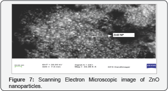
Scanning Electron microscopy of ZnO nanoparticle:The characterization of sample by SEM analysis shows the presence of ZnO nanoparticles of nm and forms the aggregates with approximate zeta potential of -3.09 (Figure 7). The results revealed where spherical structure of Zinc nanoparticle with diameter range of 11-25 nm forms an aggregates [19].
Spectrofluorescence of ZnO nanoparticles with NaF solution
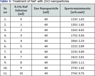
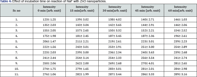
Experimental method was designed for adsorption of fluoride, where sodium fluoride (100 mg/100 ml) was taken as standard fluoride source and treated with zinc oxide nanoparticles with constant volume (40 ml). Fluorescence intensity showed that the as the nanoparticle reacts with fluoride thus fluorescence intensity increases. The nanoparticle was able to react with 8mg/ml of fluoride in solution after which the intensity remains constant (Table 3). The results of different incubation time show there was continuous increase in the fluorescence intensity up to 60 min, beyond which there was no difference in fluorescence intensity and Table 4 reported that a peak in a UV peak difference was found in the UV range is associated due to large binding energy of ZnO and reaction time [20].
Values are mean ± SD of three sample in each group.
Values are mean ± SD of three sample in each group.
Adsorption of fluoride from waste water
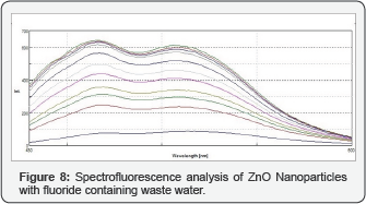
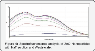
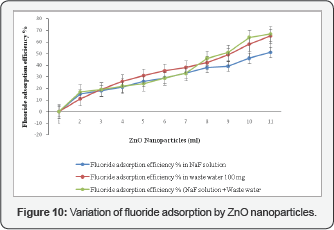
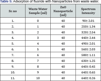
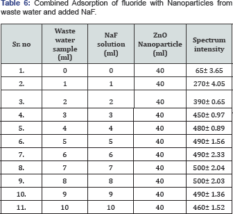
Waste water containing fluoride was collected as sample from the chemical manufacturing industry located in Ahmedabad (Gujarat, India). This sample was used for the adsorption study with ZnO nanoparticle. The sample mixture with nanoparticles was prepared with the above developed method. Spectrofluorescence intensity was recorded (Figure 8) and an evolved result indicates that the intensity increases with the increase in the fluoride concentration in the sample (Table 5). The combined adsorption of Fluoride from waste water and added 0.1% NaF solution was also studied (Figure 9) and found that with increasing fluoride concentration the nanoparticles still effectively react and get adsorb fluoride (Table 6). A typical peak at 553 nm was observed in nanoparticles, when exited at 380 nm [13]. Florescence peak of nanoparticles in water phase was reported at 465 nm [21]. The effective adsorption of fluoride was depicted in Figure 10, where the rate of fluoride adsorption was rapid throughout the difference aliquots. The adsorptive removal of fluoride was also shown by where initial adsorption was quick and then slowed due to the attainment of equilibrium, this could be due to the specific functional group and active sites [14].
Values are mean ± SD of three sample in each group.
Values are mean ± SD of three sample in each group.
Conclusion and Future Aspects
Aim of green chemistry is to minimize the use and generation of harmful chemicals and implementation of sustainable process with the adaption of various protocols suitable for environment. We have used the fungus, A. terrus to synthesize ZnO nanoparticles through biological route. These nanoparticles can be used for both chemical fluoride adsorption and inhibition of biological contamination. The antibacterial study of nanoparticles against Gram positive (Bacillus subtilis, Bacillus cereus, Bacillus megaterium, Micrococcus luteus and Staphylococcus aureus) and Gram negative (E.coli and P. aeruginosa) was studied, which shows the inhibition against the bacteria and thus can be exploited for the toxicity of other cells. The crucial outcome of the work includes removal of fluoride with the assistance of ZnO nanoparticles. Effective adsorption of fluoride was noticed from standard NaF solution (50%) and fluoride containing waste water (65%). From the above interaction experiments it can be concluded that, Fluoride gets absorbed by the ZnO nanoparticles, With the use of ZnO nanoparticles, this principle application can be further exploited to treat the waste water samples in contact time less than minutes, for adsorption of fluoride.
Acknowledgement
We are thankful to Department of Forensic Science, Gandhinagar for their assistance in SEM analysis and UGC-MANF for their financial assistance.
References
- Kumar A, Gupta MK, Kumar M (2012) l-Proline catalysed multicomponent synthesis of 3-amino alkylated indoles via a Mannich- type reaction under solvent-free conditions, Green Chem 14: 290-295.
- Xia Y, Yang P, Sun Y, Wu Y, Mayers B, et al. (2003) One-Dimensional Nanostructures: Synthesis , Characterization and Applications. Adv Mater 15(5): 353-389.
- Mason C, Vivekanandhan S, Misra M, Mohanty AK, Switchgrass (Panicum virgatum) (2012) Extract Mediated reen Synthesis of Silver Nanoparticles, World J Nano Sci Eng 2: 47-52.
- Welton T (1999) Room -Temperature Ionic Liquids. Solvents for Synthesis and Catalysis - Chemical Reviews (ACS Publications), Chem Rev 99: 2071-2083, doi: 10.1021/cr1003248.
- Szabo T, Nemeth J, Dekany I (2003) Zinc oxide nanoparticles incorporated in ultrathin layer silicate films and their photocatalytic properties, Colloids Surfaces A Physicochem Eng Asp 230: 23-25.
- Shamsuzzaman, Mashrai A, Khanam H, Aljawfi RN (2013) Biological synthesis of ZnO nanoparticles using C. albicans and studying their catalytic performance in the synthesis of steroidal pyrazolines, Arab J Chem doi:10.1016/j.arabjc.2013.05.004.
- Chanaewa, Juárez BH, Weller H, Klinke C (2012) Oxygen and light sensitive field-effect transistors based on ZnO nanoparticles attached to individual double-walled carbon nanotubes. Nanoscale 4(1): 251256.
- Premanathan M, Karthikeyan K, Jeyasubramanian K, Manivannan G (2011) Selective toxicity of ZnO nanoparticles toward Gram-positive bacteria and cancer cells by apoptosis through lipid peroxidation, Nanomedicine Nanotechnology. Biol Med 7(2): 184-192.
- Haeed A, Hasan MM, Raffi M, Hussain F, Bhatti TM, et al. (2008) Antibacterial Characterization of Silver Nanoparticles against E.coli ATCC-15224. J Mater Sci Technol 24(2): 192-196.
- Stoimenov PK, Klinger RL, Marchin GL, Klabunde KJ (2002) Metal Oxide Nanoparticles as Bactericidal Agents. Langmuir 18(17): 66796686.
- Baruah S, Pal SK, Dutta J (2012) Nanostructured Zinc Oxide for Water Treatment. Nano sci Technol 2: 90-102.
- Bianco- Prevot, Fabbri D, Pramauro E, Morales-Rubio A, Guardia M de la (2001) Continuous monitoring of photocatalytic treatments by flow injection. Degradation of dicamba in aqueous TiO2 dispersions. Chemosphere 44(2): 249-255.
- Stan M, Popa A, Toloman D, Dehelean A, Lung I, et al. (2015) Enhanced photocatalytic degradation properties of zinc oxide nanoparticles synthesized by using plant extracts, Mater Sci Semicond Process 39: 23-29.
- Ramanaiah SV, Mohan SV, Sarma PN (2007) Adsorptive removal of fluoride from aqueous phase using waste fungus (Pleurotus ostreatus 1804) biosorbent : Kinetics evaluation 31(1): 47-56.
- Vigneshwaran N, Ashtaputre NM, Varadarajan PV, Nachane RP, Paralikar KM, et al. (2007) Biological synthesis of silver nanoparticles using the fungus Aspergillus flavus. Mater Lett 61(6): 1413-1418.
- Hong R, Pan T, Qian J, Li H (2006) Synthesis and surface modification of ZnO nanoparticles. Chem Eng J 119(2-3): 71-81.
- PJP Espitia, N de FF Soares, JS dos R Coimbra, NJ de Andrade, RS Cruz, et al. (2012) Zinc Oxide Nanoparticles: Synthesis, Antimicrobial Activity and Food Packaging Applications. Food Bioprocess Technol 5(5): 1447-1464.
- Soares JW, Whitten JE, Oblas DW DW, Steeves DM (2008) Novel photoluminescence properties of surface-modified nanocrystalline zinc oxide: Toward a reactive scaffold. Langmuir 24(2): 371-374.
- Vidya C, Hiremath S, Chandraprabha MN, Antonyraj M a L, Venu I (2013) Green synthesis of ZnO nanoparticles by Calotropis Gigantea 2012-2014.
- Ha B, Ham H, Lee CJ (2008) Photoluminescence of ZnO nanowires dependent on O2 and Ar annealing. J Phys Chem Solids 69(10): 24532456.
- Jiang HL, Kwon JT, Kim YK, Kim EM, Arote R, et al. (2007) Galactosylated chitosan-graft-polyethylenimine as a gene carrier for hepatocyte targeting. Gene Ther 14(19): 1389-1398.


