An Unusual Complication of Height Loss in an Elderly Patient
Mala Anna Jones, Ashok Chaudhari, and Shobhana Chaudhari*
Department of Medicine, Metropolitan Hospital Center, NYC H+H/New York Medical College, USA
Submission: March 5, 2022; Published: March 14, 2022
*Corresponding author: Shobhana Chaudhari, Chief of Geriatrics/Department of Medicine Metropolitan Hospital Center/New York Medical College, 1901 First Avenue, New York, USA
How to cite this article: Mala Anna J, Ashok C, Shobhana C. An Unusual Complication of Height Loss in an Elderly Patient. OAJ Gerontol & Geriatric Med. 2022; 6(4): 555691. DOI: 10.19080/OAJGGM.2022.06.555691
Abstract
A tunneled dialysis catheter is used for intermediate to long term hemodialysis vascular access in End Stage Renal Disease (ESRD) patients without Arterio-Venous Fistula (AVF). The tip of Tunneled Dialysis Catheters (TDC) should ideally be positioned at the caval-atrial junction or within the upper right atrium to ensure optimal blood flow [1]. Here we present an unusual case of an ESRD patient whose catheter tip was seen in the right ventricle on radiological examination one year after placement. The migration was thought to have occurred because of shortening of the thoracic cavity due to a reduction in vertebral height.
Keywords: Tunneled dialysis catheter tip migration, Right ventricle, End stage renal disease, Elderly, Osteoporosis, Kyphoscoliosis, Loss of vertebral height, Arterio-Venous Fistula, secondary hyperparathyroidism. Osteoporosis
Abbreviations: ESRD: End Stage Renal Disease, AVF: Arterio-Venous Fistula, TDC: Tunneled Dialysis Catheter, SVC: Superior Vena Cava
Introduction
Up to a third of ESRD patients on hemodialysis use TDC usually as a bridge to a more permanent dialysis access [2,3]. Various complications are known to occur with dialysis catheter placement including hemothorax, pneumothorax, catheter thrombosis, venous thrombosis, infection and catheter tip migration. Catheter tip migration is commonly peripheral and less commonly central towards the base of right atrium or into the ventricle. Central migration can give rise to complications including atrial mural thrombus, perforation, arrhythmias, cardiac tamponade, pleural effusion and Superior Vena Cava (SVC) obstruction [4-6]. There has been a previous case report of delayed catheter tip migration into right ventricle causing SVC obstruction [7]. Here we describe an asymptomatic incidentally discovered case of dialysis catheter tip migration into the right ventricle caused by a loss vertebral height.
Case Presentation
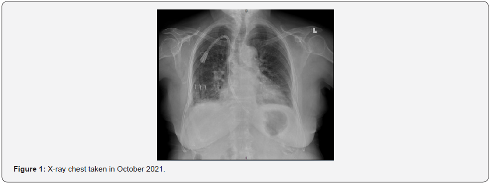
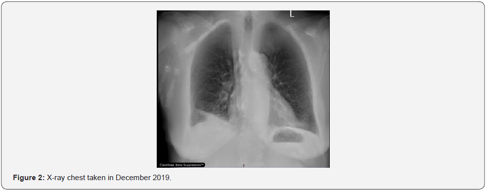
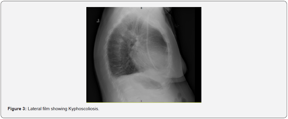
A 78-year-old female patient with mild kyphoscoliosis and ESRD on dialysis three times a week via right sided internal jugular tunneled catheter since January 2020 was admitted for evaluation of chest pain in October 2021. Work up for the chest pain was negative and it resolved with conservative measures. A chest X-ray taken during the work up demonstrated the tip of the catheter to be in the right ventricle [Figure1]. The catheter was inserted in December 2019. Review of radiological films from December 2019 showed the catheter tip to be in right atrium [Figure 2]. The distance between the point of insertion and the tip of catheter was measured by the radiologist and found to be the same in both instances demonstrating that the anchoring of the catheter by fibrosis into the cuff was intact. It was suggested that increase in Scoliosis and loss of vertebral column height could have shortened the thoracic cavity and caused the catheter tip to move downwards [Figure 3]. Accelerated bone loss due to old age and bone mineral disease associated with ESRD could have contributed. Patient’s height was measured and found to be 142cm against 149cm in 2019. She was otherwise stable and had no symptoms that could be attributed to catheter tip migration. She was advised to have her catheter removed but declined as she had no symptoms and opted to wait till her recently created AVF was matured.
Discussion
Loss of height due to osteoporosis is a part of natural aging process. With advancing age, osteoporosis weakens the bony structures and facilitates bone remodeling and rotatory deformities. Finally, aging of bone, discs, facets, ligaments, and muscles may ultimately lead to kyphoscoliosis and destabilization [8,9]. This process is accelerated in ESRD. In addition to the well described high-turnover bone disease caused by secondary hyperparathyroidism and low-turnover disease in the form of osteomalacia. Osteopenia also is present in end-stage renal disease patients. In contrast to abnormalities in the ability of bone to remodel, osteopenia is a deficiency in bone mass or volume [10]. Osteoporosis in ESRD is associated not only with increased fracture risk but also all-cause mortality [11]. Female gender is an additional risk factor [8]. In the above case, it is proposed that the catheter tip moved into right ventricle due to loss of height; an unusual complication of osteoporosis seen in an elderly patient. Timely replacement or repositioning of the dialysis catheter is the treatment of choice.


Conflict of interest:
No financial conflict of interest exists.
References
- Clinical Practice Guidelines for Vascular Access: update 2000. Am J Kidney Dis. (1 Suppl 1): S137-181.
- Rayner HC, Pisoni RL (2010) The increasing use of hemodialysis catheters: evidence from the DOPPS on its significance and ways to reverse it. Seminars in dialysis 23(1): 6-10.
- Aitken EL, Stevenson KS, Gingell-Littlejohn M, Aitken M, Clancy M (2014) The use of tunneled central venous catheters: inevitable or system failure. The journal of vascular access 15(5): 344-350.
- Paw H (2002) Bilateral pleural effusions: unexpected complication after left internal jugular venous catheterization for total parenteral nutrition. British journal of anesthesia 89(4): 647-650.
- Thomas CJ, Butler CS (1999) Delayed pneumothorax and hydrothorax with central venous catheter migration. Anesthesia 54(10): 987-990.
- Collier PE, Goodman GB (1995) Cardiac tamponade caused by central venous catheter perforation of the heart: a preventable complication. Journal of the American College of Surgeons 181(5): 459-463.
- Muhammad U Sharif, Kottarathil A Abraham (2020) Central Migration of Tunneled Dialysis Catheter into Right Ventricle causing Positional Superior Vena Cava Obstruction: A Case Report. Annal Urol & Nephrol 2(2).
- Shimizu M, Kobayashi T, Chiba H (2020) Adult spinal deformity and its relationship with height loss: a 34-year longitudinal cohort study. BMC Musculoskelet Disord 21(1): 422.
- Benoist M. (2003) Natural history of the aging spine. Eur Spine J 12: S86–S89.
- Lindberg JS, Moe SM (1999) Osteoporosis in end-state renal disease. Semin Nephrol 19(2): 115-122.
- Iseri K, Dai L, Chen Z, Qureshi AR, Brismar TB et al. (2020) Bone mineral density and mortality in end-stage renal disease patients. Clin Kidney J 13(3):307-321.


