Epidemiological, Clinical, Biological and Etiological Aspects of Hypochromic Microcytic Anemia in Aged Persons
Mzabi A, Marrackchi W*, Alaya Z, Ben Fredj F, Anoun J, Rezgui A and Kechrid Laouani C
Service de médecine interne CHU Sahloul Sousse, Tunisie
Submission: August 28, 2018; Published: September 12, 2018
*Corresponding author:Wafa Marrakchi, Medical doctor, Sousse, Tunisia, Sahloul hospital Tunisia, Africa.
How to cite this article: Mzabi A, Marrackchi W, Alaya Z, Ben Fredj F, Anoun J, Rezgui, Kechrid Laouani C . Epidemiological, Clinical, Biological and Etiological Aspects of Hypochromic Microcytic Anemia in Aged Persons. OAJ Gerontol & Geriatric Med. 2018; 4(4): 555643. DOI: 10.19080/OAJGGM.2018.04.555643
Abstract
Purpose: Hypochromic microcytic anemia is a common hematological abnormality in the older people. The aim of this study is to determine the epidemiological, clinical and biological characteristics and causes of the hypochromic microcytic anemia in elderly patients.
Patients and Methods: We performed a retrospective study of 124 patients aged 65 years and older who were hospitalized for hypochromic microcytic anemia in the internal Medicine Department.
Result: Sixty nine women and fifty five men were included in the study. The mean age was 75 years and 4 months. Iron deficiency anemia was diagnosed in 72 cases (58.1%). A gastrointestinal bleeding was the most common etiology (59.7 %). Anemia was poorly tolerated in 30.5 % of cases. Serumiron and ferritin were low in the majority of cases. An inflammatory origin of anemia diagnosed in 52 cases (41.9%). Cancers and thromboembolic causes were the most frequent etiology in these cases. The multifactorial anemia was in 15 of the patients (12%).
Conclusion: A comprehensive history, physical examination, and laboratory evaluation are required for an elderly person found to have anemia.
Keywords: Hypochromic Microcytic Anemia; Elderly; Etiology; Clinical Characteristics
Mini Review
Aging is the set of organic, physiological and psychological changes that occur with advancing age. The World Health Organization (WHO) defines the elderly subject (ES) as a person whose age is greater than or equal to 65 years old [1]. Aging is related to several health problems affecting different organs and making the geriatric pathology varied and difficult to manage. These problems include anemia, which remains the most common hematologic problem in geriatrics [2]. Literature data shows the increase in the prevalence of anemia with age and especially after 65 years old. In fact, 11% of men and 10.2% of women over 65 are anemic [3,4]. Hypochromic Microcytic Anemia (HMA) is the most common type in ES. The objective of this work is to determine, the epidemiological, clinical and biological profile according to the etiology of the HMA of elderly patients hospitalized in the department of internal medicine of the University Hospital Center (UHC) Sahloul during the period from January 1999 to January 2015.
Patients and Methods
A cross-sectional study including patients whose age was greater than or equal to 65 years old, hospitalized for HMA in the internal medicine department of UHC Sahloul de Sousse during the study period from January 1999 to January 2015 regardless of their medical and surgical history. The data was collected from patients’ medical records using a form for this study, which included epidemiological, clinical, biological and etiological data. In our study, anemia is defined according to the WHO by a rate of hemoglobin (Hb) less than 13g/dl in men and 12 g/dl in women [1]. Anemia is called microcytic hypochromia if the Mean Hemoglobin Corpuscular content (MHCC) and the Mean Corpuscular Volume (MCV) are respectively less than 27 pg and 80 fl / l.
Results
Between January 1999 and January 2015, the total number of elderly, anemic subjects hospitalized in the internal medicine department of CHU Sahloul; was 625. Among them, 124 patients (19.8%) had an HMA. It was 7.5% of all ES hospitalization grounds in our department. The average age of patients was 75.4 years old (65-97 years old). The most common age range was between 76 and 80 years (60%). Our study included 55 men (44.4%) and 69 women (55.6%), the sex ratio was 0.8.
Iron Deficiency Anemia
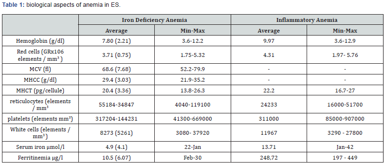
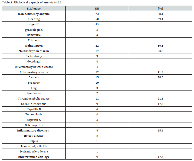
Iron Deficiency Anemia (IDA) was observed in 72 patients (58% of cases). Among them, there were 42 women (58%) and 30 men (42%); the sex ratio was 0.76. Anemic syndrome was observed in 96% of cases. The most common clinical signs were cutaneo-mucous pallor (84.7% of cases) and asthenia (66.7% of cases). The most common signs of intolerance for anemia were poorly tolerated tachycardia (37.5% of cases) followed by rest dyspnea (19.4% of cases) and cardiac arrhythmias (19.4%). Anemia was poorly tolerated in 30.5% of patients. Of the latter, 18 had a history of cardiorespiratory pathology (Table 1). summarizes the biological characteristics of patients with IDA. Blood spoliation was the most common cause of IDA (69.4% of AF cases). The different etiologies are grouped in Table 2.


Inflammatory Anemia (IA)
Anemia was of inflammatory origin in 52 cases (41.9% of microcytic hypochromic anemias). The average age of patients with inflammatory anemia was 76 years with extremes of 65 and 97 years. Anemic syndrome was present in 7 patients (13.7% of cases). The most common signs were cutaneo-mucous pallor (6 cases) and asthenia (5 cases). Table 1 summarizes the biological characteristics of IA patients. An etiology of IA was noted in 43 cases (82.7% of cases).
Multifactorial Anemia
It was caused by different comorbidities and / or acute intercurrent pathologies (infection, malnutrition, bleeding). It was present in 15 patients (12% of cases) including 7 men and 8 women.
Discussion
Our study has established the clinical, biological and epidemiological profile of anemia in elderly patients hospitalized for a period of 15 years. This period represents a strong point of the study, making it possible to list the main types of anemia as well as their clinical, biological and etiological characteristics. However, there are some limitations to be noted such as iron self-medication before hospitalization, which can influence the results in some cases. Anemia remains the most common hematologic problem in geriatrics and is responsible for significant morbidity and mortality [1]. The evaluation of the frequency of anemia in the elderly population varies widely according to the studies. This variability can be explained by the heterogeneity of the populations studied in terms of age, social conditions, co-morbidities and ethnicities. Added to this, the variability is also related to the definition of anemia in ES [1,2]. In our series, the number of ES with an HMA was 124 cases, which is 7.5%. Several studies [4]. have shown that the frequency of HMA increases proportionally with age, especially after age. In a study conducted in 2009, including 126 patients over the age of 65 with an HMA; Patiakas found an average age of 78 years and 10 months [5]. In addition, several authors have described the action of testosterone on hematopoietic organs. Indeed, testosterone exerts a stimulating effect on normal erythropoiesis by acting either directly on the stem cells, or indirectly by stimulating the production of erythropoietin [6]. In our series, there was a female predominance (69 women: 55.6%). Iron deficiency anemia is the main cause of deficiency anemia in ES [7]. The incidence of IDA in ES was 3.5 to 5.3% in Anglo-Saxon series [8].
In our study, the frequency of IDA was 11.2% in relation to all the etiologies of SA anemias. Our population was relatively young compared to those described in the literature. Indeed, in a French study conducted in 2011 [9]. the average age of patients with IDA was 79.5 years. In addition, a predominance of women has been noted in our series. Regarding clinical signs, the anemic syndrome may be difficult to diagnose in some cases, especially when anemia gradually appears with better tolerance even at a very advanced stage. In our study, cutaneous-mucous pallor was the most frequently observed sign (84.7% of cases) followed by asthenia (66.7% of cases). In the Chebbi et al. [10] Series, the most common clinical signs were cutaneo-mucous pallor and asthenia [10]. The coexistence of other pathologies plays an important role in the tolerance of anemia. The latter may be a decompensation factor for another cardiovascular, neurovascular or neuropsychiatric pathology [11]. In our study, anemia was poorly tolerated in 30.5% of IDA cases. Regarding biological characteristics, the average Hb level was 7.8 g / dl in our study. In a series including 104 elderly anemic subjects [12]. the mean value of Hb was slightly higher.
The number of reticulocytes is important for assessing the regenerative or non-regenerative nature of anemia [13]. The determination of serum ferritin is the best μ indicator of iron deficiency. μ Szymanowicz [13]. defines hyperreninemia as serotonemia less than 30 g/l in men and less than 20g/l in women. In ES, almost all iron deficiency types of anemia are associated with chronic bleeding, most often digestive. Other bleeds may be less frequently involved such as menu-metrorrhagia, gross hematuria, abundant and recurrent epistaxis [14]. In our series, digestive bleeding was the most common cause of blood spoliation (86% of cases). It was essentially related to a peptic ulcer. Gastroduodenal ulcers and gastritis accounted for the most common etiologies of digestive bleeding in the Chebbi et al. [10] and in that of Serraj et al. [7] In the literature, chronic bleeding at the origin of IDA in ES is rarely extra digestive. In our series, extra digestive bleeding was associated with gynecologic bleeding (3 cases), hematuria (one case) and recurrent epistaxis (one case). Iron deficiency anemia due to lack of iron absorption is most often related to gastrectomy’s, pelvic resections and more rarely to geophagy. Nutritional deficiency of nutritional origin remains rare in the elderly [7].
Inflammatory Anemia
Inflammation is the leading cause of anemia in ES [15,16]. Its frequency with respect to all anemia varies from 11.1% to 43.3% of anemia [3,4]. In our study, anemia was of inflammatory origin in 41.9% of cases. IA is well tolerated, and the functional signs of anemia are uncommon except in advanced forms or in cases of very severe causal pathology. It is initially normocytic norm chromium. At a more advanced stage, during a prolonged inflammatory syndrome, it becomes hypochromic microcytic [15]. In our study, the MCV ranged from 70.07 to 79.9 fl with an average of 74.7%. Anemia is usually moderate; the Hb level is inversely correlated with the intensity and duration of the inflammatory syndrome [15]. In our series, the average Hb level was 9.97g / dl. In inflammatory anemia, reticulocytes can be normal or decreased [16]. The decline in the serum iron is moderate and early onset during the was IA [17]. Serotonemia μ mol/lumen is usually ferritin level sated were during inflammatory .μg/l.Incurrentanemiapractice,.outstudy, the mean serum iron elevated sedimentation rate (SR), C-reactive protein (CRP) and fibrinogen are sufficient to establish the diagnosis of the inflammatory syndrome that accompanies IA in ES [18]. In our study, the etiologies of IA were dominated by neoplastic causes (15 cases) followed by thromboembolic causes (11 cases). Concerning neoplastic pathology, similar results were observed in the Khaldun series [3].
Conclusion
The HMA is responsible for significant morbidity and mortality in the ES. IDA remains the most common etiology. It is essentially related to digestive bleeding. The clinical picture is dominated by cutaneo-mucous pallor and asthenia. The IA holds the second place. The clinical picture is dominated by signs of causal pathology. The neoplastic cause remains the most frequent cause of IA. HMA can be as multifactorial. A martial assessment as well as an inflammatory assessment remain fundamental to guide the etiological investigation.
References
- Lemaire P, Belleville T, Robinet S, Pautas E, Siguret V (2013) L’anémie chez le patient âgé de plus deans des particularités à connaître pour le biologiste. Feuillets de Biologie vol live pp. 315.
- Den Elzen WP, Gussekloo J (2011) Anaemia in older persons. The Netherlands journal of medicine 69.
- Renevert260-7. A. L’anémie du sujet très âgé
- Khaldouni I Lesanémies dusujetâgé: étudetypographiqueàpr oposde120cas. Thèse de doctoratenmé decine, sousse, 2008: revuedelalittératureetétuderétrospectivede244 dossiers. Akritopoulou K, Hoursalas I (2009) Causes of hypochromic microcytic anemia in senior patients. Eur J Int Med 20S: 115.
- Beghé C, Wilson A, Ershler WB (2004) Prevalence and outcomes of anemia in geriatrics: A systematic review of the littérature. Am J Med 116: 1-9.
- Serraj K, Federiei L, Kaltenbach G (2008) Anémies carentielles du sujet âgé. Presse Med 37: 1319-1326.
- Diptarup M, Kanthaya M (2002) Iron deficiency anemia in older people: Investigation, management and treatment. Age ageing 31: 87- 91.
- Khung Sunera (2011) L’Anémie ferriprive du Sujit ago de plus de 65 ans et demande de -coloscopie par les plusmédecins: Etudegénéralistesd’une. Thèse de doctorate in médecine. university Paris Diderot, Paris.
- Chubby W, Arfa S, Santorum B, Sfar MH (2014) Anémies ferrierites chez les personness âgées de 65 ans et cohort de 102 patients CHU Taher Sfar, Mahdia. Rev Med Brux pp. 405-410.
- Camaschella C, Engl N (2015) Iron-deficiency anemia. Med J 372: 1832-1843.
- Gendrin V, Gehin M, Maignan M (2009) Etiologies des anémies ferriprives du sujet âge dans une série consécutive de 104 patients. Rev Med Int 30S: 476-477.
- Szymanowicz A (2013) Diagnostic des anémies. Feuillets deBiologie pp. 312.
- Provan D (1999) Mechanisms and management of iron deficiency anemia. Br J Haematol 105: 19-26.
- Eisenstaedt R, Pennix BW, Woodman RC (2006) Anemia in the elderly: Current understanding and emerging concept. Blood rev 20(4): 213- 226.
- Balducci L, Ershler WB, Krantz S (2006) Anemia in the elderly: Clinical finding and impact on healthcare rev ExpérienceoncolHematold’un. 2006service; 58de: 156gériatrie-65. et recommendations. La revue de médecine interne.
- Roth S, Obrecht V, Putetto M, Miniconi Z Anémies du sujet âgé.
- PivaE, Sanzari MC, Servidio G (2001) Length of sedimentation reaction in undiluted blood. (erythrocyte sedimentation rate) variation with sex and age and reference limits. Clin Chem Lab Med 39(5): 451-454.


