


Modeling of Human Inflammatory Bowel Disease in Rodents
AG Anikina1*, VS Tsympilov1, NV Turchinskii1, MA Oganova1, AA Alexandrov1, Ya Yu Ustiugov1, DM Kuzmina2 and MS Guseva2
1JSC Biocad, St. Petersburg, Russia
2Privolzhskiy Research Medical University, Nizhny Novgorod, Russia
Submission:August 04, 2025; Published: August 14, 2025
*Corresponding author:AG Anikina, JSC Biocad, St. Petersburg, Russia Adv Biotechnol
How to cite this article:AG Anikina, VS Tsympilov, NV Turchinskii, MA Oganova, AA Alexandrov, et.al. Modeling of Human Inflammatory Bowel Disease in Rodents. Adv Biotech & Micro. 2025; 19(1): 556004.DOI:10.19080/AIBM.2025.19.556004
Abstract
Introduction: Inflammatory bowel diseases (IBD) such as ulcerative colitis and Crohn’s disease are chronic pathologies of the gastrointestinal tract characterized by inflammatory infiltration of the intestinal wall and damage to its mucosa. The search for new therapeutic approaches for the treatment of IBD remains relevant, as the available drugs do not cover the needs of patients in effective treatment. The study aimed to comparatively evaluate the approaches in modeling human inflammatory bowel diseases on rodents by the severity of pathological changes.
Materials and methods: Four studies were conducted:
i. Induction of chronic pathology in rats (n = 10) by 4-fold rectal administration of TNBS (2,4,6-trinitrobenzenesulfonic acid).
ii. Induction of pathology in rats (n = 9) with 7-day intragastric administration of 5.5% DSS (dextran sodium sulfate) solution.
iii. Induction of pathology in rats (n = 10) with 14-day replacement of drinking water with 3% DSS solution.
iv. Induction of pathology in mice (n = 8) with replacement of drinking water with 3% DSS solution for 26 days according to the scheme.
Results: Multiple dosing of TNBS to rats results in mild colitis in all animals. When 5.5% DSS aqueous solution was administered intragastrically to rats for 7 days, morphologic signs of mild and moderate colonic inflammation were observed in 2 and 7 of 9 animals, respectively. When DSS was added to drinking water (3% aqueous solution) for 14 days, mild chronic colitis was observed in 9 out of 10 animals. Using the approach of freely drinking mice with a 3% DSS aqueous solution resulted in mild colitis in only 3 of 8 animals.
Conclusion: The obtained results may serve as a basis for choosing a methodological approach to evaluate the efficacy of drug products for the treatment of inflammatory bowel diseases with anti-inflammatory and/or immunomodulatory properties.
Keywords: Inflammatory Bowel Disease; Crohn’s Disease; Ulcerative Colitis; Rats; Mice
Abbreviations: IBD: Inflammatory Bowel Diseases; CD: Crohn’s disease; UC: Ulcerative Colitis; TNBS: Trinitrobenzene Sulfonic; DSS: Dextran Sodium Sulfate
Background
Inflammatory bowel diseases (IBDs), including Crohn’s disease (CD) and ulcerative colitis (UC), occupy one of the leading places in the structure of gastrointestinal pathology [1] and are chronic inflammatory diseases characterized by marked cellular infiltration of the intestinal wall due to an increase in the number of CD4+T-lymphocytes, mast cells, neutrophils and eosinophils [2,3]. Symptomatically, IBDs are accompanied by diarrhea with blood and/or mucus [4], with microscopic evidence of edema, crypt changes, and infiltration by inflammatory cells in the intestinal wall. The search for new therapeutic approaches for the treatment of IBDs is still relevant, since currently available drug products demonstrate clinical efficacy not higher than 40%-60% [5], and a definite percentage of patients require surgical treatment due to the lack of response to the applied therapy [2,6]. Therefore, there remains a need to develop relevant in vivo models for preclinical evaluation of drug product efficacy.
The most widely used models are those with pathology induction by toxic substances such as acetic acid, formalin, indomethacin, 2,4,6-trinitrobenzene sulfonic acid (TNBS), dextran sodium sulfate (DSS), and oxazolone [7]. Damage to the colon is caused by administration of pathology inducers into the animals’ gastrointestinal tract, which can lead to dystrophic and necrotic changes in the intestinal mucosa with the subsequent development of an inflammatory response [8]. However, most models have their limitations because in some cases, chronic inflammation does not develop and rapid regeneration of the colonic mucosa is observed, which is different from the morphologic manifestations of human IBD [9].
The model of erosive ulcerative colitis in rats developed by Morris et al. using combined rectal administration of 2,4,6-trinitrobenzene sulfonic acid (TNBS) and ethanol draws attention [10]. The main advantages of this model are the simplicity of implementation and reproducibility of the pathological process in the large intestine characterized by a long course, development of inflammatory infiltration and ulcers [11,12]. Compared with other IBD models, TNBS administration is an effective method that mimics the inflammation pattern in human IBD and can be reproduced in rodents and other animal species [11-14]. On the other hand, the TNBS model of colitis has some disadvantages, such as the lack of spontaneous relapse, which is a hallmark of ulcerative colitis in humans. In addition, the reproducibility of the model depends on the TNBS dose [15].
Dextran sodium sulfate (DSS) is also used in rodent models of colitis. According to published data, the most severe inflammation of the large intestine in mice, which most resembles ulcerative colitis in humans, results from the administration of DSS with drinking water [16]. The mechanism by which DSS causes intestinal inflammation is unclear. It presumably results from damage to the epithelium lining the colon, leading to the spread of proinflammatory intestinal contents (e.g., bacteria and their waste products) into the underlying tissues. It is also noteworthy that the DSS model is independent of adaptive immunity and is therefore often used for the evaluation of the contribution of the innate immune system to the development of intestinal inflammation [17].
In this study, four experimental models of chronic intestinal inflammation using TNBS and DSS were comparatively analyzed. The obtained results may serve as a basis for choosing a methodological approach for evaluating the efficacy of drug products for IBD.
Materials and Methods
Experiments were performed on female Wistar rats weighing 50-70 g and 180-200 g, and C57Bl/6 mice weighing 18-21 g. The animals were kept under standard conditions with free access to water and feed (extruded complete feed for laboratory animals PK-20-2_46, LLC Laboratorkorm). The experiments and the selection of biological samples for the study were carried out in accordance with the international rules in the Declaration of Helsinki. Meeting of the Ethics Committee of Privolzhskiy Research Medical University, Nizhny Novgorod, Russia, Protocol No. 14 of August 21, 2022.
Induction of pathology and study design
Model of TNBS-induced colitis in rats: To induce chronic intestinal inflammation, 23-day-old rats (n = 10) were rectally injected with 40% ethanol and TNBS (Sigma-Aldrich). Immediately (20-30 minutes) before the injection, the rectal ampulla was emptied of fecal matter. For this purpose, 1 mL of saline solution was injected into the rectal ampulla. After inhalation anesthesia of animals with isoflurane (Baxter), 100 μL of 5% TNBS in 40% ethanol was injected into the rat rectum to a depth of not less than 10 mm using a urinary catheter with an external diameter of not more than 3 mm. The mode of TNBS administration is summarized in (Table 1). The control group of animals was administered saline solution in an equivalent regimen. On Day 64 of the experiment, the animals were euthanized in a CO2 chamber.

Model of DSS-induced colitis in rats with 7-Day administration of DSS: Pathology in 4- to 6-week-old rats (n = 9) was induced by daily intragastric administration of 5.5% DSS solution (Thermo Fischer) at 100 mL/kg per day for 7 days. The amount of DSS and volume of solution were calculated based on the animals’ weight. Drinking water was administered to the control group of animals. On Day 8 of the experiment, all animals were euthanized in an CO2 chamber.
Model of DSS-induced colitis in rats with 14-Day administration of DSS: Pathology in 4-6-week-old rats (n = 10) was induced by replacing drinking water with a 3% aqueous solution of DSS for 14 days. Drinking water was administered to the control group of animals. On Day 14 of the experiment, all animals were euthanized in a CO2 chamber.
Model of DSS-induced colitis in mice: Pathology modeling in mice (n = 8) was carried out by replacing drinking water with 3% DSS aqueous solution for 26 days according to the following scheme: from day 1 to day 5 of the experiment, from day 8 to day 12 of the experiment, from day 15 to day 19 of the experiment, from day 22 to day 26 of the experiment. There were a total of four 5-day periods. On days between doses, 3% DSS aqueous solution was changed to drinking water. Drinking water was administered to the control group of animals. At the end of the observation period on day 30 of the experiment, all animals were euthanized in a CO2 chamber.
Histology
A 5-cm distal colon of rats and mice was isolated, cut with scissors along the line of mesenterium attachment along the entire length of the fragment. Fragments of the intestinal wall were fixed in 10% neutral buffered formalin solution. Subsequently, the material was dehydrated, impregnated with paraffin, and 3-4 μm thick slices were made from paraffin blocks. Sections were stained with hematoxylin-eosin. Colon wall samples were examined in longitudinal and transverse sections using an Olympus CX41 light microscope. The extent of intestinal lesions was determined for each animal in six fields of view at 200x magnification. The criteria of changes in the intestinal wall, their severity and corresponding score are summarized in (Table 2 & 3) [18,19].


The procedure for obtaining the final score for an individual animal is given in equation (1): for each criterion, the scores in the six fields of view were summed, and the resulting value was summed over all four criteria.
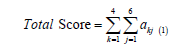
Where akj is the score in the field of view j on criterion k. The final score was used to determine the severity of colitis in the animal.
Results
Model of TNBS-induced colitis in rats
All animals treated with TNBS showed signs of mild colitis, characterized by foci with mild to moderate neutrophilic infiltration with lymphocytes and plasmocytes, with focal or complete destruction of crypts and in some cases spread of inflammation to the submucosa of the intestinal wall (Figure 1).

Model of DSS-induced colitis in rats for 7 days
Two animals had a pattern of mild colitis, which was characterized by foci with mild and moderate inflammatory infiltration represented by neutrophils, macrophages, lymphocytes, plasmocytes within the intestinal mucosa and submucosa. Seven animals developed moderate colitis with infiltrates not only in the mucosal and submucosal layers, but also in some cases in the muscular layer of the intestine. Also in the infiltration areas, there was destruction of crypts, mainly complete, the superficial epithelium was preserved in some places, and in some cases up to 2/3 of crypts were damaged. In areas of inflammation and lesions, the total volume of the intestinal wall lesion was more than 75% of the field of view area (Figure 2).

Model of DSS-induced colitis in rats for 14 days
Nine out of ten animals had a pattern of mild colitis mainly with minimal changes, which were characterized by foci with mild or moderate inflammatory lymphoplasmacytic infiltration with single macrophages and neutrophils within the intestinal mucosa in one or two fields of view. Destruction of up to 2/3 of crypts was observed in the infiltration areas. The total lesion volume was not more than 50% of the field of view area. One animal receiving DSS with drinking water showed no histologic evidence of colitis.
Model of DSS-induced colitis in mice
Three out of eight animals showed signs of mild colitis in the form of foci with mild inflammatory lymphoplasmacytic infiltration with single macrophages and neutrophils within the intestinal mucosa. In the infiltration areas, there was crypt destruction of varying severity, the superficial epithelium was preserved. The total lesion volume was not more than 25% of the field of view area (Figure 3). Also, in single fields of view, there were foci with pronounced inflammatory infiltration with numerous lymphocytes and neutrophils, in some places with involvement of the submucosa, with destruction of crypts and focal superficial epithelium, in some fields of view the lesion volume reached more than 75%. Histologic signs of colon inflammation were not detected in five animals.

The data in (Figure 4) are presented as total scores of 4 semiquantitative criteria (severity of inflammation, depth of intestinal wall lesion, extent of crypt damage, and lesion area) for each animal. Bars denote medians and interquartile range.

Discussion
To model intestinal inflammation in experimental animals, TNBS and DSS approaches are most often used, since they can induce pathological processes in the intestine of animals similar to those in humans with IBD. Besides, these schemes are quite simple to implement [7,11,12]. Varying the concentration and frequency of administration of toxic chemicals can result in both acute and chronic versions of the course of the disease, as well as influence the severity of inflammation in the intestine, which in some cases is critical to assess the efficacy of drugs under development.
Successful induction of pathology requires selection of the inducer, its concentration, multiplicity, and route of administration. The present study showed that not all animals were able to achieve signs of intestinal mucosal inflammation with different schemes of DSS addition to drinking water, which respectively requires adjustment of the dose and/or concentration of the substance used as an inducer. TNBS administration resulted in chronic non-erosive inflammation of the colon, whereas a pattern of erosive colitis was observed with intragastric administration of 5.5% DSS solution.
Conclusion
The variety of induction schemes allows modeling different degrees of severity of pathological changes in the intestine when evaluating the efficacy of drugs for IBD according to the area of their clinical application.
- Knyazev OV, Shkurko TV, Kagramanova AV, Veselov AV, Nikonov EL (2020) Epidemiology of inflammatory bowel disease. State of the problem (review). Dokazatel’naya gastroenterologiya = Russian Journal of Evidence-based Gastroenterology 9(2): 66-73.
- Gajendran M, Loganathan P, Jimenez G, Catinella AP, Ng N, et al. (2019) A comprehensive review and update on ulcerative colitis. Dis Mon 65(12): 100851.
- Bruner LP, White AM, Proksell S (2023) Inflammatory bowel disease. Prim Care 50(3): 411-427.
- Flynn S, Eisenstein S (2019) Inflammatory bowel disease presentation and diagnosis. Surg Clin North Am 99(6): 1051-1062.
- Pinton P (2022) Computational models in inflammatory bowel disease. Clin Transl Sci 15(4): 824-830.
- M'Koma AE (2022) Inflammatory bowel disease: clinical diagnosis and surgical treatment-overview. Medicina (Kaunas) 58(5): 567.
- Baydi Z, Limami Y, Khalki L, Zaid N, Naya A, et al. (2021) An update of research animal models of inflammatory bowel disease. Scientific World Journal pp. 7479540.
- Katsandegwaza B, Horsnell W, Smith K (2022) Inflammatory bowel disease: a review of pre-clinical murine models of human disease. IntJ Mol Sci 23: 16.
- Elson CO, Sartor RB, Tennyson GS, Riddel RH (1995) Experimental models of inflammatory bowel disease. Gastroenterology 109(4): 1344-1367.
- Morris GP, Beck PL, Herridge MS, Depew WT, Szewczuk MR, et al. (1989) Hapten-induced model of chronic inflammation and ulceration in the rat colon. Gastroenterology 96(3): 795-803.
- Silva I, Solas J, Pinto R, Mateus V (2022) Chronic experimental model of TNBS-induced colitis to study inflammatory bowel disease. Int J Mol Sci 23(9): 4739.
- Shaikh-Omar A, Murad HA, Alotaibi NM (2022) Rectal roflumilast improves trinitrobenzenesulfonic acid-induced chronic colitis in rats. Braz J Med Biol Res 55: e11877.
- Linden DR, Chen JX, Gershon MD, Sharkey KA, Mawe GM (2003) Serotonin availability is increased in mucosa of guinea pigs with TNBS-induced colitis. Am J Physiol Gastrointest Liver Physiol 285(1): G207-G216.
- Reshetnikov MN, Vinogradova TI, Zyuzya YuR, Plotkin DV, Volkov AA, et al. (2023) Choosing the optimal concentration of dextran sulfate sodium for reproducing chemically induced colitis in rabbits. Experimental and Clinical Gastroenterology (9): 125-130.
- Stevenson CS, Marshall LA, Morgan DW (2006) In vivo Models of Inflammation. 2nd Berlin: Birkhauser Verlag pp. 150-151.
- Riemschneider S, Hoffmann M, Slanina U (2021) Indol-3-carbinol and quercetin ameliorate chronic DSS-induced colitis in C57BL/6 mice by AhR-mediated anti-inflammatory mechanisms. Int J Environ Res Public Health 18(5): 2262.
- Dieleman LA, Ridwan BU, Tennyson GS (1994) Dextran sulfate sodium-induced colitis occurs in severe combined immunodeficient mice. Gastroenterology 107(6): 1643-1652.
- D’Errico A, Malvi D (2019) Diagnosis of ulcerative colitis: morphology and histopathological characteristics. Updates Surg pp. 61-92.
- Kellermann L, Riis LB (2021) A close view on histopathological changes in inflammatory bowel disease, a narrative review. Digestive Medicine Research 4(3): 1-15.


