




 AIBM Home



Comparision of ERIC-PCR, OUT- PCR and Spoligotyping Methods to Diagnose of Cross-Contamination with Mycobacterium tuberculosis
Gülnur Tarhan1*, Fatih Ocak2 and Ismail Ceyhan3
1Department of Medical Microbiology, Adiyaman University, Turkey
2Yenimahalle State Hospital, Microbiology Laboratory, Turkey
3Atatürk Chest Diseases and Thoracic Surgery Education and Research Hospital, Turkey
Submission: September 30, 2016; Published: October 14, 2016
*Corresponding author: Gülnur Tarhan, Department of Medical Microbiology, Adiyaman University, Faculty of Medicine, Siteler Mahallesi, Atatürk Bulvar, No: 411 Adiyaman, Turkey, Tel:+90-416-223-16-90/1448 Email: gulnur.tarhan@yahoo.com
How to cite this article: Gülnur Tarhan*, Fatih Ocak and Ismail Ceyhan. Comparision of ERIC-PCR, OUT- PCR and Spoligotyping Methods to Diagnose of Cross-Contamination with Mycobacterium tuberculosis. Adv Biotech & Micro. 2016; 1(3):555564.DOI:10.19080/AIBM.2016.01.555564
Abstract
The culture of Mycobacterium tuberculosis from clinical specimens is considered to be the gold standard for the diagnosis of tuberculosis. However, false-positive cultures can occur due to cross contamination. Molecular typing methods are a useful tool for quick diagnosis of false-positive culture. In this study, we aimed to comperative evaluation of false-positive culture results by using ERIC-PCR, OUT PCR and spoligotyping methods. In the presentinvestigation, we evaluated a total of 29 culture isolates obtained from five groups samples processed on the same day during 5 different time periods.
By using spoligotyping, 5 spoligotype family including LAM9, LAM7-TUR, T and an unidentified spoligotype were identified according to SITVIT2 database. With the OUT-PCR, 4 different band pattern was determined at Nusieve agarose jel electrophoresis. For ERIC-PCR method, 2-3 band pattern were obtained at Nusive agarose jel and 6-7 band pattern were determined at polyacriylamide gel electrophoresis. All theisolates were collected in two groups by ERIC-PCR, but this created groups were not efficient to determine the degree of separation between isolates. In the study, spoligotyping and OUT-PCR groups modeling were compared with each other revealed compliance rate of 96.55% between the two tests. The application of OUT-PCR has been serve as a rapid and efficient method to investigate cases of possible cross-contamination with M. tuberculosis.
Keywords: Cross contamination;ERIC-PCR; OUT-PCR; spoligotyping; Mycobacterium tuberculosis
Introduction
The isolation of Mycobacterium tuberculosis complex organisms from clinical specimens collected from suspected patients serves as the gold standard for the proper diagnosis of tuberculosis [1]. The correct isolation with cultre is the most important step for definitive diagnosis of tuberculosis. False-positive cultures occur due to contaminated clinical equipment, clerical errors or cross-contamination of specimens.
The cross-contamination frequency has been reported between 0.1% to 3% of M. tuberculosis in the published studies. Indications of cross-contamination include culture results inconsistent with the patient’s clinical course, unexpected drug resistance, single culture-positive specimens and solid-medium cultures with low-colony-number counts [2-7].
A single positive culture is the most commonly reported indicator of false positivity. False-positive results for tuberculosis have been a matter of concern because of the clinical, therapeutic, and social impacts of the misdiagnosis of tuberculosis. At the same time, these false-positive cultures lead to an overestimation of the incidence and prevalence of tuberculosis in humans [8-10].
The false positivity due to laboratory cross-contamination is suspected when
a. Mycobacterium tuberculosis (MTB) is cultured from only one of the serial specimens of a patient
b. the bacterial yield in the culture is low and
c. the suspected sample has been processed together (or in a short period of time apart) with at least one other from a patient with a high bacterial load.
Molecular genotyping methods are widely used to determine cross-contamination depend on M. tuberculosis in the laboratory.
A definitive demonstration of cross-contamination requires the application of molecular tools to prove that the MTB isolates from the co-processed specimens share identical genotypic patterns. IS6110 RFLP, spoligotyping, MIRU-VNTR and MST are the most commonly used molecular methods for cross contamination [11-16]. Within the these methods, IS6110 RFLP is accepted as gold standard. But, this method requires well-grown cultures and takes a long time to provide an answer, usually longer than is acceptable for microbiologists and clinicians to make decisions. Moreover, the latter method requires an advanced laboratory equipment and substantial amount of time due to the fastidious nature of M. tuberculosis.
More importantly from, a strictly technical perspective, IS6110-RFLP analysis does a poor job of indicating the presence of M. tuberculosis when these organisms contain only a few copies of the IS6110 sequence. Recently, PCR-based techniques have proven to be rapid methods for the resolution of crosscontamination events [17-19]. In our study, 29 culture isolates suspected with cross-contamination occuring at five separate periods have been evaluated for the appropriateness of routine applications of two different easy to use PCR methods comparision with spoligotyping in the genotyping of Mycobacterium tuberculosis. One of the PCR based typing methods is OUT-PCR that is specific for amplifying IS6110 in using outward direction primer (OUT-primer), other is ERIC-PCR. ERIC (enterobacterial repetitive intergenic concensus) is specific genomic sequence which was found in Gram negative bacterial genom.
Materials and Methods
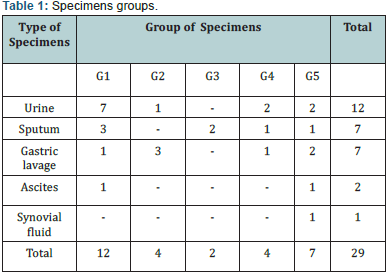
In our study, 5 groups specimens (totaly 29 samples) conducted routine laboratory procedures on the same day in 5 separate periods (sample acceptance, smear microscopy, culture) was be evaluated. 29 specimen was consisted of 12 urine, 7 sputum, 7 gastric lavage, 4 ascites and 3 synovial fluid (Table 1). All the applications related to clinical material (smear, culture cultivation and microscopic preparations, etc.) has been performed in a class II biosafety cabinet in P3 laboratory. Clinic material processing, smear microscopy and culture cultivation are carried out in accordance with the standards defined by the World Health Organization [20-22].
DNA extraction from the isolates and the control strains was performed according to solid phase absorption method (QIAamp DNA Mini Kit, Qiagen, Valencia, CA) . The DNA extract of each sample was stored at –20 oC. For OUT-PCR; CGG GGG TTC CCG GAC III (I: Inosine) (İontek, Bursa) single primer enabled to DNA amplification at outer region of IS6110 was used [17,19]. The PCR reaction was subjected to 40 cycles of amplification ( 3 min at 94 oC, 1 min at 94 oC, 1 min at 62 oC ) ; this was follewed by 1 min of extension at 72 oC.
For ERIC-PCR; ERIC1R(Tb-2) 5’ATG TAA GCT CCT GGG GAT TCA C and ERIC2(Tb-3) AAG TAA GTG ACT GGG GTG AGC G (Iontek-Bursa) primer pairs were used. The composition of the PCR mixture (50μl ) was 5μl 10x Taq PCR buffer (500mM KCl, 100mM Tris-HCl, pH:8.3) , Master AmpTM, 5μl 10x PCR Enhancer (Epicenter, Technologies), 3μl MgCl2 (25mM), 5μl dNTP(mix- 2mM), 0,75μl ERIC1R (87pmol/μl) primer, 0,9μl ERIC2 (73pmol/ μl) primer and Taq polimerase 1U (0.2μl) and 10μl template DNA. The PCR reaction was performed to 35 cycles of amplification (2 min at 94 oC, 45 sec at 94 oC, 1 min at 52 o C 1 min at 52 oC) and 20 min at 72 oC final extension. The presence of amplified product was confirmed by 1.4% NuiSieve agarose gel (Sigma, St Louis, MO, USA) [23].
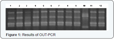
For DNA band analysis; UVI soft,UVI band, Windows Application V99.06 (BioRad,England) programme was used. Four main groups revealed by OUT- PCR method. Four main grup was detected in OUT-PCR. The samples which 100% compatible each other were collected in the same group, genotypically, (Figure 1).
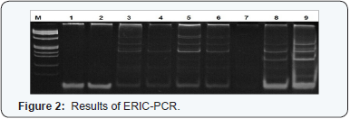
With the ERIC-PCR, 2-3 band pattern was detected on Nusieve agarose gel electrophoresis for all isolates.For better distinction, poliacrylamide gel electrophoresis was performed. According to this test results, 6-7 bant patern was obtained. The isolates were collected in two group according to two electrophoresisresults. However, these groups were not enough to determined the degree of separation between isolates (Figure 2).
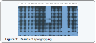
The spoligotyping was carried out with a commercial kit (Isogen Bioscience BV, Maarssen, The Netherlands) according to the manufacturer’s instructions. The 43 spacers between the direct repeats in the target region were amplified by using DRa biotinylated at the 59 end and DRb primers. The PCR product was hybridized to a membrane containing 43 oligonucleotides derived from the spacer sequences of M. tuberculosis H37Rv and M.bovis BCG P3 by reverse line blotting. M. tuberculosis H37Rv and M. bovis BCG P3 were used as control for spoligotyping. Spoligotypes results were converted into octal code and in the SITVIT2 proprietary database of the Pasteur Institute of Guadeloupe, which is an updated version of the previously released SpolDB4 database.
At the time of the present study, the database contained genotyping information on about 67,000 M. tuberculosis clinical isolates from 160 countries of origin. In this database, spoligotype international type (SIT) designates a spoligotyping pattern shared by two or more patient isolates. Major spoligotypingbased phylogenetic clades were assigned according to signatures provided in SpolDB4 [24] (Figure 3).
Sample group
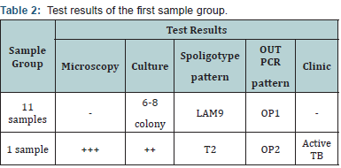
The evaluations of the 3 microscopic samples carried out in successive days, shows that 10 clinical samples (sputum, urine, gastric lavage fluid) out of 12 were negative. A sample (acid liquid) evaluated in one day had the negative microscopy result. Of these samples thought to be cross-contamination were determined less than 8 colony in the culture tubes. The clinical results of all the samples were negative. In another sample processed in the same day, the microscopy was three positive (3+) and in the culture tubes two positive (2+) reproduction was observed.
The patient had clinically active tuberculosis. When these groups OUT-PCR band patterns and spoligotypes were evaluated in themselves, among 12 samples in a sample, which had positive microscopy and culture results, different band patterns were detected. The band patterns of other 11 samples were the same. 11 samples belonged to spoligotype LAM9 family and in one sample a different spoligotype belonging to T2 family was detected. In OUT PCR and spoligotyping methods 100% compatibility was detected (Table 2).
Sample group
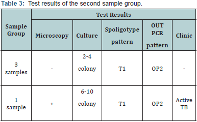
4 samples processed in the same day were negative. In this group the contamination cause couldn’t be defined. In the second sample group, 3 samples out of 4 had negative microscopic and clinical results. In the culture tubes of these samples a reproduction which is less than 5 colonies, was observed. The microscopy result of one sample had a negative result and 6-8 colonies were detected in its culture. This sample was clinically positive at the same time. With OUT-PCR method the same band pattern was detected in all 4 samples.The spoligotype of all samples were identified as T1. In OUT PCR ve spoligotipleme methods 100% compatibility was detected (Table 3).
Sample group
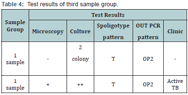
The microscopic results of two samples in the third sample group were defined negative and in the culture tubes two colonies were observed. The OUT-PCR band pattern and spoligotype of these samples were the same. In OUT PCR ve spoligotipleme methods 100% compatibility was detected.It was detected that the contamination was resulted from the positive sample (Table 4).
Sample group
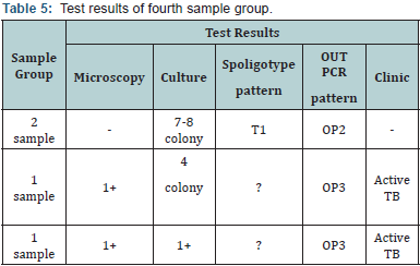
2 out of 4 samples in the fourth group had negative microscopic results and in their culture tubes 7-8 colony were observed. The patients were clinically negative. The microscopic results of the other 2 samples were 1+ positive. The microscopic results of both was 1 positive. Among two Kültür positifliği detected samples, one belonged to a patient who had been receiving tuberculosis treatment and the other belonged to a new tuberculosis patient. Two of the samples belonged to T1 spoligotype family but unidentified spoligotype was detected in two samples. In OUT PCR ve spoligotipleme methods 100% compatibility was detected (Table 5).
Sample group
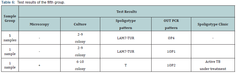
5 out of 7 samples in the fifth group had negative microscopic results and in their culture tubes 2-9 colonies were observed. Other 2 samples were detected as microscopically positive, one of them was 3 positive and the other was 1 positive. The culture of these samples were 1-4 positive. The two samples which were detected as culture positive were both belonged to patients who were undergoing tuberculosis treatment. In the OUT-PCR result the same band pattern was detected in 6 samples but one sample had a different band pattern. It was determined that 6 samples belonged to LAM7-TUR family and 1 belonged to T family. In 5 samples belonging to LAM7-TUR family the same OUT-PCR pattern was detected but one sample had a different pattern. In the 5th group betweenOUT PCR ve spoligotipleme methods 85.71% compatibility was detected (Table 6).
Discussion
The molecular typing methods have been used as an important indicator in the tuberculosis laboratories in determining the source of transmission in cases where there is a doubt of cross-contamination [2-5]. The most important expectation for these methods is the determination of some similar genotypes which cause a false increase in the group and the identification of the episdemiological relationship [11,12,24]. Despite the acceptance of IS6110- RFLP as a primary standard, the need for high quantity of pure DNA, the long duration of the test procedure, the complexity of the process, the high amount of the workload, and the necessity for advanced computer softwares for the analysis of the restriction pieces restrains the area of routine use.
Also with this test the typing of the origins which have less copies than six and the IS6110-RFLP profiles are similar is not adequate [12]. On the other hand Spoligotyping is known as a cheaper, faster, easier and highly sensitive method. For marked oligonucleotide absorbed membranes the evaluation of the test results in the method which is based on the hybridization of PCR product, doesn’t require computer hardware [24].
The expected results with these methods are the high distinction capacity between the origins, repeatability and easy application. In the study during 5 different routine application time, the cross contamination possibility of the clinical samples processed in the same day, PZR based ERIC PCR, OUT-PCR and spoligotyping methods are relatively evaluated. In 5 sample groups which were assumed to have cross contamination, with OUT-PCR method 4 , with spoligitiplendirme 6 different band patterns were detected out of 29 samples. Since the ERIC-PCR method is not standardized it couldn’t be evaluated. 96.55% compatibility was detected between spoligotyping and OUT-PCR group modelling tests when they were compared with eachother.
When the demographic and clinical data of the samples in all groups were analysed, it was detected that there were no epidemiological relation between the patients. The ERICPCR method was found inadequate in terms of its difficulty in standardization and lack of capacity to provide enough distinction, on the other hand OUT-PCR method was determined to be adequate by means of its easy application and its ability to show the distinctions during the typing of the isolates which are few in number.
To determine the proximity degree of a large number of isolates, the use of a program is necesary. It was detected that OUT-PCR method is very sufficient for the distinction of different origins unlike ERIC-PCR method. As a result, it is determined that OUT-PCR method is very efficient for the detection of laboratory resulted cross contaminations in terms of being practical and fast but can not be used alone as method in epidemiological studies.
- (1990) Diagnostic Standards and Classification of Tuberculosis. Am Rev Respir Dis 142(3): 725-735.
- Cook VJ, Stark G, Roscoe DL, Kwong A, Elwood RK (2006) Investigation of suspected laboratory cross-contamination: interpretation of single smear-negative, positive cultures for Mycobacterium tuberculosis. Clin Microbiol Infect 12(10): 1042-1045.
- de Boer AS, Blommerde B, de Haas PE, Sebek MM, Lambregts-van Weezenbeek KS, et al. (2002) False-positive Mycobacterium tuberculosis cultures in 44 laboratories in The Netherlands (1993 to 2000): incidence, risk factors, and consequences. J Clin Microbiol 40(11): 4004-4009.
- Burman WJ, Stone BL, Reves RR, Wilson ML, Yang Z, et al. (1997) The incidence of false-positive cultures for Mycobacterium tuberculosis. Am J Respir Crit Care Med 155(1): 321-326.
- Fitzpatrick L, Braden C, Cronin W, English J, Campbell E, et al. (2004) Investigation of Laboratory cross-contamination of Mycobacterium tuberculosis Clin Infect Dis 38(6): e52-e54.
- de C Ramos M, Soini H, Roscanni GC, Jaques M, Villares MC, et al. (1999) Extensive cross-contamination of specimens with Mycobacterium tuberculosis in a reference laboratory. J Clin Microbiol 37(4): 916-919.
- Carroll NM, Richardson M, van Helden PD (2003) Criteria for identification of cross-contamination of cultures of Mycobacterium tuberculosis in routine microbiology laboratories. J Clin Microbiol 41(5): 2269-2270.
- CDC (1997) Multiple misdiagnoses of tuberculosis resulting from laboratory error—Wisconsin, 1996. MMWR Morb Mortal Wkly Rep 46(34): 797-801.
- Miller AC, Sharnprapai S, Suruki R, Corkren E, Nardell EA, et al. (2002) Impact of genotyping of Mycobacterium tuberculosis on public health practice in Massachusetts. Emerg Infect Dis 8(11): 1285-1289.
- Burman, WJ, Reves RR (2000) Review of false-positive cultures for Mycobacterium tuberculosis and recommendations for avoiding unnecessary treatment. Clin Infect Dis 31(6): 1390-1395.
- Kanduma E, McHugh TD, Gillespie SH (2003) Molecular methods for Mycobacterium tuberculosis strain typing: A users guide. J Appl Microbiol 94(5): 781-791.
- Durmaz R, Zozio T, Gunal S, Allix C, Fauville-Dufaux M, et al. (2007) Population-based molecular epidemiological study of tuberculosis in Malatya,Turkey. J Clin Microbiol 45(12): 4027-4035.
- Kamerbeek J, Schouls L, Kolk A, Van Agterveld M, Van Soolingen D, et al. (1997) Simultaneous detection and strain differentiation of Mycobacterium tuberculosis for diagnosis and epidemiology. J Clin Microbiol 35(4): 907-914.
- Martin A, Herranz M, Lirola MM, Fernandez RF, Bouza E, et al. (2008) Optimized molecular resolution of cross-contamination alerts in clinical mycobacteriology laboratories. BMC Microbiol 8: 30-33.
- Martinez M, Garcia V, Alonso M, Andres S, Bouza E, et al. (2006) Impact of laboratory cross-contamination on molecular epidemiology studies of tuberculosis. JClin Microbiol 44(8): 2967-2969.
- Small PM, McClenny NB, Singh SP, Schoolnik GK, Tompkins LS, et al. (1993) Molecular strain typing of Mycobacterium tuberculosis to confirm cross-contamination in the mycobacteriology laboratory and modification of procedures to minimize occurrence of false-positive cultures. J Clin Microbiol 31(7): 1677-1682.
- Ross B, Dwyer B (1993) Rapid, simple method of typing isolates of Mycobacterium tuberculosis by using the polymerase chain reaction. J Clin Microbiol 31(2): 329-334.
- Gutierrez M, Samper S, Gavigan JA, Marin JFG, Martin C (1995) diffrentiation by molecular typing of Mycobacterium bovis straıns causing tuberculosis in cattle and goats. J Clin Microbiol 33(11): 2953-2956.
- Sechi LA, Zanetti S, Dupre I, Delogu G, Fadda G (1998) Enterobacterial repetitive intergenic consensus sequences as molecular targets for typing of Mycobacterium tuberculosis J Clin Microbiol 36(1): 128-132.
- Kent PT, Kubica GP (1985) Public Health Mycobacteriology, A Guide For The Level III Laboratory. Centers for Disease Control Atlanta, Georgia, USA.
- Salfinger M, Pfyffer GE (1994) The new diagnostic mycobacteriology laboratory. Eur J Clin Microbiol Infect Dis 13(11): 961-979.
- Kubica GPW, Dye E, Cohn ML, Middlebrook G (1993) Sputum digestion and decontamination with N-acetyl-L-cysteine-sodiumhydroxide for culture of Mycobacteria. Am Rev Respir Dis 87: 775-779.
- Tarhan G, Ceyhan I, Şimşek H, S.Tuncer (2009) Evaluation of the efficacy of the five DNA extraction methods for the detection of Mycobacterium tuberculosis DNA in direct and processed sputum by in-House PCR Method. Turk J Med Sci 39(2): 253-257.
- Kısa O, Tarhan G, Gunal S, AlbayA, Durmaz R, et al. (2012) The distribution of spoligotyping defined genotypic lineages among drug-resistant tuberculosis complex clinical ısolates in Ankara, Turkey. Plos One 7(1): e30331.

























