




 OAJS Home



Abstract
Focal therapy (FT) for localized prostate cancer seeks to eradicate the dominant clinically significant lesion while preserving uninvolved prostate tissue and adjacent functional structures. Interest in FT has increased with improvements in imaging (multiparametric magnetic resonance imaging (mpMRI), Micro Ultrasound prostate-specific membrane antigen (PSMA) positron emission tomography); biopsy technology (fusion biopsy, transperineal sampling) and delivery technology (High-intensity focused ultrasound (HIFU), cryotherapy, irreversible electroporation, focal laser ablation, vascular-targeted photodynamic therapy, and brachytherapy.) However, the optimum candidate remains narrowly defined as long-term randomized comparative evidence is limited. This narrative review summarizes recent consensus statements, guideline positions, and outcome studies to define the patient most likely to benefit from FT. The best candidate is a well-informed, treatment-naive man with clinically significant cancer, adequate life expectancy, a strong preference for preservation of urinary and sexual function, and clinically localized MRI-visible lesion with concordant targeted and systematic biopsy findings, and disease biology most consistent with favorable or selected unfavorable intermediate-risk cancer. Very-low-risk or low-risk Grade Group 1 disease is better managed with active surveillance, while high-risk, locally advanced, nodal, metastatic, MRI-invisible, or extensive bilateral clinically significant disease are considered unsuitable to pursue this management strategy. The optimal patient must also accept rigorous surveillance with prostate-specific antigen testing, mpMRI, and protocol biopsy, as well as accepting that retreatment or salvage whole-gland therapy may be required.
Keywords:Prostate Cancer; Focal Therapy; Partial Gland Ablation; High-Intensity Focused Ultrasound; Cryotherapy; Irreversible Electroporation; Patient Selection; mpMRI; Fusion Biopsy
Abbreviations:FT: Focal Therapy; PSMA: Prostate-Specific Membrane Antigen; HIFU: High-Intensity Focused Ultrasound; IRE: Irreversible Electroporation; FALCON: FocAL Therapy CONsensus; GG: Grade Group
Introduction
Significant advances in prostate cancer detection and treatment have improved risk stratification and local control, but treatment-related quality-of-life burdens remain clinically important. Radical prostatectomy and radiation therapy are the time tested and established curative options, yet they may impair erectile function, urinary continence, and bowel function. Focal therapy (FT), also termed partial gland ablation, aims to bridge the gap between active surveillance and whole-gland treatment by focusing on a focal area, dubbed the ‘index lesion’, for the purpose of minimizing damage to non-cancerous tissue, the neurovascular bundles, urethral sphincter, rectum, and bladder neck when feasible. It aims to balance maximizing treatment outcomes while minimizing morbidity. Available modalities include high-intensity focused ultrasound (HIFU), cryotherapy, irreversible electroporation (IRE), focal laser ablation, focal brachytherapy, and vascular-targeted photodynamic therapy, among others [1,2].
The first question to address when considering FT is which patient with clinically significant, localized prostate cancer is an appropriate candidate for this treatment strategy. The answer requires integration of tumor grade, lesion visibility, biopsy mapping, tumor distribution, anatomy, life expectancy, patient preferences, and willingness to undergo surveillance. The original FocAL therapy CONsensus (FALCON) research letter introduced an international effort to standardize partial-gland ablation practice, and the full FALCON project, published in 2025, reported a modified Delphi process involving 246 initial participants, of whom 148 completed all three rounds [3,4]. The University of California Urologic Collaborative, or UC-Squared, consensus statement was published in 2024, offering a concise contemporary position statements from high-volume academic centers [5]. Together with recent reviews and outcomes studies, these publications permit a clearer description of optimal candidate for FT.
Despite this progress, FT recommended in major guidelines as an accepted option as is radical prostatectomy or radiation therapy. The AUA/ASTRO guideline states that patients with intermediate-risk disease considering whole-gland or focal ablation should be informed that high-quality comparative data with surgery and radiation therapy, are lacking, and that focal or whole-gland ablation should not be recommended for high-risk disease outside a clinical trial [6,7]. The EAU recommends focal HIFU or cryotherapy only within a prospective registry and other ablative modalities only within well-designed prospective trials [8]. NCCN guideline is similarly cautious, reserving focal and ablative approaches within the context of clinical trial settings in intermediate-risk disease and discouraging their use in low-risk or high-risk disease outside prospective clinical trials [9].
A literature search was performed using PubMed, ClinicalTrials.gov, and major guideline websites and terms included “prostate cancer focal therapy,” “partial gland ablation,” “HIFU,” “cryotherapy,” “irreversible electroporation,” “patient selection,” “FALCON consensus,” “UC-Squared consensus,” “localized prostate cancer guidelines,” “surveillance after focal therapy,” and “genomic classifier focal therapy.” Recent consensus statements, guidelines, systematic reviews, prospective studies, and retrospective studies focused on candidacy, oncologic outcomes, functional outcomes, and follow-up after FT were included. Historical consensus statements were included when they influenced current selection criteria. Salvage-only FT studies, non-prostate studies, nonclinical technical papers without patient-selection relevance, and non- English-language articles were excluded. A narrative review was then completed with the equal participation from the authors with a total of over 20 years’ experience with focal therapy.
Defining the Optimal Candidate
The optimal patient for FT is best understood by distinguishing him from the risk groups where he is not a candidate: Very lowrisk or low-risk Grade Group (GG) 1 disease. For (GG) 1 disease, active surveillance is the preferred strategy for such patients as it avoids treatment morbidity and preserves the opportunity for curative treatment if progression occurs. Patient with highrisk, locally advanced, nodal, metastatic, or extensive bilateral clinically significant disease risks undertreating biologically aggressive or multifocal disease [6-9]. The best candidate is a treatment-naive man with clinically localized, MRI-visible, biopsyconfirmed intermediate-risk prostate cancer, most commonly ISUP GG2, with selected GG3 cases considered only after thorough staging. An example: a man with Gleason 3 + 4 disease, a discrete lesion visible on high-quality mpMRI, concordant targeted and systematic biopsy findings, no clinically significant contralateral disease, no extraprostatic extension, and an ablation zone that can include an adequate margin without unacceptable functional risk. A more cautious but still possible candidate is a man with limited Gleason 4 + 3 disease, if imaging, biopsy mapping, and clinical risk assessment do not suggest multifocal high-grade disease or high probability of extracapsular disease [2,3].
Lesion distribution is critical. FT is most effective for unifocal or unilateral clinically significant disease. Multifocal MRI-visible disease is not an automatic exclusion criteria in every consensus statement, but it is less ideal as it weakens the index-lesion rationale and may require wider ablation, thereby reducing the functional advantage of FT. Low-grade GG1 disease outside the treatment zone may be acceptable if the patient is otherwise suitable and understands that untreated tissue must remain under surveillance. Life expectancy and patient’s values are equally important. Most consensus statements recommend a life expectancy of at least 8 to 10 years as the patient should live long enough to benefit from local control [5]. Age alone should not determine eligibility. A fit older man with favorable anatomy and a strong desire to preserve function may be suitable, while a younger man with diffuse bilateral disease may not. Baseline erectile dysfunction, moderate lower urinary tract symptoms, and larger prostate volume are not absolute exclusions, although each affects counseling, technical feasibility, and expected functional benefit [3,4].
Imaging and Biopsy Requirements
High-quality mpMRI is foundational for FT patient selection. It defines the target, informs the ablation margin, identifies lesions near the sphincter, urethra, rectum, neurovascular bundles, or bladder neck, and helps exclude extraprostatic extension [4,5]. A patient with biopsy-positive clinically significant cancer but negative or nondiagnostic MRI is a poor candidate because accurate lesion targeting becomes unreliable. FALCON and UC-Squared both emphasize MRI-visible disease, and FALCON does not recommend PSMA PET/CT as a substitute for highquality mpMRI contrasting the recommendation from the UCSquared Consensus which does recommend PSMA PET scan for unfavorable intermediate risk disease (4+3) [3,5].
Biopsy should combine targeted and systematic sampling [10]. Targeted cores confirm the MRI-visible lesion, while systematic cores reduce the risk of missing contralateral, multifocal, or highergrade disease. Transperineal biopsy is increasingly preferred because it improves anatomic mapping and lowers infection risk. FALCON recommended rigorous sampling, with multiple targeted cores of the lesion and systematic cores across the gland [3]. The clinical importance of systematic biopsy was supported by Jang et al., [10] who applied FALCON criteria to a large MRIguided, biopsy-proven cohort. Approximately one-quarter of all men with localized prostate cancer met broad selection criteria, while only a much smaller proportion represented ideal functional-preservation candidates. Systematic biopsy was a major determining factor in this reduction, revealing high grade disease (GG4 or 5) in up to 52% cores. The authors endorsed the importance of the FALCON requirements, including a high-quality positive mpMRI with ≥ 3 targeted and ≥10 systematic biopsy to best detect patients requiring higher levels of care and not pursue FT. Importantly, systematic biopsy favored men who appeared eligible based on targeted cores alone, supporting combined targeted and systematic sampling before FT [10].
PSMA PET/CT is a supplemental staging tool rather than a replacement for mpMRI. It may be most useful for unfavorable intermediate-risk disease, discordant MRI and biopsy findings, suspected anterior or multifocal disease, or cases in which detection of nodal or metastatic disease would change management. UC-Squared recommends PSMA PET in selected unfavorable intermediate-risk cases, while FALCON is more conservative [4,5]. The optimal candidate should have no evidence of nodal, metastatic, seminal vesicle, or locally advanced disease on staging [5,8].
Tumor Biology and Biomarkers
Histology remains the main determinant of suitability. ISUP GG2 disease is the best fit, especially when pattern 4 is limited and cribriform or intraductal carcinoma is absent [3,4]. Selected GG3 disease may be considered, but recurrence risk appears higher, and counseling should be more cautious. Disease greater than GG3, extensive pattern 4, intraductal carcinoma, suspicious extraprostatic extension, or high-risk clinical features should direct the patient toward whole-gland or multimodal therapy rather than focal ablation [3,4]. PSA should influence selection but should not be consider alone. Earlier consensus criteria often used PSA thresholds less than 10 or 15 ng/mL. Contemporary reviews suggest PSA below 20 ng/mL, ideally below 10 ng/mL, for most candidates [2, 5]. FALCON allowed more flexibility by emphasizing overall localization and PSA density rather than a strict PSA cutoff; candidates with PSA above 15 ng/mL may still require careful consideration when PSA density is low and staging is reassuring [3]. A rising PSA, high PSA density, or PSA discordant with apparently limited MRI disease should trigger further evaluation before FT.
Genomic classifiers are promising but not yet validated for routine FT selection. Shee et al. [11] studied men treated with robotic HIFU who underwent 1-year post-HIFU MRI-fusion biopsy [11]. In that cohort, pre-HIFU GG3 or higher disease and high genomic risk were associated with in-field recurrence, while GG3 or higher disease and PSA greater than 10 ng/mL versus less than 6 ng/mL were associated with overall biopsy-proven recurrence [11]. These findings support the concept that tumor biology should refine selection, but they do not yet establish a genomic cutoff for routine candidacy decisions. Germline highrisk features, including BRCA-related mutations, should prompt caution and shared decision-making because such patients may harbor more aggressive biology than imaging alone suggests [3,4].
Anatomic and Technical Considerations
In practice, an optimal lesion should be discrete, reachable by the intended technology, and treatable with a safety margin. A margin of at least 5 millimeters is typically desired as microscopic disease can extend beyond MRI-defined borders. Lesions close to the external urinary sphincter may increase incontinence risk, and lesions abutting the urethra, bladder neck, neurovascular bundle, or rectum require technology-specific planning. FALCON considered lesion proximity to the sphincter more concerning than proximity to the rectum stating that lesions < 5mm from the rectum are eligible, but < 5mm from the sphincter are not, but this must be interpreted in light of device type, operator experience, rectal safety mechanisms, and patient-specific anatomy [3]. Prostate volume alone should not automatically exclude a patient, although large glands may impair access, increase treatment time, reduce energy delivery to anterior lesions, and complicate follow-up interpretation. Different energy sources have specific strengths and limitations. HIFU and cryotherapy have the largest evidence and are specifically addressed in guideline and registry recommendations. IRE, focal laser ablation, photodynamic therapy, radiofrequency ablation, and focal brachytherapy require prospective evidence and standardized follow-up before broad adoption. Device availability should not drive patient selection; rather, a specific technology should be selected after the patient and lesion are deemed appropriate for an organ-sparing approach.
Expected Outcomes and Why Selection Matters
A recent systematic review and meta-analysis of established FT modalities, including HIFU, cryotherapy, and IRE, reported favorable short- to intermediate-term survival and functional outcomes, but highlighted residual clinically significant cancer after treatment, variation in biopsy protocols, inconsistent definitions of recurrence, and the need for longer follow-up [12]. These limitations highlight why rigorous selection is essential. FT has not proven oncologic equivalence to radical prostatectomy or radiotherapy. The expected functional advantage is real but not absolute. The untreated prostate, urethra, sphincter, neurovascular bundles, and rectum are spared to varying degrees, so continence and erectile outcomes are often favorable compared with whole-gland treatment. Nonetheless, urinary symptoms, erectile dysfunction, urinary retention, infection, stricture, fistula, or rectal injury may occur [12-16]. Patients should counseled that the tradeoff of tissue preservation is oncologic uncertainty in untreated tissue and the need for structured surveillance. The ideal patient values functional preservation to accept this tradeoff but must be aware of the possibility of recurrence and the potential need for salvage treatment [17-21].
Post-Treatment Surveillance as a Selection Criterion
A patient who cannot or will not comply with surveillance is not an adequate candidate for FT. PSA alone is insufficient after partial-gland ablation because untreated prostate tissue continues to produce PSA and PSA kinetics post-treatment are variable. Contemporary surveillance reviews recommend multimodal follow-up using PSA, mpMRI, and biopsy, with many protocols including first post-treatment MRI at approximately 6 to 12 months and protocol biopsy at approximately 12 months, including targeted biopsy of suspicious or treated regions and systematic sampling of untreated tissue [13]. Long-term surveillance is necessary as recurrence may occur in-field from incomplete ablation or out-of-field from missed or de novo clinically significant cancer. The patient should also accept that future salvage treatment may be required. Repeat focal therapy, radical prostatectomy, radiotherapy, or systemic therapy may be required if clinically significant recurrence, progression, metastasis, or treatment failure occurs. Pre-treatment counseling should therefore include not only immediate risks but also the downstream consequences of salvage therapy.
Future Directions
The Jupiter Registry, first posted on ClinicalTrials.gov in January 2025, aims to prospectively collect standardized data from multiple European centers for patients undergoing FT for intermediate-risk localized prostate cancer. Its criteria include life expectancy greater than 10 years, prior targeted plus systematic MRI-ultrasound fusion biopsy, mpMRI-visible ISUP GG2 or GG3 disease, acceptance of GG1 outside the treatment area, and 5-year follow-up of oncologic, functional, and salvagetreatment outcomes [14]. This registry framework closely reflects the practical direction of the field: FT should be evaluated in intermediate-risk patients using standardized definitions and mandatory follow-up. Genomic classifiers, PSMA PET, artificial intelligence-assisted MRI interpretation, radiomics, and improved biopsy mapping may eventually identify patients whose disease is biologically unifocal and organ-confined. Until these tools are validated for FT-specific endpoints, they should supplement rather than replace careful mpMRI, targeted and systematic biopsy, conventional risk stratification, and shared decision-making.
Practical Optimal Patient Profile
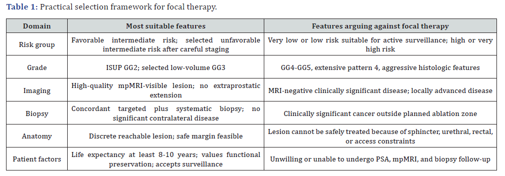
In our interpretation, of the current literature, the strongest candidate for FT is a well-informed, treatment-naive man with life expectancy of at least 8 to 10 years with localized disease, an MRI-visible and biopsy-confirmed clinically significant lesion, most commonly ISUP GG2 and selected GG3, limited disease, no clinically significant cancer outside the planned treatment area, no high-risk histologic or genomic features, and favorable anatomy for safe ablation with an adequate margin. PSA is ideally less than 10 ng/mL, and less than 20 ng/mL when PSA density, MRI findings, biopsy distribution, and risk category are considered. The optimal patient prioritizes functional preservation, understands that FT is not yet a guideline-equivalent standard of care, and agrees to PSA testing, mpMRI, protocol biopsy, and the potential for additional retreatment. [Table 1]
Conclusion
Optimal patient selection for prostate cancer FT has evolved from broad enthusiasm for organ preservation toward a more disciplined, evidence-informed profile. The ideal candidate is not simply any man with localized prostate cancer. The optimal candidate is a carefully staged, well-informed patient with MRIvisible, biopsy-concordant, localized clinically significant disease, most often ISUP GG2 and selected GG3, whose anatomy permits safe partial-gland ablation and whose preferences strongly favor functional preservation. Until long-term randomized comparative evidence matures, FT should be offered through clinical trials, prospective registries, or experienced programs with standardized imaging, biopsy, outcome reporting, and surveillance.
References
- Hopstaken JS, Bomers JGR, Sedelaar MJP, Valerio M, Futterer JJ, et al. (2022) An updated systematic review on focal therapy in localized prostate cancer: what has changed over the past 5 years? Eur Urol 81(1): 5-33.
- Ghoreifi A, Gomella L, Hu JC, Konety B, Lunelli L, et al. (2025) Identifying the best candidate for focal therapy: a comprehensive review. Prostate Cancer Prostatic Dis 28(3): 684-692.
- Rodriguez-Sanchez L, Cathelineau X, De Reijke TM, Stricker P, Emberton M, et al. (2025) Refining partial gland ablation for localised prostate cancer: the FALCON project. BJU Int 135(6): 1000-1009.
- Rodriguez-Sanchez L, Reiter R, Rodriguez A, Emberton M, Reijke TD, et al. (2024) The FocAL therapy CONsensus (FALCON): enhancing partial gland ablation for localised prostate cancer. BJU Int 134(1): 50-53.
- Javier-DesLoges J, Dall'Era MA, Brisbane W, Chamie K, Washington SL III, et al. (2024) The state of focal therapy in the treatment of prostate cancer: the University of California Collaborative (UC-Squared) consensus statement. Prostate Cancer Prostatic Dis 27(4): 579-581.
- Eastham JA, Auffenberg GB, Barocas DA, Chou R, Crispino T, et al. (2022) Clinically localized prostate cancer: AUA/ASTRO guideline. J Urol 208(3): 505-507.
- Eastham JA, Barocas DA, Chu CE, Morgans AK, Rodrigues G, et al. (2026) Clinically localized prostate cancer: AUA/ASTRO guideline amendment (2026). J Urol.
- (2026) European Association of Urology. EAU-EANM-ESTRO-ESUR-ISUP-SIOG guidelines on prostate cancer. 2026 edition. Arnhem, The Netherlands: EAU Guidelines Office.
- (2026) National Comprehensive Cancer Network. NCCN Clinical Practice Guidelines in Oncology: Prostate Cancer. Version 5.2026. Plymouth Meeting, PA: National Comprehensive Cancer Network.
- Jang JW, Handa N, Alam R, Neill C, Ross AE, et al. (2025) Incidence of ideal candidates for focal therapy: a scoping study following the FALCON consensus statements. BJUI Compass 6(12): e70121.
- Shee K, Pace WA, Liu AW, Cowan JE, Subramanyam V, et al. (2025) Determining optimal patient selection for high-intensity focused ultrasound for prostate cancer. Eur Urol Focus 11(6): 869-875.
- Tay KJ, Fong KY, Stabile A, Dominguez-Escrig JL, Ukimura O, et al. (2025) Established focal therapy-HIFU, IRE, or cryotherapy-where are we now? A systematic review and meta-analysis. Prostate Cancer Prostatic Dis 28(3): 693-706.
- Marra G, Marquis A, Suberville M, Woo H, Govorov A, et al. (2025) Surveillance after focal therapy: a comprehensive review. Prostate Cancer Prostatic Dis 28(3): 662-671.
- (2025) ClinicalTrials.gov. Jupiter Registry: Prospective Registry for Patients Undergoing Focal Therapy for Localized Prostate Cancer. NCT06772116.
- De la Rosette J, Ahmed H, Barentsz J, Johansen TB, Brausi M, et al. (2010) Focal therapy in prostate cancer: report from a consensus panel. J Endourol 24(5): 775-780.
- Van Den Bos W, Muller BG, Ahmed H, Bangma CH, Barret E, et al. (2014) Focal therapy in prostate cancer: international multidisciplinary consensus on trial design. Eur Urol 65(6): 1078-1083.
- Donaldson IA, Alonzi R, Barratt D, Barret E, Berge V, et al. (2015) Focal therapy: patients, interventions, and outcomes - a report from a consensus meeting. Eur Urol 67(4): 771-777.
- Tay KJ, Scheltema MJ, Ahmed HU, Barret E, Coleman JA, et al. (2017) Patient selection for prostate focal therapy in the era of active surveillance: an international Delphi consensus project. Prostate Cancer Prostatic Dis 20(3): 294-299.
- Tan WP, Rastinehad AR, Klotz L, Carroll PR, Emberton M, et al. (2021) Utilization of focal therapy for patients discontinuing active surveillance of prostate cancer: recommendations of an international Delphi consensus. Urol Oncol 39(11): 781.e17-781.e24.
- Von Hardenberg J, Borkowetz A, Siegel F, Kornienko K, Westhoff N, et al. (2021) Potential candidates for focal therapy in prostate cancer in the era of magnetic resonance imaging-targeted biopsy: a large multicenter cohort study. Eur Urol Focus 7(5): 1002-1010.
- Pompe RS, Kuhn-Thoma B, Nagaraj Y, Veleva V, Preisser F, et al. (2018) Validation of the current eligibility criteria for focal therapy in men with localized prostate cancer and the role of MRI. World J Urol 36(5): 705-712.

























