




 OAJS Home



Abstract
Background: Tuberculosis (TB) remains a major global health challenge, most commonly affecting the lungs. Whereas abdominal tuberculosis is uncommon, tuberculous peritonitis in low-incidence countries such as the United Kingdom (UK) is exceedingly rare, especially when there are no coexisting symptoms or radiological features of active lung disease.
Case presentation: We report the case of a 32-year-old man with no symptoms or radiological evidence of TB, who presented with progressive abdominal pain and computed tomography (CT) findings highly suggestive of hollow viscus perforation. The clinical and radiological concerns prompted emergency surgical exploration. Intraoperatively, there was a large volume of ascites, diffuse miliary nodularity involving the visceral and parietal peritoneum, and marked omental thickening, initially raising suspicion of disseminated intra-abdominal malignancy. No perforation or enteric contamination was identified.
Histopathological examination of omental biopsy subsequently demonstrated acid-fast bacilli (AFB), confirming tuberculous peritonitis.
Discussion: This case highlights the protean nature of extrapulmonary TB and the diagnostic difficulty posed by isolated peritoneal disease in low-incidence settings such as UK. The positive tissue diagnosis prompted anti-tuberculous therapy, which is highly effective.
Conclusion: Clinicians in low TB incidence countries should consider tuberculous peritonitis in seemingly TB naïve patients presenting with acute abdomen, even when CT suggests hollow viscus perforation. Early tissue diagnosis avoids delay in definitive medical management.
Keywords:Abdominal Tuberculosis; Tuberculous Peritonitis; Extrapulmonary Tuberculosis
Abbreviations:TB: Tuberculosis; CT: Computed Tomography; AFB: Acid-Fast Bacilli; A&E: Accident and Emergency; NEWS2: National Early Warning Score; CRP: C- Reactive Protein; PA: Posterior-Anterior; NELA: National Emergency Laparotomy Audit
Introduction
Tuberculosis (TB) remains one of the leading infectious causes of morbidity and mortality worldwide, with an estimated 10.6 million new cases reported annually [1]. Although pulmonary TB is the most common and most readily recognized form, in 2023 extrapulmonary tuberculosis accounted for approximately 16% of notified TB cases globally [2]. Extrapulmonary presentations may affect virtually any organ system and frequently mimic inflammatory, neoplastic or acute surgical conditions [3]. Abdominal tuberculosis is an uncommon but well-recognized manifestation of extrapulmonary TB and may involve the gastrointestinal tract, peritoneum, mesenteric lymph nodes or solid intra-abdominal organs [3]. Among these, tuberculous peritonitis is particularly rare, especially in low-incidence countries such as the United Kingdom (UK). Patients typically present with non-specific symptoms including abdominal pain, weight loss, fever, ascites and constitutional upset [4].
The condition may closely resemble peritoneal carcinomatosis, inflammatory bowel disease, or intra-abdominal sepsis often leading to delayed or incidental diagnosis [4,5]. The absence of pulmonary symptoms or thoracic imaging abnormalities may further lower clinical suspicion and contribute to diagnostic uncertainty. In such circumstances, definitive diagnosis often depends on tissue sampling and histopathological confirmation [6]. We present a rare case of isolated tuberculous peritonitis in the UK presenting as an acute surgical emergency, with cross-sectional imaging strongly suggesting hollow viscus perforation. This case is notable not only for the absence of pulmonary involvement but also for the striking radiological and operative discordance.
Case Presentation
A 32-year-old man presented to the Accident and Emergency (A&E) department at Royal Preston Hospital in 2025 with a one- week history of progressively worsening abdominal pain on a background of approximately five months of intermittent lower abdominal discomfort. Associated symptoms included anorexia, nausea and intermittent vomiting. There was no reported history of cough, hemoptysis or night sweats. On examination, he had a tachycardia of 110 beats/min, hypotension of 89/65 mmHg, reduced urine output and a National Early Warning Score (NEWS2) of 6. There was tenderness with guarding, mostly in the lower quadrants. Laboratory investigations showed a white cell count of 13.0 × 10⁹/L and a C- Reactive Protein (CRP) of 146 mg/L.
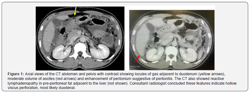
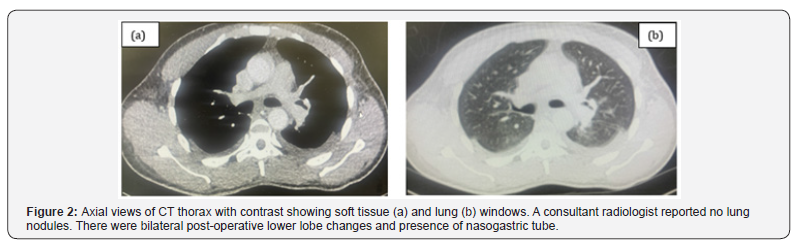
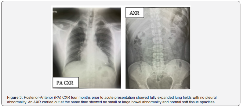
He was previously fit and well and had no personal or family history of TB or malignancy. He denied smoking and alcohol use. He had relocated to the UK within the preceding 12 months from a region with higher TB prevalence. CT of the abdomen and pelvis (Figure 1) demonstrated moderate volume ascites, locules of free intraperitoneal gas adjacent to the duodenum, and radiological features consistent with peritonitis. These findings were interpreted by a consultant radiologist as highly suspicious for hollow viscus perforation, most likely of duodenal origin. CT imaging of the thorax immediately post-operatively revealed no pulmonary nodules, cavitation, lymphadenopathy or other evidence of active intrathoracic disease (Figure 2). A chest radiograph carried out four months earlier similarly demonstrated clear lung fields (Figure 3). Furthermore, an abdominal X ray (AXR) requested by the patient’s general practitioner four months earlier was also normal.
Treatment
Given the concerning clinical and radiological findings, the patient underwent urgent diagnostic laparoscopy, which was subsequently converted to laparotomy. Pre-operative National Emergency Laparotomy Audit (NELA) assessment categorized the patient as moderate-to-high risk, warranting consultantled operative management and peri-operative optimization. Intraoperative exploration revealed large volume ascites and extensive nodular deposits involving both the parietal and visceral peritoneum. There was marked omental thickening, with nodular involvement extending to the diaphragmatic and pelvic peritoneal surfaces. These appearances were initially highly suspicious for disseminated intra-abdominal malignancy, particularly peritoneal carcinomatosis.
Importantly, no definitive perforation, bile or enteric contamination were identified. The discrepancy between the pre-operative imaging and operative findings prompted reconsideration of the working diagnosis. Peritoneal and omental biopsies were therefore obtained, together with ascitic fluid for cytological and microbiological analysis. Histopathological examination of omental tissue demonstrated granulomatous inflammation with acid-fast bacilli (AFB), establishing the diagnosis of abdominal tuberculosis with tuberculous peritonitis. Peritoneal biopsy and ascitic fluid analysis showed no evidence of malignancy. Repeated sputum cultures were negative, and there was no microbiological or radiological evidence of concomitant pulmonary disease (Figure 2).
Outcome and follow up
The patient was referred to the Infectious Diseases and commenced on standard anti-tuberculous therapy comprising Rifampicin, Isoniazid, Pyrazinamide and ethambutol once he was able to tolerate oral diet. He was in hospital for twelve days and discharged with a clear outpatient follow up plan. At five months follow up, he continues to do well on his regularly adjusted antituberculous medications.
Discussion
Tuberculous peritonitis is a rare but important manifestation of extrapulmonary Mycobacterium tuberculosis infection. Although abdominal TB remains more prevalent in endemic regions, it is encountered far less frequently in low-incidence countries, wherein diagnostic suspicion may be lower with priority given to alternative diagnoses such as malignancy or perforated viscus. This contributes to delays in diagnosis and may result in unnecessary surgical intervention [3,4]. The present case illustrates several clinically significant diagnostic challenges. First, the patient presented with an acute abdomen and systemic inflammatory response, a scenario in which urgent surgical pathology must appropriately be excluded. Second, CT findings demonstrated locules of free gas adjacent to the duodenum, together with ascites and peritoneal inflammatory change, strongly suggesting hollow viscus perforation. Such radiological appearances understandably prompted emergency operative exploration. However, at surgery no perforation was identified, and the operative findings instead revealed diffuse miliary peritoneal disease with omental thickening and ascites, appearances that closely resembled peritoneal carcinomatosis. This radiological/ operative discordance is unusual and underlines the protean nature of abdominal TB [3,5].
Peritoneal tuberculosis is known to mimic peritoneal malignancy both radiologically and macroscopically. Shared features include ascites, peritoneal thickening, omental caking and nodular peritoneal deposits. Imaging alone therefore lacks sufficient specificity to reliably distinguish between malignant and infectious etiologies [5,7]. In such cases, tissue diagnosis remains the cornerstone of definitive diagnosis [6,8]. In our patient, histopathological demonstration of AFB on omental biopsy was decisive, while ascitic fluid and peritoneal sampling excluded malignancy. A further unique feature of this case is the complete absence of pulmonary involvement. The patient had no respiratory symptoms, recent CXR was normal (Figure 3), CT thorax demonstrated no evidence of active thoracic disease (Figure 2) and sputum cultures were repeatedly negative. Although isolated extrapulmonary TB is well described, the absence of pulmonary disease substantially lowers clinical suspicion, particularly in a low-incidence setting [6,9]. This emphasizes that normal thoracic imaging does not exclude TB and that extrapulmonary disease may present in isolation.
The rarity of tuberculous peritonitis in the UK and other low-incidence settings has been highlighted in scattered case reports, but most published cases describe subacute or chronic presentations characterized by abdominal distension, weight loss, fever or unexplained ascites. Fewer reports describe presentation as a true acute surgical emergency, and even fewer studies describe cross-sectional imaging that is highly suggestive of hollow viscus perforation [9]. Therefore, the combination of isolated peritoneal disease without pulmonary involvement, radiological suspicion of duodenal perforation, intraoperative appearances mimicking peritoneal carcinomatosis and the low-incidence UK setting make this case particularly unusual and educational. Early diagnosis is crucial because prompt initiation of combination anti-tuberculous therapy is associated with excellent outcomes and may prevent complications such as bowel obstruction, fistulation, perforation, chronic adhesions or unnecessary repeated intervention [4,6,8].
Conclusion
Tuberculous peritonitis is an uncommon but important differential diagnosis in patients presenting with acute abdomen and ascites, even in low-incidence countries such as the United Kingdom. On imaging, it may closely mimic hollow viscus perforation thus creating significant diagnostic uncertainty. This case is particularly unique for its isolated acute peritoneal presentation, absence of pulmonary radiological or microbiological evidence of TB, and the marked discordance between preoperative imaging and intraoperative findings. Histopathological tissue diagnosis remains essential for diagnosis and timely initiation of definitive anti-tuberculous therapy.
References
- World Health Organization (2024) Global Tuberculosis Report 2024. World Health Organization, Geneva, Switzerland.
- World Health Organization (2024) TB diagnosis and treatment: Case notifications. In: Global Tuberculosis Report 2024. World Health Organization, Geneva, Switzerland.
- Da Rocha EL, Pedrassa BC, Bormann RL, Kierszenbaum ML, Torres LR, et al. (2015) Abdominal tuberculosis: a radiological review with emphasis on computed tomography and magnetic resonance imaging findings. Radiol Bras 48(3): 181-191.
- Manohar A, Simjee AE, Haffejee AA, Pettengell KE (1990) Symptoms and investigative findings in 145 patients with tuberculous peritonitis diagnosed by peritoneoscopy and biopsy over a five-year period. Gut 31(10): 1130-1132.
- Ha HK, Jung JI, Lee MS, Choi BG, Lee MG, et al. (1996) CT differentiation of tuberculous peritonitis and peritoneal carcinomatosis. AJR Am J Roentgenol 167(3): 743-748.
- Koff A, Leuenberger D, Mouton Y, Mégarbane B, Braun E, et al. (2020) Diagnosing peritoneal tuberculosis. BMJ Case Rep 13(2): e232792.
- Aslan B, Ustuner E, Gün F, Aydinli B, Dural AC, et al. (2019) Tuberculous peritonitis mimicking carcinomatosis peritonei. Case Rep Gastrointest Med 2019: 4970836.
- Abdelaal A, Alfkey R, Abdo A, Mansour A, Shaheen K, et al. (2014) Role of laparoscopic peritoneal biopsy in the diagnosis of peritoneal tuberculosis. Ann R Coll Surg Engl 96(2): 145-149.
- Nonaka M, Shibuki T, Kanda K, Takada T, Kanno Y, et al. (2024) Tuberculous peritonitis diagnosed following laparoscopic examination for suspected advanced ovarian cancer. BMJ Case Rep 17(3): e257973.

























