



 OAJS Home



Abstract
Recent advancements in diagnostic imaging, pathology and informatics have enabled precision surgery; Minimally invasive surgery had been mostly practiced for non-malignant conditions - however, these recent advancements have enabled minimally invasive surgical applications to procure tissues for cancer diagnosis. This article reviews how advancement in genetics and other ancillary diagnostic methods have enabled tissue-based diagnosis; illustrates the limitations of minimal tissue procurement in cancer diagnosis. A multi-disciplinary approach is important with quality monitoring and communication around tissue adequacy for diagnosis to avoid errors.
Keywords:Tumor; Surgical Pathology; Minimally Invasive Surgery; Laparoscopy; Cancer Diagnosis; Onco-surgery; General Surgery
Abbreviations:FISH: Fluorescence In-Situ Hybridization; PCR: Polymerase Chain Reaction; NGS: Next Generation Sequencing; FNA: Fine Needle Aspiration
Introduction
Recent developments in diagnostic medicine as well as technological advancements in surgery is dynamically changing the way we procure tissue for diagnostic work of cancer. Imaging modalities [1] makes it possible to precisely target even small lesions with minimally invasive surgical procedures and obtain tissues for pathologists to arrive at a precise diagnosis. This is further enabled by recent progress in diagnostic pathology, incorporating genetic and genomic technologies [2]. Digital pathology and Informatics further enhance this ability to make diagnosis of cancer with precision and accuracy [3].
Developments in minimally invasive onco-surgery
Laparascopic surgery followed by robotic surgical tools are used in practice as minimally invasive surgical procedures [4]. While useful in many general surgical scenarios, there are limitations in their use in cancer related surgical procedures [5]. In addition, many of these invasive (minimal or not) are operator dependent (skills and training), needs a team effort (radiology, onsite adequacy assessment, para-surgical staff and pre-procedure preparation etc.,).
Progress in pathology
Unlike removal of tissues in general surgery, such as removal of a gall bladder or appendectomy, obtaining adequate and appropriate cancer tissue for diagnostic purposes, is a key component of surgical procedures in a cancer patient. This tissue adequacy is needed for appropriate diagnostic classification of tumors, grading tumors and in surgical resections, staging of cancers (example., for TNM staging).
Minimally invasive surgical methods are useful in these contexts as well and is promoted heavily in many institutions. Small core needle biopsies and aspiration cytology are the two nonexcisional methods for obtaining tissue [6]. Addition of ancillary methods beyond morphological cytopathology or histopathology has revolutionized the field of diagnostic onco-pathology. Thus, flow cytometry, cytogenetics, Fluorescence In-Situ Hybridization (FISH), Polymerase Chain Reaction (PCR) based genetic testing and Next Generation Sequencing (NGS) based genetics and Genomic testing have made it possible to diagnose, classify and further sub-classify tumors, in a precise way. Circulating tumor cells, tumor DNA testing and cell-free DNA from blood is enabling tumor identification from blood samples, but this methodology is severely limited, to the extent that primary tumor diagnosis still heavily relies only on tissue tumor sampling obtained by surgical means.
Issues with tissues
Without a multi-modal approach (imaging, surgery, pathology), obtaining tumor samples is fraught with difficulties and challenges. This will include identifying tumor locations anatomically, knowing viable areas of tumors to sample, and when possible, arrange for tissue adequacy by cytologic or frozen sections when the patient is still in the procedure, so that if the tissue is inadequate the surgeon may sample appropriate tissue in the same setting.
Tissue adequacy in the diagnosis of cancer
Assessing adequacy of tissue for onco-pathology is a very important step; this unfortunately is not done with care or precision universally. Only a few limited tissue or cytology procedures have carefully defined “adequacy assessment” as part of their diagnostic protocol [7]. For example, due to the lack of uniformity of cytologic adequacy assessment, Pap smear cytology was refined through the Bethesda criteria for adequacy. Similarly, thyroid Fine Needle Aspiration (FNA) cytology specimen have defined rules for adequacy measurement. If these cytologic assessment criteria are not met, then the diagnostic tissue is considered inadequate. Ancillary techniques and Molecular Pathology does not completely overcome inadequacies of tissue procurement [8].
Even though many of the ancillary studies are utilized because of their improved analytic sensitivity compared to visual microscopic examination [9], if the tissue sampling is sub-optimal many of these procedures become redundant and inaccurate. Flow cytometry methods for hematolymphoid malignancies can be very sensitive to identify 1 in 10,000 events or more for minimal residual disease detection and measurement. However, if the tissue (bone marrow or lymphoid) sample is inadequate, these sensitive methods are of no utility. Of note, flow cytometry is widely useful only in hematolymphoid malignancies, thus limited their use in the diagnosis of solid tumors (epithelial carcinoma or soft tissue sarcoma). Extending the same principle though, molecular methods (PCR, FISH and NGS for instance) still require adequate tumor sampling. While some of these methods can work with partially viable / non-viable sub-optimal sample, as long as intact Nucleic acid is present, most times, they are fraught with tissue artifacts and should be interpreted with caution.
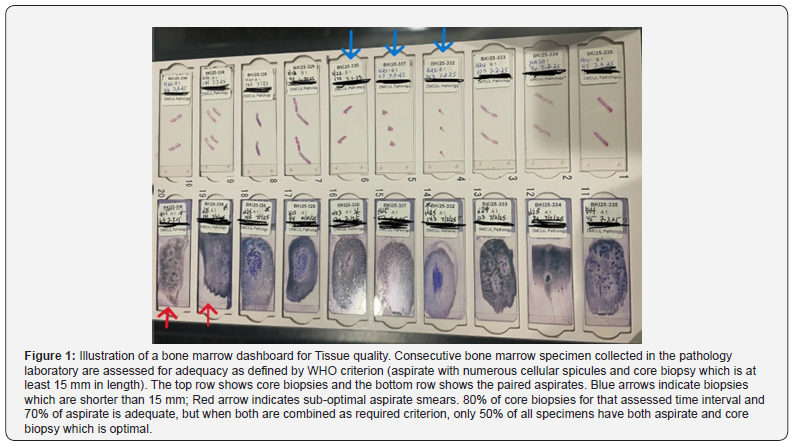
Bone marrow tissue adequacy for example is assessed based on adequately obtaining spicules (bone marrow intertrabecular particles) as well as a long core biopsy. The WHO defines the length of core biopsy tissue obtained to be at least 15 mm and that the particles should be adequate spread well in slides to make adequate morphologic assessment. Laboratories used to have laboratory-based technologists to assist in the procurement of bone marrow samples and they will make bedside assessment of adequacy. Unfortunately, shortage of staff and increasing expenses in the US healthcare systems, have precluded such ‘luxury’ in many institutions. This adequacy assessment, especially for precious tissue samples, should still be practiced to enable accurate diagnosis.
We utilize a “running” dashboard of 10 samples of bone marrow, to rapidly and visually assess bone marrow sampling quality. In this model, the best slide (aspirate and core biopsy) from each case is collected after sign-out. The 10 samples give a visual indicator of how many cases fulfill the adequacy criteria. This adequacy rate may go up or down, and is an useful feedback to the operator to look at causes (new trainee or new staff) if this is persistent and causes issues with tumor tissues (Figure 1).
Multi-disciplinary team communication - Tissue adequacy in open conversations - “Tumor is a rumor”
Unfortunately, good tissue adequacy criteria frequently gets lost in the ‘handing-over’ of tissue from surgeon-pathologist- (molecular) laboratory. Unless the end expert (in this case molecular) laboratory makes sure that they also incorporate tissue adequacy as part of their tumor tissue testing, their findings, in particular their negative findings should be interpreted with caution (False Negative results). Similar to the communication between pathologist and the molecular laboratory, a much wider tumor tissue adequacy communication between the radiologist and surgeon, surgeon and pathologist is also very important [10]. This may be enabled by multi-disciplinary tumor boards, where adequacy of tissue should be brought up when pertinent or if there is a surprising finding [11].
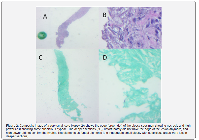
Such an example is illustrated in Figure 2. A Suspicious nodule from a patient with a history of large cell lymphoma was biopsied based on the radiologist impression and sent to pathologist. Unfortunately, the tumor was very small and did not have lymphoma in the sample. However, at the edge of the tumor sample, there appeared to be fungal like elements. A GMS stain was attempted but since the biopsied specimen was too small, the tissue did not show the suspicious focus in the GMS-stained slide. This inadequacy to rule in or rule out a lymphoma was communicated in a tumor board setting (pathologist presented it to oncologist, radiologist and surgeon) along with the suspicion of fungi but inability to confirm it. The tumor board forum was also utilized to decide the area(s) to biopsy optimal tissue samples. This approach leads to improvement of diagnostic tissue yields and better patient outcomes, since “good tissue is a no issue” in diagnostic oncopathology (Figure 2 & 3).
Conclusion
The fields of surgery, imaging, and pathology are undergoing tremendous progress. When tissue is procured for cancer diagnostics, adequacy is an important criterion, and systematic assessment of adequacy and communication of this adequacy to the multi-disciplinary team is very important in order to coordinate care, so that diagnostic accuracy is enhanced.
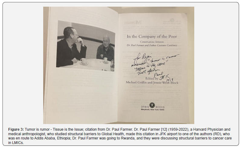
The title of this article is cited from Dr. Paul Farmer’s autographed book to one of the authors (RD). Paul Farmer was a Global Health leader from Harvard Medical School, working in several Low- and Middle-Income Countries (LMICs). Tissue inadequacy and artifacts were a problem in cancer diagnosis in these countries.
“For Rajan, Remember, Tumor is Rumor, tissue is the issue. With affection. Paul. JFK. 10/13”
The quotation is about the need for adequate tissue to make cancer diagnosis possible. He emphasizes the need for good quality Pathology, in the context of cancer diagnosis, in reference to Low- and Middle-Income Countries (LMIC). The authors emphasizes that this statement holds true for oncopathology everywhere, with illustrated examples in this article.
References
- Ishizawa T, Saiura A (2019) Fluorescence Imaging for Minimally Invasive Cancer Surgery. Surg Oncol Clin N Am 28(1): 45-60.
- Nacchio M, Palladino R, Vigliar E, Pisapia P, Salatiello M, et al. (2023) Evaluating local thyroid cytopathology practices by molecular quality metrics: A multi-institutional study on 4651 FNAs with a focus on the role of the interventional cytopathologist. Cancer Cytopathol 131(12): 772-780.
- Ahmed MS, Klippel-Almaraz D, Amin SE, Sura GH, Kundu UR, et al. (2025) Utility of whole-slide imaging for rapid evaluation of thyroid FNA: A multireader prospective study. Cancer Cytopathol 133(9): e70046.
- Melstrom KA, Kaiser AM (2020) Role of minimally invasive surgery for rectal cancer. World J Gastroenterol 26(30): 4394-4414.
- Melamed A, Eisenhauer EL (2024) Minimally invasive interval cytoreductive surgery for advanced ovarian cancer. J Surg Oncol 129(1): 126-127.
- F, Smith MA, Lane MC, Pantanowitz L, Dacic S, et al. (2015) Adequacy of core needle biopsy specimens and fine-needle aspirates for molecular testing of lung adenocarcinomas. Am J Clin Pathol 143(2): 193-200.
- Da Cunha Santos G, Saieg MA (2015) Preanalytic parameters in epidermal growth factor receptor mutation testing for non-small cell lung carcinoma: A review of cytologic series. Cancer Cytopathol 123(11): 633-643.
- Roy-Chowdhuri S, Stewart J (2016) Preanalytic Variables in Cytology: Lessons Learned from Next-Generation Sequencing-The MD Anderson Experience. Arch Pathol Lab Med 140(11): 1191-1199.
- Stoy SP, Segal JP, Mueller J, Furtado LV, Vokes EE, et al. (2018) Feasibility of Endobronchial Ultrasound-guided Transbronchial Needle Aspiration Cytology Specimens for Next Generation Sequencing in Non-small-cell Lung Cancer. Clin Lung Cancer 19(3): 230-238.
- Grosu HB (2018) EBUS-TBNA for the Diagnosis of Lymphoma: Time to Give In? J Bronchology Interv Pulmonol 25(3): 165-166.
- Clementsen PF, Konge L (2018) EUS-B-guided Biopsies of Lung Tumors. J Bronchology Interv Pulmonol 25(1): e3-e4.

























