




 OAJS Home



Abstract
Background: Methods: Results: Conclusions:
There are many techniques in rhinoplasty to perform the tip-plasty. The suture of the Fusion Sling, an embryologic curved Y-shaped structure that consists of dense connective tissue fibers and perichondrium-like material that connects LLC (Lower Lateral Cartilages) to the corresponding ULC (Upper Lateral Cartilages) and the contralateral LLC to the Anterior Septal Angle (ASA), it is a preservation method to support the nose tip. With this technique it is possible to control the projection and rotation of the nose tip. This retrospective study, which included patients in whom this suturing was the only technique used to support the nose tip, was performed on a total of 40 patients between May 2023 and May 2024 with a one-year follow-up of tip projection and rotation. As a result, we consider the Sling Suture is a useful technique to provide support for the nose tip that appears to allow control of the tip projection and rotation with results that last over time.
Keywords:Sling Suture; Fusion Sling; Deep Pitanguy Ligament; Tip-plasty; Tip Support; Tip Projection; Tip Rotation; Preservation Rhinoplasty
Abbreviations:LLC: Lower Lateral Cartilages; ULC: Upper Lateral Cartilages; ASA: Anterior Septal Angle; SMAS: Superficial Musculoaponeurotic System; ANSA: Anterior Nasal Septal Angle
Introduction
Pitanguy described in 1965 a ligament originating on the undersurface of the dermis and running down to and between the alar cartilages [1]. Letourneau and Daniel contributed with the first description of the nasal Superficial Musculoaponeurotic System (SMAS) [2]. Posteriorly, Saban et al in 2008, demonstrated that the medial SMAS divides into a superficial and a deep layer at the level of the internal nasal valve. The deep medial layer of the SMAS runs above the anterior septal angle and beneath the interdomal ligament and then runs in the membranous septum downward toward the anterior nasal spine, between the caudal border of the septum and the medial crura of the LLC. Saban concluded that the deep medial SMAS could correspond to the ligament described by Pitanguy [3].
Daniel RK described in 2011 the Tip Position Suture, a simple transverse suture between the anterior dorsal septum and the infralobular mucosa, to control the tip rotation and increase projection [4]. He further described it in 2018, given previous anatomical studies, as a simple transverse suture between the anterior dorsal septum and the deep portion of the Pitanguy ligament, naming it “THE MOST POWERFUL OF THE TIP SUTURES” [5]. Cakir et al in 2012 agreed with Saban that the deep medial layer of the SMAS is the Pitanguy ligament, and although this is not a true dermocartilaginous ligament, to acknowledge the contributions of Pitanguy, they used the term Pitanguy’s midline ligament and described its reconstruction during rhinoplasty to maintain the desired tip projection and rotation which also helps prevent the formation of a fluid-collecting dead space and stabilizes the supratip skin [6].
In 2024 Eskalen and Cakir described the Neo-interdomal Ligament Suture, in this technique the Pintaguy ligament is sutured to the WASA segment, which is located between the W point and the ASA. In open approach cases the Pitanguy ligament is first repaired by suturing between the medial crura soft tissue and the Pitanguy Ligament. This technique also uses a columellar strut graft to support the tip [7]. In 2024, Neves et al for the first time explain the Fusion Sling is “an embryologic structure that connects the cephalic border of the LLC to their vicinity, consists of perichondrium-like material in the scroll and supratip regions and dense connective tissue fibers between the medial crura, intercrural fascia”, and described how to preserve, during dissection in the midline, the Fusion Sling of the LLC along their entire length to then reconstruct it on the Anterior Septal Angle, this suture will serve as an anchor for the nasal tip, holding and defining its position. And concluded that for enhanced definition of the supratip it is necessary to resuture the vertical ligaments of the supratip [8].
In a recent study conducted by Neves et al., which included surgical dissections and cadaveric analyses followed by histological evaluation, particular attention was given to the Fusion Sling and associated anatomical compartments to assess the consistency of the suspensory structures. One of the principal histological findings, was the absence of identifiable ligamentous structures as discrete histological entities. Instead, at the level of the vertical and intercrural densifications, the observed tissue was predominantly fibroadipose in nature. These results indicate that what has been referred to as the “Pitanguy Ligament” is likely not an actual anatomical or histological structure, but rather a surgical construct is an artifact created during dissection.
In this last study the Fusion Sling is described as a curved Y-shaped structure that consist of dense connective tissue fibers perichondrium-like material formed during the embryologic development of the nose by the fusion of the lateral and medial olfactory processes. This structure extends from the pyriform aperture toward the nasal tip and connects the upper and lateral portions of the nasal cartilages, especially the cephalic border of the LLC, through a network of connective tissues that functions as a suspensory mechanism that stabilizes the nasal tip and divides the nose into anatomical compartments. It is composed of vertical densifications (connective fibers that connect the ULC and the LLC perpendicularly) and a horizontal perichondriumlike structure (extends along the cartilage surfaces as a supportive layer). Concluding that “the identification of the Fusion Sling as a distinct suspensory structure redefines conventional theories of nasal tip support. By providing a stable framework for both the medial and lateral compartments, it introduces new opportunities for rhinoplasty procedures that prioritize the preservation and enhancement of nasal tip stability” [9].
If it is understood that the fusion sling between the medial crura of the LLC is a structure of great importance for the support of the tip, it can be understood that its reconstruction/resuture is of utmost importance for the anchorage of the tip, and that according to the authors mentioned previously, it is a tool to achieve the desired projection and rotation. However, since there is no literature to support this above, we have decided to perform the following study in order to evaluate whether there is improvement in nasal tip projection and rotation with the use of the Fusion Sling suture alone (without another technique as Columellar Strut Graft, Anterior Nasal Septal Angle (ANSA Banner) Extension Graft, Septal Extension Graft, among others).
Surgical Technique
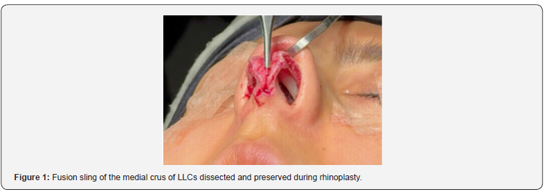
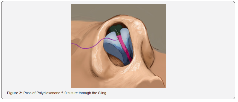
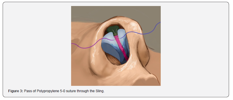
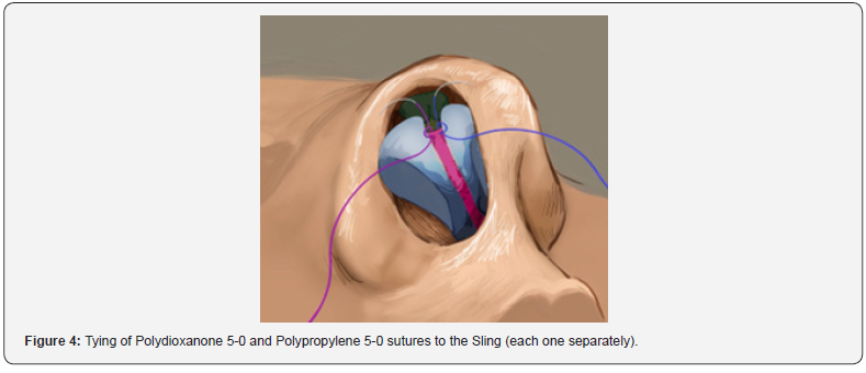
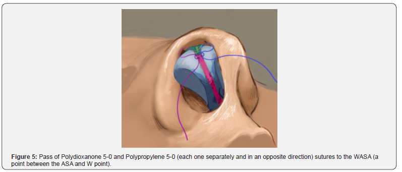
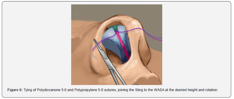
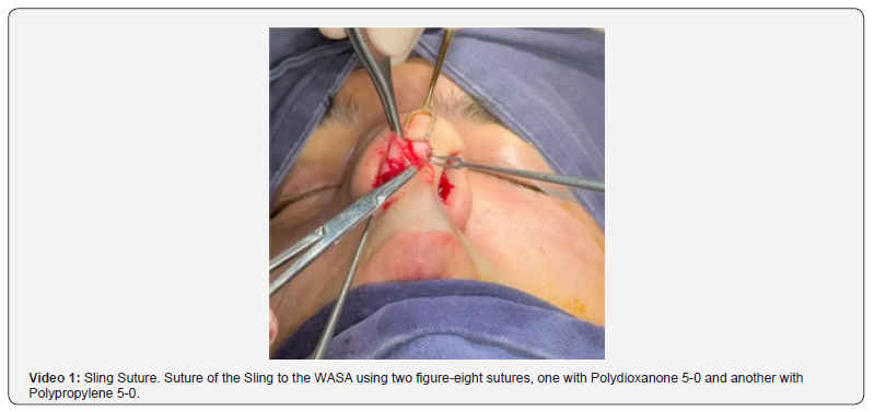
The Sling Suture is a preservation technique that can be performed using either an open or closed approach. A supraperichondrial dissection is carried out across the nasal tip cartilages and the ASA, preserving the Fusion Sling between the medial crura along its entire length. In Figure 1 you can see how the Fusion Sling preserved is held between the Adson forceps. When the surgeon wants to perform the tip-plasty the Fusion Sling is taken with forceps and is sutured to the WASA (a point between the ASA and W point) according to the desired projection and rotation, using two figure-eight sutures, one with Polydioxanone 5-0 and another with Polypropylene 5-0. Then the transdomal and interdomal suture is performed (Figure 1-6).
Materials and Methods
This observational, retrospective and descriptive study was approved by a local ethics committee and was performed in accordance with the Declaration of Helsinki. A total of 40 patients were included in this study, all adults (≥18 years old) who underwent Primary Rhinoplasty by the same Facial Plastic Surgeon (Nicolas Heredia, MD) using only the Sling Suture technique for tip-plasty in the period from May 2023 to May 2024 who authorized the use of their audiovisual material for scientific purposes by signing an informed consent and were followed for one year postoperatively. Exclusion criteria included patients who underwent Primary Rhinoplasty using another technique for tip support (such as Columellar Strut Graft, Anterior Nasal Septal Angle (ANSA Banner) Extension Graft, Septal Extension Graft, among others), Revisional Rhinoplasty, those who did not consent to the use of their images, and those who could not be followed for one year.
Measurements
A Nikon D700 camera with a Nikkor AF 52 mm lens was used to obtain standardized photographic images of all patients. Profile photographs were taken in the Frankfurt plane to perform profilometry and evaluate tip projection (measured from the alar crease to the nasal tip) and tip rotation (measured as the nasolabial angle). The same technique was applied preoperatively and at one year postoperatively: the camera positioned 2 meters from the patient, with the patient upright and the Frankfurt plane parallel to the floor, and the patient placed 1 meter away from the background (Table 1).

Results
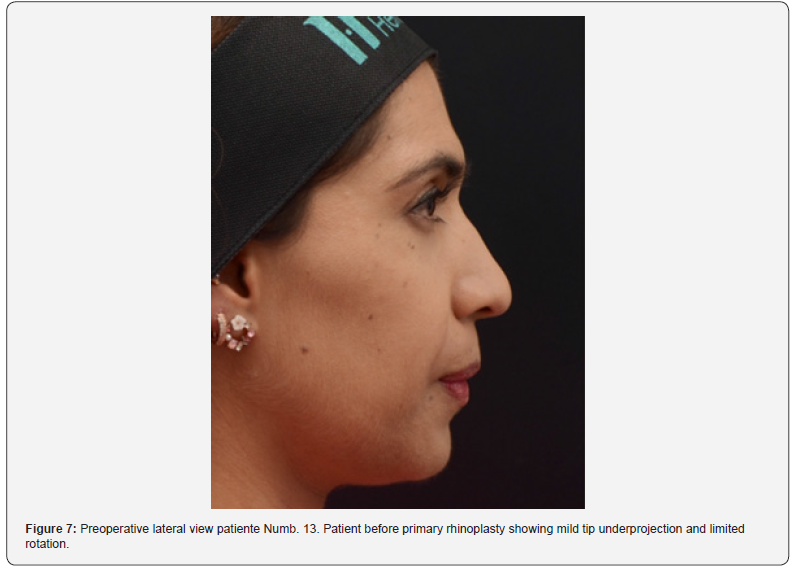
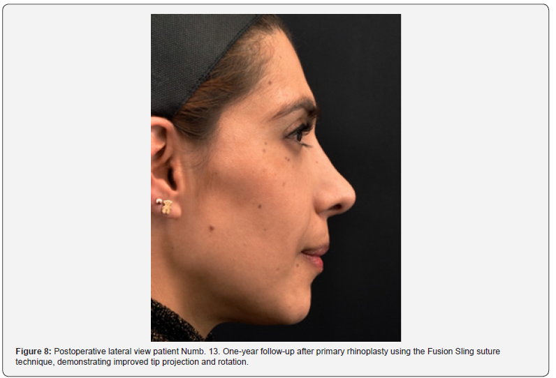
There were a total of 40 patients included in this study. The mean age of the patients was 30 years with an age range from 18 to 51 years. There were 30 females and 10 males with a sex ratio 3:1. 57.5% (23 cases) were performed with a closed approach and 42.5% (17 cases) with open approach. 5 patients needed revision surgery, 3 were performed in patients who underwent open approach and 2 in patients who underwent close approach. Three due to a recurrence of the hump, one due a loss of tip definition and one due a deviation of the tip and nostril asymmetry. No revision surgeries were required due to loss of projection or rotation of the nasal tip (Figure 7 & 8).
Discussion
The findings of this study support that the Fusion Sling suture, when used as the sole method for nasal tip support in primary rhinoplasty, provides reliable and lasting control over nasal tip projection and rotation. This technique, based on a preservation philosophy, utilizes the Fusion Sling, preserves and re-sutures it to the ASA, providing an effective and minimally invasive alternative to more traditional structural grafting methods such as columellar strut or septal extension grafts. The anatomical understanding of the Fusion Sling has evolved in recent years. Our findings support the idea proposed by Saban, Daniel RK, Cakir and Neves that this midline ligamentous structure plays a significant role in nasal tip support and can be harnessed to control tip dynamics. Importantly, this study builds on the work of Neves et al by being the first to evaluate clinical outcomes in a series of patients using the Fusion Sling suture technique alone.
Over a one-year follow-up, none of the patients presented with a loss of nasal tip projection or rotation, despite the absence of additional tip support structures. This underscores the mechanical strength and anchoring ability of the Fusion Sling suture. Furthermore, although five patients underwent revision surgery, none were due to failures in maintaining tip support, highlighting the functional reliability of this method. The surgeries were performed using both open and closed approaches, reinforcing that this technique is versatile and suitable for less invasive rhinoplasty methods. While aesthetic refinements or deviations required revision in a small subset, these outcomes were not related to the performance of the Fusion Sling itself, but rather to other structural or surface irregularities.
Although this study provides encouraging results regarding the use of the Fusion Sling suture as a sole method for nasal tip support, further research is required to confirm these findings. One important limitation is that only photographic measurements were used to evaluate tip projection and rotation. Future studies should incorporate intraoperative measurements of nasal tip position and compare them with postoperative and long-term follow-up data. This approach would allow a more precise understanding of the dynamic changes in tip support and stability over time. Prospective studies with larger cohorts, standardized measurement protocols, and comparative groups using alternative tip support techniques will be essential to establish the reproducibility and generalizability of our results.
Conclusion
The Fusion Sling Suture appears to be a reliable and effective technique for nasal tip support in primary rhinoplasty. When used as the sole method for tip stabilization, it allows for predictable control of tip projection and rotation, with durable results over a one-year follow-up. This preservation-based approach minimizes the need for additional grafts and may reduce operative complexity and donor site morbidity. Based on our experience, the Fusion Sling suture represents a valuable tool in the armamentarium of modern rhinoplasty, especially for surgeons seeking to maintain nasal tip dynamics with minimal structural alteration.
References
- Pitanguy I (1965) Surgical importance of a dermocartilaginous ligament in bulbous noses. Plastic and Reconstructive Surgery 36(2): 247-253.
- Letourneau A, Daniel RK (1988) The superficial musculo-aponeurotic system of the nose. Plastic and Reconstructive Surgery 82(1): 48-57.
- Saban Y, Andretto Amodeo C, Hammou JC, Polselli R (2008) An anatomical study of the nasal superficial musculoaponeurotic system: Surgical applications in rhinoplasty. Archives of Facial Plastic Surgery 10(2): 109-115.
- Daniel RK (2011) Rhinoplasty: Open tip suture techniques: A 25-year experience. Facial Plastic Surgery 27(2): 190-203.
- Daniel RK, Palházi P (2018) Rhinoplasty: An anatomical and clinical atlas.
- Çakir B, Öreroglu AR, Doğan T, Akan M (2012) A complete subperichondrial dissection technique for rhinoplasty with management of the nasal ligaments. Aesthetic Surgery Journal 32(4): 564-574.
- Eskalen A, Çakir B (2024) A suture technique for nasal tip support: 'Neo-interdomal ligament suture.' Aesthetic Plastic Surgery 49: 2439-2445.
- Neves JC, Toriumi DM, Göksel A (2024). Dorsal preservation rhinoplasty. Facial Plastic Surgery Clinics of North America 32(4): 585-602.
- Neves JC, Rijo-Cedeño J, Teixeira Rodrigues JL, Filgueiras H, Sa Sodré H, et al. (2025) The Fusion Sling-Revisiting the nasal tip suspensory anatomy. Facial Plastic Surgery Clinics of North America 33(2): 143-157.

























