




 OAJS Home



Intracranial Reticular Hemangioendothelioma: Report of a Rare Case
Haibing Xiong1, Jianhong Huo1, Chunfang Guo1, kangyi Liu2, Minghai Yu3 and Yi Li3*
1Affiliated Banan Hospital of Chongqing Medical University, Department of Neurosurgery, China
2Zunyi Medical University, Department of Stomatology, China
3Chengkou County People’s Hospital, Department of Neurosurgery, China
Submission:March 17, 2025;Published:March 24, 2025
*Corresponding author:Yi LI, Chengkou County People’s Hospital, Department of Neurosurgery, China.
How to cite this article:Haibing X, Jianhong H, Chunfang G, kangyi L, Minghai Y, et al. Intracranial Reticular Hemangioendothelioma: Report of a Rare Case Open Access J Surg. 2025; 16(3): 555938.DOI: 10.19080/OAJS.2025.16.555938.
Abstract
As this rare case of acute renal infarction in a long-failed renal graft illustrates, evaluation of abdominal pain in transplant recipients requires a very broad differential diagnosis. Acute renal infarction, often due to thromboembolic events, results in tissue necrosis with varying signs and symptoms that often include local pain, fever, reduced renal function, and systemic inflammation. The 65-year-old subject of this case report is a healthy and fit recipient of two kidney transplants on long-term immunosuppressive medications. He presented with right lower quadrant abdominal pain, intermittent moderate fevers, anorexia, and malaise. Laboratory testing most prominently showed systemic inflammation with significantly elevated C-reactive protein level and erythrocyte sedimentation rate. Abdominal CT scanning showed fat stranding and small bowel obstruction. After consideration of typical surgical etiologies; viral, bacterial, and parasitic infections; autoimmune disease, and kidney rejection, he was diagnosed with an inflammatory reaction to acute infarction of his first 40-year-old non-functioning renal graft. A month-long prednisone taper, held earlier due to concerns about worsening an infectious process, produced dramatic pain relief and a reduction in systemic inflammation. The subject resumed both work and vigorous exercise with only occasional, mild right lower quadrant pain likely from continuing renal necrosis. This case emphasizes: 1) the potential longevity and quality of life possible with kidney transplant, 2) common and uncommon causes of abdominal pain that must be considered in renal transplant recipients, 3) the diverse symptoms attributable to inflammation, and 4) the unique features of renal infarction occurring in a non-functioning renal graft.
Key words: Renal Transplant, Acute Abdomen; Renal Infarction; Systemic Inflammation; Retransplantation; Failed Renal Graft
Abbreviations: Abs IG: Absolute Immature Granulocyte Count; Abs LMC: Absolute Lymphocyte Count; Abs NTC: Absolute Neutrophil Count; ALT: Alanine Transferase; BUN: Blood Urea Nitrogen; COVID-19: Corona VIrus Disease of 2019; CT: Computed Axial Tomography; CMV: Cytomegalovirus; DNA: Deoxyribonucleic Acid; EBV: Epstein-Barr Virus; EMD: Emergency Medical Department; ESR: Erythrocyte Sedimentation Rate; HSV: Herpes Simplex Virus; HZV: Herpes Zoster Virus; Kg: Kilogram; NF: Nonfasting; PCR: Polymerase Chain Reaction; RNA: Ribonucleic Acid; Rx: Prescription for; SII: Systemic Immune-Inflammatory Index; Sx: Symptoms; WBC: White Blood Cell Count
Introduction
Reticular hemangioendothelioma is a rare intermediate vascular tumor. Only about 70 cases have been reported in the literature. The tumor develops in the skin and subcutaneous tissues, and there are no reports of RH with intracranial origin. In this article, we report the clinical data of a patient with intracranial reticular hemangioendothelioma and summarize the diagnostic and therapeutic features of intracranial reticular hemangioendothelioma in the light of the literature.
Key words:Reticular; Vascular Endothelioma; Intracranial; Rare
Abbreviations:RH: Retiform Hemangioendothelioma; CT: Computed Tomography; MRI: Magnetic Resonance Imaging; WHO: World Health Organization
Introduction
Reticular hemangioendothelioma (retiform hemangioendothelioma RH) is a rare intermediate-type tumor of vascular origin. The tumor consists mainly of an elongated, branching testis-like vascular network with peg-like endothelial cells. It was first reported by Calonje et al. in 1994 [1], and only about 70 cases have been reported in the national and international literature [2]. The tumor is most commonly found in the skin and subcutaneous tissues, with a few reports of occurrence in rare sites such as the pleura [3]. There are no reports of RH with intracranial origin. The aim of this study is to analyze the pathological features, clinical manifestations, imaging presentation and treatment of this disease based on previous reports in the literature.
Case Reports
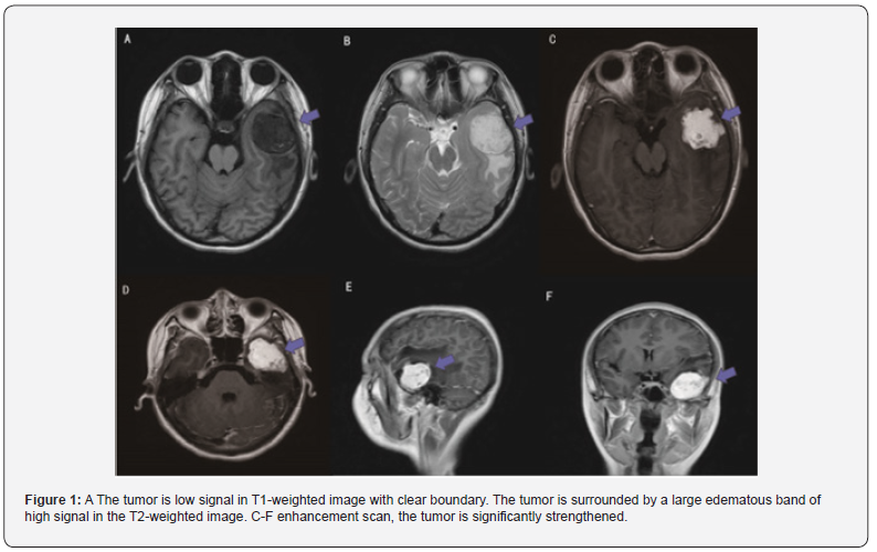
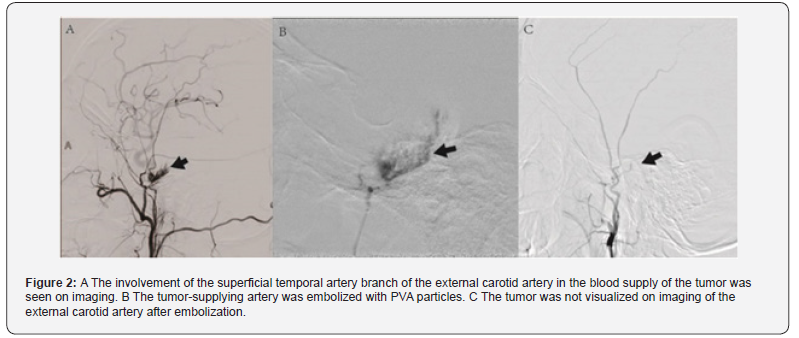
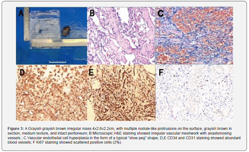
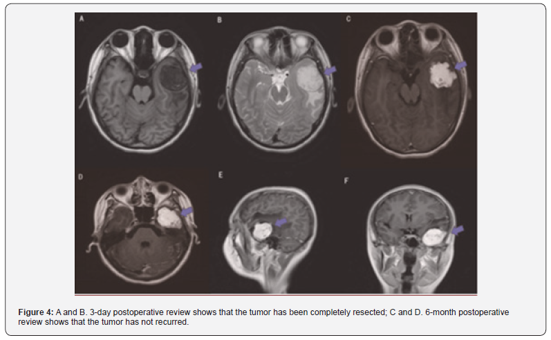
The patient, a 19-year-old female, was admitted to the Department of Neurosurgery, Banan Hospital of Chongqing Medical University on September 8, 2023, the patient started to have headache 2 weeks ago, which was mainly characterized by left-sided head swelling and pain. The patient did not pay attention to it and did not go to the hospital. Four hours before admission, the patient had a sudden convulsion with loss of consciousness in front of his home, which stopped on its own after a few minutes. The ambulance sent to the local hospital after the examination of cranial CT suggests left temporal lobe occupational lesion possibility, bleeding to be drained. Re-transferred to our hospital. Admission examination showed no obvious neurological abnormalities. After admission levetiracetam antiepileptic and mannitol decongestive therapy were given. Examination of MR showed that a mass of about 44x34mm in size was seen in the left temporal region (Figure 1), which was considered to be more likely to be an angioepithelioma, with meningiomas to be drained. From the imaging, the intracranial tumor blood supply was very rich, and the surgery might bleed a lot. The treatment plan was chosen to perform preoperative angiography to clarify the blood-supplying arteries of the tumor, and then interventional embolization of the blood-supplying arteries to reduce intraoperative bleeding before total resection of the intracranial tumor. Cerebral angiography + embolization of tumor blood-supplying arteries was performed under local anesthesia at 09:00 on August 25, 2023 (Figure 2). After successful embolization, craniectomy for left temporal lobe occupying lesion was performed on August 26, 2023, at 1005 hours under general anesthesia. The operation went well and the tumor was seen to originate from the meninges at. Surgical total resection of the tumor was performed, and the tumor envelope is intact (Figure 3A). After surgery, levetiracetam antiepileptic treatment was continued. Postoperative follow-up cranial MRI showed that the tumor was completely resected (Figure 4A and B). Postoperative pathology showed extensive vascular proliferation within the lesion with lobulated structures, significant anastomosis of the proliferating vessels, and papillary proliferation of endothelial cells in some areas. A fibrous peripheral envelope was seen around the lesion, which was clearly demarcated from the brain tissue. Immunohistochemistry: EMA(-), Vim(+), PR(-), GFAP(-), S100(-), SOX10(-), CD34(+), hCD31(+), ERG(+), Desmin(-), SMA(weakly +), actin(weakly +), Calponin(-);EMA(-), CK(-) , Vim(+), PR(-), ER(-), S100(-), Ki67 (~2%+) (Figure 3B-F). No postoperative radiotherapy was performed because the surgery had been total excision. The tumor did not recur during the six-month follow-up after surgery (Figure 4C and D).
Discussion
The 5th edition of the WHO Soft Tissue Tumor Classification released in 2020 defines RH as a rare intermediate vascular tumor that rarely metastasizes [4]. Intermediate (rarely metastasizing). RH was first reported to occur in all age ranges with no apparent gender predilection [1]. However, recent reports have shown that RH occurs predominantly in young and middle-aged people, with more females than males [3,5,6], which is consistent with the case reported herein.
RH occurs in the skin and subcutaneous tissues throughout the body, mostly as exophytic or patchy skin lesions, usually as solitary nodules or plaques of blackish red to brownish, purple color with pain, or the skin may show only thickening, elevation, and swelling, and there may be fusion of the nodules, thickening of the skin, or formation of vesicles or ulcers [7,8]. There have been 2 cases reported to occur in the pleura and both patients had dry cough and dyspnea [9,10]. There are no reports of RH with intracranial origin, and only two cases of RH originating from the skull have been reported worldwide [11,12]. These two cases originated from the parietal bone and the pterygoid bone, respectively, and both of them had bone destruction. The tumor in the case reported in this paper originated in the meninges, with destruction of the meninges, without affecting the skull, and the tumor compressed the nerves causing epileptic symptoms.
Imaging manifestations of RH vary depending on the origin. CT manifestations of pleural RH are mainly irregular masses [9,10]. The imaging manifestations of RH of bone origin all have osteolytic bone destruction [11-13]. The present case is the first reported case of intracranial RH, which has some of the features of benign and malignant intracranial tumors. The CT presentation of RH was a well-defined slightly low-signal mass. Like other intracranial tumors, the main preoperative diagnosis relied on MRI. The mass was low signal in T1-weighted images and high signal in T1-weighted images with clear borders. Due to the slightly rapid growth of RH, mixed signal shadows may appear within the tumor. Due to the very rich blood supply of tumors of vascular origin, obvious enhancement of the tumor can be seen on enhancement scan. MRS can be used as an auxiliary diagnostic test, and RH tumors have low NAA wave peaks and no neuronal markers, suggesting non-neuronal brain tumors. Decreased Cho and increased ML peaks are in line with the characteristics of intermediate tumors.
The diagnosis of RH is mainly based on pathological examination, and its histological features mainly include Extensive blood vessels in the tumor are irregularly proliferating and anastomosing with each other to form a reticulum. Vascular endothelial cells are uniform in size, with little cytoplasm, and some endothelial cells have protruding nuclei, which appear as “boots nails” into the lumen of the vessels. There is no nuclear heterogeneity or mitosis. Immunohistochemistry showed positive expression of vascular endothelial cell markers CD34, CD31 and ER G [13,14]. This case showed typical reticular vascular hyperplasia and the “boot nail” sign, which, combined with immunohistochemistry, was consistent with the pathologic diagnosis of RH.
Surgical total resection is the treatment of choice for RH. Surgery is challenging because of the very rich blood supply of RH and the relative difficulty of intracranial hemostasis. Preoperative cerebral angiography is recommended, and if branches of the external carotid artery can be found to be involved in the blood supply of the tumor, these blood-supplying arteries should be embolized immediately. Postoperative adjuvant therapy needs to be decided according to the type of pathology and surgery. Local radiotherapy combined with cisplatin chemotherapy is recommended for patients with incompletely resected tumors and with lymph node metastases [7]. Postoperative pathology should focus on Ki67. For patients with completely resected tumors, local radiotherapy is recommended if Ki67 is >15%. For patients with Ki67>30%, local radiotherapy combined with chemotherapy is recommended. Metastasis of RH is rare, but recurrence is possible, and strict follow-up is needed after surgery. For recurrent RH, surgery combined with postoperative radiotherapy is recommended.
Conclusion
In summary, RH matters a rare intermediate-type vascular tumor, and intracranial RH is very rare. It is difficult to diagnose clinically, and the diagnosis should be confirmed by pathology. Surgical resection is the first choice of treatment, and patients can benefit from preoperative embolization of the bloodsupplying arteries because of the rich blood supply of the tumor. Radiotherapy is effective in postoperative adjuvant treatment of RH, depending on the type of pathology and the surgical situation, and RH has the potential to recur, requiring strict longterm follow-up after surgery. Nowadays, the number of reports on intracranial RH in the world literature is low, and the human exploration of the diagnosis and treatment of this disease is still continuing, which requires more case experiences and persistent follow-up by scholars around the world.
Consent for Publication
The patient has given written consent to the publication of this case report and any accompanying images.
Author’s Contribution
BXH and KYL performed literature review and drafted the manuscript. JHH helped to collect and review radiological data. CFG performed the histological and immunohistochemical evaluation.LY operate on the patient and revised the manuscript. All authors contributed to this paper and approved the submitted version.
Funding
This study was supported by a grant from the Chongqing Science and Health Joint Medical Research Program (2021MSXM261).
Acknowledgement
We would like to thank Professor Yi Tang of Chongqing Medical University for pathological diagnosis of this case.
Conflict of Interest
The authors declare that this article does not contain any commercial or financial relationships with potential conflicts of interest.
References
- E Calonje, CD Fletcher, E Wilson-Jones (1994) Retiform hemangioendothelioma. a distinctive form of low-grade angiosarcoma delineated in a series of 15 cases. Am J Surg Pathol 18(2): 115-125.
- Qurratulain Chundriger, Muhammad Usman Tariq, Shabina Rahim, Jamshid Abdul-Ghafar, Nasir Ud Din (2021) Retiform hemangioendothelioma: a case series and review of the literature. J Med Case Rep 15(1): 69.
- Khade Shalaka, Rao Meenakshi, Vishnoi Jeewan R, Soudamini Arsha Balakrishnan, Aasma Nalwa (2022) Retiform Hemangioendothelioma: A Rare Tumor in the Medial Canthus. Journal of Cutaneous and Aesthetic Surgery 15(3): 319-322.
- Fletcher CDM, Bridge JA, Hogendoorn PCW, Mertens F (2020) WHO classification of tumors of soft tissue and bone. 5th ed Lyon IARC.
- Jasmine S Saleh, Carli P Whittington, Scott C Bresler, Rajiv M Patel (2023) Cutaneous vascular tumors: an updated review. Hum Pathol 140: 53-65.
- Folpe Andrew L (2024) Vascular tumors of intermediate malignancy: An update. Hum Pathol 147: 114-128.
- Zhang Mingyue, Yin Xiaojiao, Yao Weikai, Ning Liu, Ying Yue (2021) Retiform hemangioendothelioma: a rare lesion of the vulva. J Int Med Res 49(8): 3000605211027783.
- Jiang J, Li X, Zhu F, Yue He (2021) Retiform hemangioendothelioma of the mandible: A case report. Oral Oncology 115: 105120.
- Ullah Hidayat, Khan Imran, Khan Aria, Apoorva Tangri, Saral Lamichhane, et al. (2024) The Pleural Origin of Retiform Hemangioendothelioma: An Unusual Origin of a Rare Diagnosis. Case Rep Oncol 17(1): 741-746.
- Liu Q, Ouyang R, Chen P, Rui Zhou (2015) A case report of retiform hemangioendothelioma as pleural nodules with literature review. Diagnostic Pathology 10: 1-5.
- Kim I K, Cho H Y, Jung B S, Sang-Pill P, Hyun-Woo C, et al. (2016) Retiform hemangioendothelioma in the infratemporal fossa and buccal area: a case report and literature review. Journal of the Korean Association of Oral and Maxillofacial Surgeons 42(5): 307-314.
- GS Aditya, Vani Santosh, TC Yasha (2004) Epithelioid and retiform hemangioendothelioma of the skull bone--report of four cases. Indian J Pathol Microbiol 2003 46(4): 645-649.
- Jiang Xiaoguang, Li Fengshuo, Zhu (2020) Retiform hemangioendothelioma of the mandible: a case report. Oral Oncol 115: 10.
- DJ Jr, Papke JL Hornick (2022) Recent advances in the diagnosis, classification and molecular pathogenesis of cutaneous mesenchymal neoplasms. Histopathology 80(1): 216-232.

























