Torso Tractotomy – A Viable Approach to Thoracic Impalement Injury
Madhur Uniyal*, Rupesh Pakrasi and Agniva Mukhopadhyay
Department of Trauma Surgery and Critical Care, All India Institute of Medical Sciences Rishikesh, India
Submission: May 19, 2024; Published: August 05, 2024
*Corresponding author: Madhur Uniyal, Department of Trauma Surgery and Critical Care, All India Institute of Medical Sciences Rishikesh, India
How to cite this article: Madhur Uniyal*, Rupesh Pakrasi and Agniva Mukhopadhyay. Cerebral Hyperperfusion Syndrome: A Mini-Review. Open Access J Surg. 2024; 15(4): 555919. DOI: 10.19080/OAJS.2024.15.555919.
Abstract
Objective: Impalement injuries are considered one of the most severe forms of penetrating injuries. Individuals with penetrating injuries who reach hospital in time have a chance at survival. In the following case we approach the patient via torso tractotomy which has rarely been discussed in literature.
Material and Methods: In the following case report, we describe the presentation of the patient and approach via torso tractotomy incision after which the outcome and recovery of the patient has been described
Result: We present the successful use of the Tractotomy approach in a case of impalement injury to the right thorax treated with removal of metallic rod along with repair of right lung laceration with repair of muscles and soft tissue over back and right hemithorax.
Conclusion: Tractotomy is a viable approach to treat thoracic impalement injuries as per literature and can be considered as a standard approach in case of similar injuries in the future.
Keywords: Tractotomy; Impalement Injury
Abbreviations: ATLS: Advanced trauma life support; CXR: Chest EX-ray; OR: Operating room
Case Report
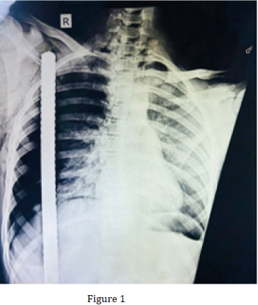
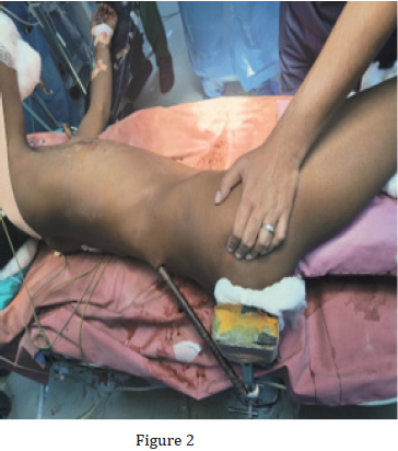
A 17-year-old male suffered an impalement injury with a large iron rod when the vehicle he was driving, fell over a construction site. It was a through and through penetrating injury over his right hemithorax and back. The iron bar pierced the right flank region and was seen coming out through the right chest wall. The patient was immediately rushed to the local hospital where initial resuscitation was done and then patient was referred to AIIMS Rishikesh for definitive management. On presentation, patient was evaluated as per ATLS protocol and following findings were noted. His airway was patent, and he was breathing spontaneously and maintaining SpO2 of 98% on 5lit/min oxygen support via face mask with right sided ICD in situ. On further examination, his right chest wall movement was found to be absent and there was diminished breath over right lung field. However, patient was hemodynamically stable, and rest of the primary survey was normal. A CXR was done in the previous hospital when patient arrived, and it showed the metallic bar traversing through the right hemithorax (Figure 2). Patient was in left lateral decubitus position due the impaling rod and was unable to change his position (Figure 1).
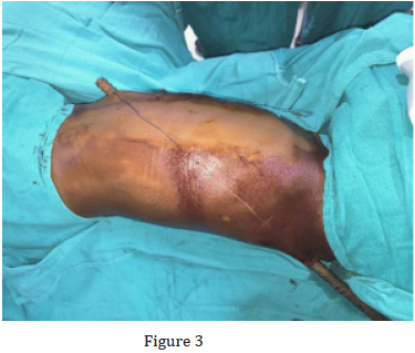
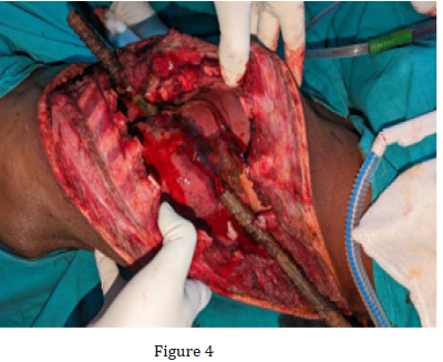
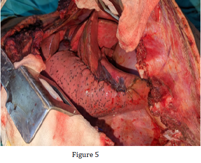
The patient was taken to the operating room (OR) after obtaining an informed consent. The relatively fixed position of the patient posed significant challenge to the anaesthesia team while intubation. The patient had to be intubated in the left lateral decubitus position only and a double lumen tube of no. 39 was used. Patient was then positioned properly for right posterolateral thoracotomy and a tractotomy incision was then made extending from the entry point to the exit point (Figure 3). The incision was deepened and the right-side Latissimus dorsi muscle was then divided. Upon reaching the thoracic cage, right 4th-10th ribs were divided for proper exposure. Intraoperatively, the iron rod was seen to have traversed through subcutaneous and muscular tissue of right posterior abdominal wall and posterior chest wall for almost 8cm before entering the thoracic cavity. Within the thoracic cavity, the rod penetrated the right lower lobe (Figure 4). Rest of the lung tissue appeared normal. The muscles and soft tissue over right flank with right side of back and chest wall were also injured along the tract. After complete assessment of all the injuries, decision was taken to remove the foreign body followed by definitive repair. The Lung tissue over the iron rod was divided using 60mm white (vascular) endo-stapler. Soft tissue over the tract of the iron rod was divided and it was then carefully removed. The residual lung laceration was repaired with prolene 3-0 continuous suture (figure 5). Thorough debridement if all the devitalized tissue was done and the thoracic cavity was irrigated with copious warm saline. Satisfactory expansion of right lung under positive pressure was ensured intra-operatively. Through saline lavage was also given to the injured soft tissue and the incision was then closed in layers. Around 3cm of the incision near the entry site was kept open as this area was grossly contaminated. Regular dressing was done, and the defect was closed with secondary suturing on POD 5. Patient was kept intubated for around 18hour and was successfully weaned on POD 1. Patient had an otherwise uneventful postoperative recovery and was discharged on POD 7.
Discussion
Impalement injuries are defined as injuries sustained due to penetration with fixed elongated objects1. In today’s date these injuries are uncommon and happen mostly due to fall on a sharp object from height 2. In these types of injuries, manipulation or any attempts of extraction of the foreign object in the prehospital setting should be avoided, as very often it acts as a tamponade. Attempt at removal of this object in an uncontrolled environment might lead to exsanguinating haemorrhage and death. CT scan is difficult in these cases due to peculiar positioning of the patient as well as the metal rod interfering with the movement of the machine. Further Metallic objects in CT scan produce artefacts which appear as bright and dark streaks and lower the diagnostic value of the scan. Impalement through the right chest can cause severe injury to the lung, great vessels and heart however it is comparatively less common when compared to right lung3. In our case, the iron rod pierced the right back and then entered the Costo diaphragmatic recess [1-3].
The rod exited through the right chest just above the right diaphragm, thus narrowly missing the liver and right kidney. There are no universal protocols for impalement injuries due to inadequate data and hence management of such patients pose significant challenge. It is usually recommended that a diverse team consisting of CTVS surgeon, GI surgeon, Plastic surgeon and orthopaedic surgeon manage the case, however in our case the entire operation was successfully managed by trauma surgeons [4]. Anaesthesia is extremely challenging in these patients due the peculiar and fixed position of the patient. Hence should be handled by and experienced anaesthetist [5]. Torso tractotomy is a quick incision and allows lifting off impaled objects. It allows good exposure of the areas of interest and allows easy haemostasis hence can be considered as a standard approach for impalement injuries of the chest and abdomen [6].
Though impalement injuries may appear difficult to manage, proper and timely management can result in successful outcome. Proper planning of both anaesthesia and surgery are key to management. VATS approach is not a favourable approach in impalement injury as slight manipulation of the impaled object may lead to torrential haemorrhage, and we would lack the exposure to control it. Torso tractotomy provides the best exposure hence increases the spectrum of surgical options and enhanced safety and thus appears to be most viable approach to such injuries.
References
- Robicsek F, Daugherty HK, Stansfield AV (1984) Massive chest trauma due to impalement. J Thorac Cardiovasc Surg 87(4): 634-636.
- M Bemelman, ER Hammacher (2005) Rectal impalement by pirate ship: A case report. Injury Extra 36(11): 508-510.
- Robicsek Francis, Harry K Daugherty, Alan V Stansfield (1984) Massive chest trauma due to impalement. The Journal of thoracic and cardiovascular surgery 87(4): 634-636.
- Anthony J Cartwright, Karel O Taams, M Jonathan Unsworth-White, Nilofer Mahmood, Peter M Murphy (2001) Suicidal nonfatal impalement injury of the thorax. The Annals of Thoracic Surgery 72(4): 1364-1366.
- Sawhney C, D'souza N, Mishra B, Gupta B, Das S (2009) Management of a massive thoracoabdominal impalement: a case report. Scand J Trauma Resusc Emerg Med 17(50).
- Biplab M, Sushma S, Subdoh K, Mohit J, Chhavi S. et al. (2016) ‘Torso-Tractotomy’- A New and Apt Terminology for a Novel Surgical Incision and Approach for Managing Penetrating Torso Injuries, Particularly Impalement or Trans fixation Injuries: Naming and Report of a Technique. Open Access J Surg 2(1): 555577


