Application of Pressure Injury Classification
Kanav Gupta1, Ravi Kumar Chittoria2*, Jacob Antony C3 and Padmalakshmi BM4
1,3Department of Plastic Surgery, MBBS, MS General Surgery, DNB General Surgery, Senior Resident, JIPMER, Puducherry, India
2Senior Professor and Associate Dean (Academic), Head of IT Wing and Telemedicine, Department of Plastic Surgery and Telemedicine, JIPMER, Puducherry, India
3Assistant professor, Department of Plastic Surgery, JIPMER, Puducherry, India
Submission: May 12, 2024; Published: May 22, 2024
*Corresponding author: Ravi Kumar Chittoria, Professor and Associate Dean (Academic), Head of IT Wing and Telemedicine, Department of Plastic Surgery and Telemedicine, JIPMER, Puducherry, India, Email: drchittoria@yahoo.com
How to cite this article: Kanav Gupta, Ravi Kumar Chittoria*, Jacob Antony C and Padmalakshmi BM. Application of Pressure Injury Classification. Open Access J Surg. 2024; 15(3): 555915. DOI: 10.19080/OAJS.2024.15.555915.
Abstract
Pressure sores are a serious complication of multi morbidity and lack of mobility. Pressure injuries are commonly encountered in geriatric patients. Certain well-recognized risk factors, such as immobility and incontinence, may predispose to the development of pressure injury (PI); consequently, risk factor modification is an important aspect of prevention and treatment. One of the fundamental documentation details when describing a wound is the use of the appropriate staging system. Each staging system is unique to a wound’s type, should align with your description of the wound, and be integrated into your documentation details. For research purpose more improvements in staging with inclusion of infection, progression of ulcer and major risk factors on existing stages of only depth has been suggested by Kumar. This will have bearing on the prognosis of the disease and management protocol and hence better outcome of research work. The most extensive published experiences with pressure sore treatment are those of Conway and Griffith (1000 cases) and Dansereau and Conway’s update of the Bronx Veterans Administration Hospital data (2000 cases). But with new technologies of wound care more research is required to standardize the prevention and management of pressure injury. There is a need to reduce cost of the care for long term compliance. For research purposes more improvements in staging with inclusion of infection, progression of pressure injury and major risk factors in existing staging (four stages) of depth only has been suggested by Kumar. This will have bearing on prognosis of the disease and management protocol and hence, better outcome of research work. The classification has been updated by inclusion of suspected deep tissue injury. This study highlights the role Kumar classification applied on a case of pressure injury.
Keywords: Kumar; Classification; Pressure; Injury; Management
Abbreviations: PI: Pressure Injury; DS: Depth Stages; IS: Infection Stages; PS: Progression Staging; RC: Risk Category
Introduction
Pressure injuries pose a significant risk for individuals dealing with multiple health conditions and limited mobility. They are frequently observed among elderly patients. Factors such as immobility and incontinence are well-known contributors to their development, underscoring the importance of addressing these risk factors in prevention and management efforts [1,2]. Proper documentation of wounds is crucial, with classification systems tailored to each wound type playing a central role in accurate description and treatment planning. Kumar P proposes a new classification for pressure injuries (PI), outlined as follows:
Depth Stages (DS)
1. S. Suspected deep tissue injury.
2. Pre-ulcer stage –redness over pressure point.
3. Epidermal/ superficial dermal desquamation, abrasion, blistering, ruptured blister with pink floor.
4. Deep dermal- white/ purple floor.
5. Subcutaneous- exposed fat/ granulation forms the floor.
6. plus - Undeterminable Depth: covered with black coloured dead tough tissue.
7. Ligament/bone exposed with or without granulation tissue.
Infection Stage (IS)
1. Wound Culture negative.
2. Wound Culture positive; blood culture negative.
3. Systemic signs positive for Sepsis (Blood culture +).
Progression Staging (PS)
1. On first examination.
2. Regressing/ healing (on two consecutive inspection after 3 days interval).
3. Stationary (status co on two consecutive inspection after 3 days interval).
4. Progressing (progressively increasing soakage/slough/ necrotic tissue on two consecutive inspection after 3 days interval).
Risk Category (RC)
• Ambulatory (a)
• Bed ridden (b)
1. Protective sensation present.
2. Protective sensation absent/ altered consciousness.
3. Incontinence presents with or without protective sensation/altered consciousness.
4. Deformity of spine/ extremity joints/spasticity/ multiple pressure ulcers causing abnormal prominence of pressure points and or changing the position of the patient is difficult due to some or other reason [3].
This study highlights the application of Kumar classification on one of the cases of pressure injury.
Materials and Methods
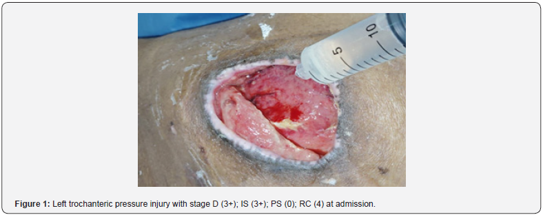
This study was conducted in a tertiary care hospital in South India after obtaining department’s scientific & ethical committee approval. Informed consent was taken from the patient & attendants. The Kumar classification was applied on a patient with left trochanter pressure injury (figure 1) and stage was D (3+); IS (3+) PS (0) RC (4) at admission. The staging was recorded whenever the dressing was changed to know the progress and response to the treatment.


Results
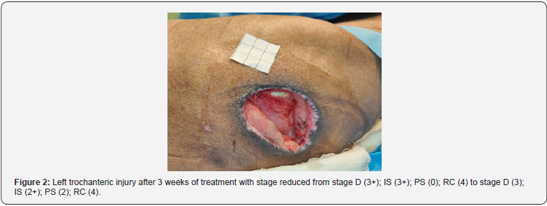
The Kumar classification for pressure injury reduced from stage D (3+); IS (3+); PS (0); RC (4) (figure1) to stage D (3); IS (2+); PS (2); RC (4) (figure2) after 3 weeks of the treatment allowing us to know the progress and response to the treatment (figure 2). The treatment is still going on till the reporting of this study. We found Kumar classification for pressure injury useful not only for documenting the findings at the time of admission but also to know the response to the treatment.
Discussion
For research purposes more improvements in staging with inclusion of infection, progression of ulcer and major risk factors on existing stages of only depth has been suggested by Kumar. This will have bearing on the prognosis of the disease and management protocol and hence better outcome of research work. The most extensive published experiences with pressure sore treatment are those of Conway and Griffith (1000 cases) and Dansereau and Conway’s update of the Bronx Veterans Administration Hospital data (2000 cases). But with new technologies of wound care more research is required to standardize the prevention and management of pressure injury [4]. There is a need to reduce the cost of the care for long-term compliance. For research purposes more improvements in staging with inclusion of infection, progression of pressure injury and major risk factors in existing staging (four stages) of depth only has been suggested by Kumar [5]. This will have bearing on prognosis of the disease and management protocol and hence, better outcome of research work. The classification has been updated by inclusion of suspected deep tissue injury. The updated classification by Kumar is as below:
Depth Stages (DS)
1. S. Suspected deep tissue injury. 2. Pre-ulcer stage –redness over pressure point. 3. Epidermal/ superficial dermal desquamation, abrasion, blistering, ruptured blister with pink floor. 4. Deep dermal- white/ purple floor. 5. Subcutaneous- exposed fat/ granulation forms the floor. 6. plus - Undeterminable Depth: covered with black coloured dead tough tissue. 7. Ligament/bone exposed with or without granulation tissue.Infection Stage (IS)
1. Wound Culture negative.
2. Wound Culture positive; blood culture negative.
3. Systemic signs positive for Sepsis (Blood culture +).
Progression Staging (PS)
1. On first examination.
2. Regressing/ healing (on two consecutive inspection after 3 days interval).
3. Stationary (status co on two consecutive inspection after 3 days interval).
4. Progressing (progressively increasing soakage/slough/ necrotic tissue on two consecutive inspection after 3 days interval).
Risk Category (RC)
• Ambulatory (a)
• Bed ridden (b)
1. Protective sensation present.
2. Protective sensation absent/ altered consciousness.
3. Incontinence present with or without protective sensation/altered consciousness.
4. Deformity of spine/ extremity joints/spasticity/ multiple pressure ulcers causing abnormal prominence of pressure points and or changing the position of the patient is difficult due to some or other reason [3].
The location of pressure injuries varies depending on the individual’s posture. In a supine position, common sites include the occiput, scapula, olecranon, sacrum, and heel. When lying laterally, pressure injury may develop on the ear, acromion process, greater trochanter, lateral condyle of the femur, and lateral malleolus. In a prone position, areas prone to pressure injuries are the zygoma, acromion process, female breasts, pubis, patella, metatarsal over the distal foot dorsum, and toes. When sitting, pressure injuries are often found on the shoulder blades, lower back, sacrum, ischial tuberosity, and heel. [4,5] Although pressure injuries typically occur over bony prominences, they can also develop in well-padded areas such as the buttocks and breasts after prolonged pressure. This study highlights the role Kumar classification applied on a case of pressure injury and found to be useful. The limitation of our study is that it is applied on a single case and large randomized double blind controlled study is required to validate our study.
Conclusion
This study highlights the role Kumar classification applied on a case of pressure injury and found to be useful. The limitation of our study is that it is applied on a single case and large randomized double blind controlled study is required to validate our study.
References
- Evans JM, Andrews KL, Chutka DS, Fleming KC, Garness SL (1995) Pressure ulcers: prevention and management. Mayo Clin Proc 70(8): 789-799.
- Hess Cathy Thomas (2020) Classification of Pressure Injuries. Advances in Skin & Wound Care 33(10): 558-559.
- Kumar P, Chittoria RK, Bajaj SP, Singh AK, Tiwari VK, et al. (2017) Guidelines for Pressure Ulcers. JSWCR 10(1): 2-22.
- Conway H, Griffith BH (1956) Plastic surgery for closure of decubitus ulcers in patients with paraplegia; based on experience with 1000 cases. Am J Surg 91(6): 946-975.
- Kumar P (2015) An improvised classification of pressure ulcer (Letter). JSWCR 8: 37-38.


