Beneath the Surface: A Case of Intussusception due to a Submucosal Lipoma in an Adult Patient
Faris Alsobyani1*, Amar Akbar1, Abdulaziz Jastaniah2, Suleiman Jastaniah3 and Rabah Khatir1
1Department of General Surgery, Al Noor Specialist Hospital, Makkah, Saudi Arabia
2Medical Student, Umm Al-Qura University, Makkah, Saudi Arabia
3Department of Surgery, Faculty of Medicine, Umm Al-Qura University, Makkah, Saudi Arabia
Submission: April 30, 2024; Published: May 08, 2024
*Corresponding author: Faris Alsobyani, Department of General Surgery, Al Noor Specialist Hospital, Makkah, Saudi Arabia. Email: FarisAlsobyani@gmail.com
How to cite this article: Faris Alsobyani*, Amar Akbar, Abdulaziz Jastaniah, Suleiman Jastaniah and Rabah Khatir. Hemosuccus Pancreaticus- A Concise Summary on Current Knowledge of this Rare Cause of Upper GI Bleed. Open Access J Surg. 2024; 15(3): 555914. DOI: 10.19080/OAJS.2024.15.555914.
Abstract
Intussusception, rare in adults, presents diagnostic challenges. We report a case of a 41-year-old male with intussusception caused by a submucosal lipoma, highlighting an unusual etiology. Clinical presentation, diagnostic workup, management, and outcome are detailed. Colonic lipomas, though often asymptomatic, can lead to complications like intussusception. CT scans are crucial for diagnosis, with surgery often necessary for larger lipomas. Delayed diagnosis can result in severe consequences. This case emphasizes the importance of recognizing uncommon causes in adult intestinal obstruction, enhancing diagnostic and management approaches in adult gastrointestinal pathology.
Keywords: Intussusception; Submucosal; Lipoma; Adult; Gastrointestinal pathology
Abbreviations: PR: Per Rectal; Hb: Hemoglobin; WBC: White Blood Cells; CT: Computed Tomography; US: Ultrasonography; MRI: Magnetic Resonance Imaging
Introduction
Intussusception, a condition typically associated with pediatric patients, is a rare occurrence in adults, accounting for less than 5% of all cases of intestinal obstruction [1,2]. We present a unique case of an adult patient who presented with intestinal obstruction, where diagnostic evaluation unveiled a submucosal lipoma as the underlying culprit of intussusception. This case serves to highlight a rare etiology in adult patients presenting with intestinal obstruction. In this report, we describe the clinical presentation, diagnostic workup, management, and outcome of this intriguing case to contribute to the expanding body of knowledge surrounding adult intussusception.
Case Report
This is a 41-year-old male, not known to have any past medical illness or surgical history, presented with a history of obstipation for 6-day duration. It was associated with multiple episodes of nausea and non-bloody vomiting of gastric secretions. The patient had a history of intermittent non-specific mild abdominal pain over the past 1 month. There was no history of per-rectal (PR) bleeding. He denied having any history of fever, weight loss, or loss of appetite. There was no history of jaundice or urinary complaints. There was no known family history of intestinal obstruction or malignancy. There were no previous upper or lower endoscopies done.
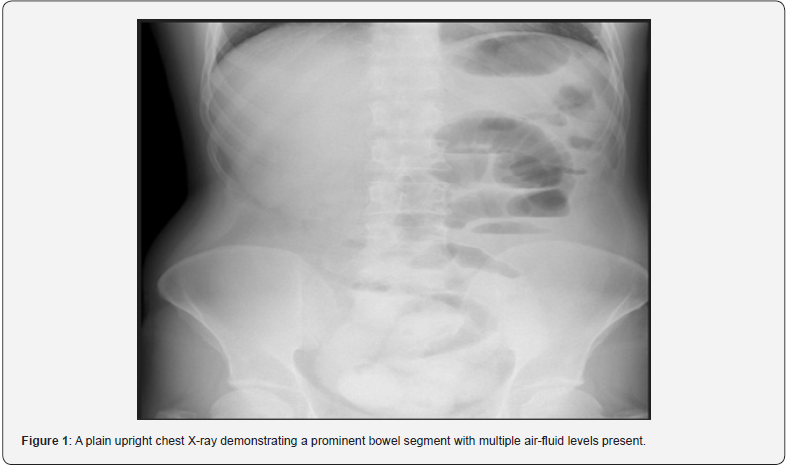
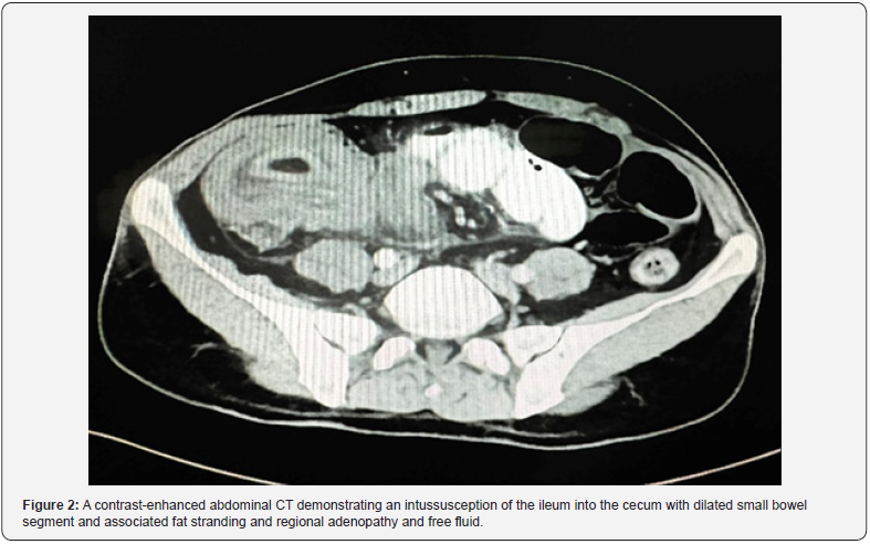
The patient was conscious, alert, and oriented. All vitals were within normal limits. Upon examination, the abdomen was mildly distended, with a vague generalized tenderness with no marked guarding or rigidity. All hernial orifices were intact. PR examination revealed an empty rectum with no palpable masses. The hemoglobin (Hb) level was 18 g/dL, with the white blood cells (WBC) count of 11x109/L. Electrolytes, liver, and renal function tests were all within normal parameters. On erect abdominal film, there were multiple air-fluid levels and prominent bowel loops as shown in Figure 1. Abdominal computed tomography (CT) with intravenous contrast was done showing a bowel-in-bowel sign in the right lower quadrant with the layering of the bowel wall and mesenteric fat, associated with fat stranding and regional adenopathy, surrounded with free fluid. There is an intussusception of the ileum into the caecum extending through to the ascending colon. Proximally, there is a dilated small bowel segment, measuring up to 3.5 cm. The remaining colon is dilated and filled with fecal materials as shown in Figure 2.


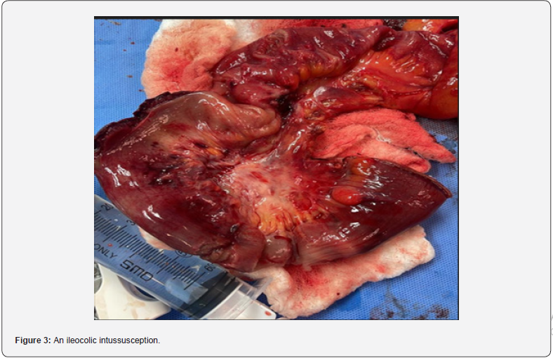
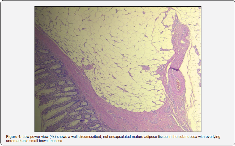
The patient was admitted as a case of intestinal obstruction due to intussusception for exploratory laparotomy. Intra-operatively, there was an ileocolic intussusception with no obvious leading point, with considerable pelvic free fluid as shown in Figure 3. A decision was made to go for a right hemicolectomy with an ileocolic anastomosis. The initial histopathology specimen yielded a submucosal lipoma as the culprit of the intussusception with twelve reactive lymph nodes as shown in Figure 4. No outpatient follow-up was made since the patient traveled to his home country shortly after discharge.
Discussion
A lesion in the bowel wall that causes invagination alters the normal peristalsis, which leads to intussusception. It can occur anywhere in the small and large intestine. Over 95% of cases of intestinal obstruction due to intussusception occur in children [1]. Adult intussusception is a rare pathology with an annual incidence of 1-2 per 1,000,000 [2]. Approximately 1-5% of adult intussusception cases involve bowel obstruction [3,4]. In adults, the average age of intussusception is 50 years, with no gender predominance [5].
Intussusception in adults can have a wide range of clinical manifestations. The most common symptom is abdominal pain, which is followed by obstruction and a palpable tumor. The most common symptom of intussusception has been reported to be abdominal pain followed by nausea and diarrhea, vomiting, and per-rectal bleeding [6]. The junctions between freely moving segments and the retroperitoneal space, as well as segments fixed by adhesions, are the most common sites of intussusception in the gastrointestinal tract [7]. Although the exact mechanism of development is unknown, it has been reported that any lesions in the intestinal wall or irritants within the lumen that disrupt normal peristalsis can cause an invagination [8,9].
The most common malignant cause of colonic intussusception is primary colonic adenocarcinoma, and the most common benign cause is colonic lipoma [5]. Lipomas are usually asymptomatic; however, lipomas larger than 2 cm in diameter may cause bowel obstruction, abdominal cramps, bleeding, diarrhea, or intussusception by forming a lead point [10]. Colonic lipomas causing intussusception are uncommon, occurring more frequently in women aged 40-70 years, and can cause abdominal pain, constipation, rectal bleeding, and diarrhea; surgical treatment varies depending on the location of the lipoma [11].
CT is the most sensitive diagnostic modality, providing information regarding lesions functioning as a lead point as well as the viability of the intestine [12-14]. The sensitivity of CT scans to correctly diagnose intussusceptions has been reported to range between 71.4% and 87.5%, whereas its specificity in adults has been reported to be 100% [15]. Ultrasonography (US) is another diagnostic tool used for intestinal intussusception. Magnetic resonance imaging (MRI) can detect benign fat lesions, such as intestinal lipomas [13,15]. This can be confirmed by colonoscopy [13]. There are various ways to manage intussusception in children, spanning from using a barium enema for reduction to resorting to surgical intervention [16].
It has been widely recommended to surgically resect lipomas larger than 2 cm, especially in adults, where intussusception is more commonly associated with malignancy [2,3,13]. Because adults appear with acute, subacute, or chronic nonspecific symptoms, the initial diagnosis is overlooked or often delayed, and the patient is only diagnosed when he or she is on the operating table [17]. Delayed treatment of intussusception in adults can have major consequences, as reported in the case of a 46-yearold man with bilateral ileoileal and ileocecocolic intussusception caused by an ileal lipoma, who suffered from acute renal failure, liver shock, and pulmonary thrombosis [18].
Conclusion
In conclusion, the case presented here underscores the rarity of intussusception in adults and the exceptional infrequency of submucosal lipomas as the inciting factor. As intussusception is primarily associated with the pediatric population, its occurrence in adults remains a diagnostic challenge. Our case highlights the importance of considering unusual etiologies when evaluating adult patients with intestinal obstruction, as early recognition is crucial for optimal management. Through our detailed examination of the clinical presentation, diagnostic investigations, treatment strategies, and ultimate patient outcome, we aim to contribute to the broader understanding of adult intussusception, shedding light on its diverse presentations and management options. This case report, therefore, provides valuable insights for healthcare professionals faced with similar diagnostic dilemmas, ultimately enhancing the body of knowledge in the field of adult gastrointestinal pathology.
Acknowledgements
The authors have received no funding for this paper and declare no conflicts of interest.
References
- Tan KY, Tan SM, Tan AGS, Chen CYY, Chng HC, et al. (2003) Adult intussusception: experience in Singapore. ANZ J Surg 73(12): 1044-1047.
- Dener C, Bozoklu S, Bozoklu A, Ozdemir A (2001) Adult intussusception due to a malignant polyp: a case report. Am Surg 67(4): 351-353.
- Begos DG, Sandor A, Modlin IM (1997) The diagnosis and management of adult intussusception. Am J Surg 173(2): 88-94.
- Eisen LK, Cunningham JD, Aufses AH (1999) Intussusception in adults: institutional review. J Am Coll Surg 188(4): 390-395.
- Marsicovetere P, Ivatury SJ, White B, Holubar SD (2017) Intestinal Intussusception: Etiology, Diagnosis, and Treatment,” Clin. Colon Rectal Surg 30(1): 30-39.
- Kim KH (2021) Intussusception in Adults: A Retrospective Review from a Single Institution. Open Access Emerg Med 13: 233-237.
- Marinis A, Anneza Y, Lazaros S, Nikolaos D, Georgios A, et al. (2009) Intussusception of the bowel in adults: a review. World J Gastroenterol 15(4): 407-411.
- Zubaidi A, Al-Saif F, Silverman R (2006) Adult intussusception: a retrospective review. Dis Colon Rectum 49(10): 1546-1551.
- Wang N, Xing-Yu C, Yu L, Jin L, Yuan HX, et al. (2009) Adult intussusception: a retrospective review of 41 cases. World J Gastroenterol 15(26): 3303-3308.
- Minaya Bravo AM, Vera Mansilla C, Noguerales F, Granell Vicent FJ (2012) Ileocolic intussusception due to giant ileal lipoma: Review of literature and report of a case. Int J Surg Case Rep 3(8): 382-384.
- Tasselli FM, Fabrizio U, Guido S, Giulia B, Francesca P, et al. (2021) Colonic Lipoma Causing Bowel Intussusception: An Up-to-Date Systematic Review. J Clin Med 10(21): 5149.
- Treppiedi E, Lorenzo C, Giuseppe Z, Alberto M, Valeria S, et al. (2020) Ileocolic invagination in adults: A totally minimally invasive endoscopic and laparoscopic staged approach. J Minimal Access Surg 16(1): 87-89.
- Cordeiro J, Cordeiro L, Pôssa P, Candido P, Oliveira A (2019) Intestinal intussusception related to colonic pedunculated lipoma: A case report and review of the literature. Int J Surg Case Rep 55: 206-209.
- Hong KD, Kim J, Ji W, Wexner SD (2019) Adult intussusception: a systematic review and meta-analysis. Tech Coloproctology 23(4): 315-324.
- Mouaqit O, Hafid H, Leila C, Abdelmalek O, Khalid M, et al. (2013) Pedunculated lipoma causing colo-colonic intussusception: a rare case report. BMC Surg 13: 51.
- Hoffman RD, Levine HA, Baram S, Tiberin E, Soroker D (1992) Painless intussusception. Giving conservative treatment another chance. Postgrad Med 91(1): 283-284.
- Haas EM, Etter EL, Ellis S, Taylor TV (2003) Adult intussusception. Am J Surg 186(1): 7576.
- Krasniqi A, Astrit RH, Lulzim MS, Gazmend SS, Besnik XB, et al. (2011) Compound double ileoilealand ileocecocolic intussusception caused by lipoma of the ileum in an adult patient: A case report. J Med Case Reports 5: 452.


