Radiation Angiosarcoma of the Breast, Early Appearance Case Report
Nathalie Mantilla1, Hassan Mashbari1*, Turkia Abbed1, Andres Acosta2, Rajyasree Emmadi2 and Michael Warso1
1General Surgery Department, Division of Surgical Oncology, University of Illinois at Chicago, USA
2Department of Pathology, University of Illinois at Chicago, USA
Submission: August 28, 2017; Published: September 06, 2017
*Corresponding author: Hassan Mashbari, General Surgery resident, University of Illinois at Chicago, Department of Surgery (MC 958), 840 S, Wood Street, Suite 376-CSN. Chicago, IL 60612, USA, Tel: 312-996-6765; Fax: 312-355-3722
How to cite this article: Nathalie M, Hassan M, Turkia A, Andres A, et al. Post-Radiation Angiosarcoma of the Breast, Early Appearance Case Report. Open Access J Surg. 2017; 5(5): 555673. DOI: 10.19080/OAJS.2017.05.555673
Abstract
Radiation-induced angiosarcoma of the breast represents a diagnostic challenge due to the clinical resemblance to radiation-induced skin changes. A high index of suspicion is of critical importance, particularly within 5 years after completion of radiation therapy. We present a case of a 70 year old woman with multiple co-morbidities who originally presented with in-situ and invasive duct carcinoma. She underwent a nippleinclusive partial mastectomy, and axillary node dissection. Surgical pathology showed multifocal in-situ and invasive duct carcinoma with 1 of 25 axillary nodes positive for metastatic tumor (pT1b (m) pN1a). Due to severe side effects from the first dose of chemotherapy, she only received 1 cycle of CMF (Cyclophosphamide, Methotrexate and 5-Fluorouracil) and was referred to Radiation Oncology. She received a final cumulative dose of radiotherapy to the lumpectomy cavity of 6080cGy in 44 days.
Four and a half years later, she returned to the clinic after noticing a cyst-like lesion in the skin of her left breast. A biopsy showed a high grade angiosarcoma of the breast and a left total mastectomy was performed. The final pathology showed a superficial high grade angiosarcoma, measuring 2.6 cm with ulceration of the skin (pT1a), and multifocal infiltrating mammary carcinoma, grade 1 of 3, with multiple small foci (2 mm) of elastosis consistent with the prior Angiosarcoma of the Breast treated tumor site. Resection margins were negative for carcinoma and sarcoma. Her postoperative recovery was slow due to complications related to her medical conditions.
Keywords: Breast cancer; Radiation; Angiosarcoma
Introduction
Radiation-induced second non-mammary malignancies are infrequent long term complications of the therapy. Angiosarcoma is particularly challenging to diagnose given its rare presentation (less than 1% of all malignancies of the breast) and clinical resemblance to radiation-induced skin changes [1]. Therefore, it is crucial to have a high index of suspicion in these patients. It is commonly seen in older patients, with a mean age of 68 years and a latency period of 5 or more years (range 3-25 years) [24]. We report a case of an early presentation of angiosarcoma of the breast following breast conservation surgery and radiation therapy for breast cancer.
Case Report
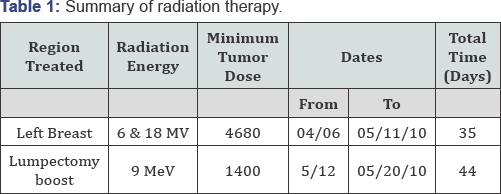
The patient is a 70 year old woman with multiple medical problems including systemic and pulmonary hypertension, diabetes, hypothyroidism, morbid obesity, obstructive sleep apnea, congestive heart failure, coronary artery disease, chronic kidney disease and lower extremity deep vein thrombosis, who presented with abnormal breast imaging and a bloody discharge from the left breast. She underwent an image-guided biopsy that showed ductal carcinoma in situ (DCIS). She underwent a nipple-inclusive partial mastectomy and axillary node dissection. Pathology showed in-situ and multifocal invasive ductal carcinoma, the largest focus measuring 0.9 cm, and 1 of 25 lymph nodes positive for metastatic carcinoma for a tumor stage of pT1b(m) pN1a. The tumor was estrogen and progesterone receptor positive, Her2/neu-negative. Due to severe side effects from the first dose of chemotherapy, she only received 1 cycle of CMF (Cyclophosphamide 600 mg/m2 = 1300 mg, Methotrexate 40 mg/m2 = 90 mg, 5- Fluorouracil 600 mg/m2 = 1300 mg) and was referred to Radiation Oncology. She received a final cumulative dose of radiotherapy to the lumpectomy cavity of 6080cGy in 44 days (Table 1).


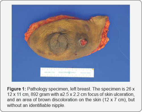
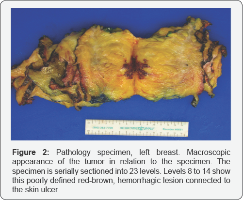
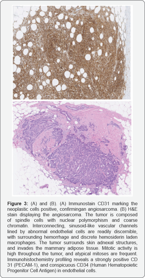
She was also started on Anastrozole. Four and a half years later, she returned to the clinic after noticing a cyst-like lesion in the skin of her left breast. It was initially tender with purulent drainage. The lesion hardened and opened up exposing a fleshy mass in the area. She underwent a biopsy that showed a high gradeangiosarcoma of the breast. Four years after the initial diagnosis of the breast cancer, she underwent a left total mastectomy. The final pathology showed an angiosarcoma, 2.6 cm, high grade, superficial, with ulceration of the skin (pT1a), and multifocal infiltrating mammary carcinoma, grade 1 of 3 [Nottingham score = 5; pT1a(m)], and multiple small foci (2 mm) of elastosis consistent with the prior treated tumor site.Resection margins were widely negative for carcinoma and sarcoma (Figures 1 & 2). Her postoperative recovery was slow due to complications related to her medical conditions (Figure 3).

Discussion
Angiosarcomas are highly aggressive tumors with poor prognosis based on the high recurrence and metastatic rates. Median time of survival ranges from 18 to 40 months, and the overall survival rate at 5 years is up to 20% [5]. Since the early 1900s, radiotherapy has been demonstrated to play a causative role in the pathogenesis of sarcomas [6]. In general, the prognosis of sarcomas is poor whether or not it is associated with radiation therapy for a primary cancer [7]. The first report of sarcoma following radiotherapy for breast cancer was by Warren and Sommer in 1936 [8]. Despite the widespread use of radiation therapy, post-radiation angiosarcoma of the breast is a rare but severe complication with a prevalence of 0.05-0.16%. Early diagnosis and treatment is crucial as these cancers are rapidly fatal if untreated. The relationship between the total radiation dose and the incidence of these tumors has not been established [5], although some studies have shown that postradiation carcinomas arise in tissues exposed to lower doses, whereas sarcomas are more commonly seen in heavily radiated tissues in or close proximity to the radiation fields [9].
A significant number of cases have been reported with a wide variation in the time of presentation of signs and symptoms (3-25 years) with a median latency period of 5 years [2-4]. Therefore, early development of post-radiation angiosarcoma of the breast is not a common complication during the first 5 years post-therapy. These lesions are difficult to diagnose due to their variable appearance, sometimes resembling radiation-induced cutaneous changes. A high index of suspicion is important in patients who develop skin lesions in irradiated areas, warranting mammography and biopsy to confirm the diagnosis. However, in many cases, biopsies are negative in early stages of angiosarcoma [5,10-12].
Fineberg published one of the few reports of a series of patients who received conventional high energy postoperative doses of external beam radiation therapy to the breast with early development of cutaneous angiosarcoma. Diagnosis was made at 3.5 years, 3.7 years, and 5.25 years following radiotherapy. He concluded that unlike other radiation therapy- induced sarcomas, cutaneous angiosarcoma often occurs within a short time interval after radiotherapy [12]. The majority of publications report high grade sarcomas, although Moskaluk and Bolin reported cases of low grade angiosacromas in 1992 and 1996 respectively [12,13]. A wide local excision should be performed whenever possible. Palliative chemotherapy should be considered in poor surgical candidates.
Conclusion
Post-radiation cutaneous angiosarcoma of the breast may represent a challenge in diagnosis, especially when it presents in the first few years after completion of treatment. Delay in the diagnosis can be detrimental to the prognosis of the patient and is often due to its initially benign appearance. Therefore, early recognition is crucial and diagnosis is confirmed with cutaneous biopsies. We recommend biopsy of any suspicious lesion arising in a previously irradiated breast.
References
- Yi A (2009) Radiation-Induced Complications aftser Breast Cancer Radiation Therapy: a Pictorial Review of Multimodality Imaging Findings. Korean J Radiol 10: 496-507.
- Seinen J, Styring E, Verstappen V, Vult von Steyern F (2012) Radiation- Associated Angiosarcoma After Breast Cancer: High Recurrence Rate and Poor Survival Despite Surgical Treatment with R0 Resection. Ann Surg Oncol 19(8): 2700-2706.
- Styring E, Fernebro J, Jonsson PE, Ehringer A, Engellau J, et al. (2010) Changing clinical presentation of angiosarcomas after breast cancer: from late tumors in edematous arms to earlier tumors on the thoracic wall. Breast Cancer Res Treat 122: 883-887.
- Aljarrah A, Nos C, Clough K, Lefrere-Belda M, Lecuru F (2014) A case report on radiation induced angiosarcoma of breast post skin-sparing mastectomy and reconstruction with transverse rectus abdominal muscle. E-cancer 8: 402.
- Cabete J, Lencastre A, Fidalgo A, Lobo L, Joao A, et al. (2014) Postradiation Cutaneous Angiosarcoma of the Breast: A Diagnosis to Keep in Mind. The Breast Journal 20(1): 89-90.
- Perthes G (1904) Zurfrage der roentgentherapie des carcinomas. Archiv fur klinischechirurgie 74: 400-405.
- Yap J, Chuba PJ, Thomas R, Aref A, Lucas D, et al. (2002) Sarcoma as a second malignancy after treatment for breast cancer. Int J Radiat Oncol Biol Phys 52(5): 1231-1237.
- Warren S, Sommer GN (1936) Fibrosarcoma of the soft parts with special reference to recurrence and metastasis. Arch Surg 33: 425-450.
- Kuttesch JF, Wexler LH, Marcus RB, Fairclough D, Weaver-McClure L, et al. (1996) Second malignancies after Ewing's sarcoma: radiation dose- dependency of secondary sarcomas. J Clin Oncol 14: 2818-2825.
- Hildebrandt G, Mittag M, Gutz U, Kunze ML, Haustein UF (2001) Cutaneous breast angiosarcoma after conserving treatment of breast cancer. Eur J Dermatol 11(6): 580-583.
- Fineberg S, Rosen PP (1994) Cutaneous angiosarcoma and atypical vascular lesions of the skin and breast after radiation therapy for breast carcinoma. Am J Clin Pathol 102(6): 757-763.
- Moskaluk CA, Merino MJ, Danforth DN, Medeiros LJ (1992) Low-grade angiosarcoma of the skin of the breast: a complication of lumpectomy and radiation therapy for breast carcinoma. Hum Pathol 23(6): 710-714.
- Bolin DJ, Lukas GM (1996) Low-grade dermal angiosarcoma of the breast following radiotherapy. Am Surg 62(8): 668-672.


