Choanal Atresia
Sushila B Ladumor*1 and HibaEsmayil2
1Consultant Radiologist, Hamad Medical Corporation, HGH, Clinical Imaging, Qatar
2R1 Radiology Resident, Hamad Medical Corporation, HGH, Clinical Imaging, Qatar
Submission: August 24, 2017; Published: August 30, 2017
*Corresponding author: Sushila B Ladumor, Consultant Radiologist, Hamad Medical Corporation,HGH, Clinical Imaging, P.O. Box 3050, Doha, Qatar, Assistant Professor in Clinical Radiology, Weil Cornel Medical College, Doha, Qatar (WCMC-Q). Email: drsbladumor@yahoo.com
How to cite this article: Sushila B L, Hiba E. Choanal Atresia. Open Access J Surg. 2017; 5(5): 555671. DOI: 10.19080/OAJS.2017.05.555671
Abstract
Congenital narrowing of the nasal airway at the posterior choanae, which can be uni- or bilateral.Choanal atresia is an abnormality of canalization during development of the nasal passages due to failure of resorption of the bucco-pharyngeal membrane during embryonic development. The atresia can be membranous or bony in nature, but is usually mixed in most cases. It is one of the commonest causes of nasal obstruction in early age. When the atresia is bilateral, it present early and newborns can have significant airway obstruction with respiratory distress and cyanosis is aggravated by crying. Bilateral congenital choanal atresia is a relatively rare anomaly of the upper airway, which may cause life-threatening respiratory emergency and require rapid diagnosis and treatment. This condition usually occurs sporadically, but has also been rarely described in siblings (7). Bilateral choanal atresia is managed with an oropharyngeal airway. Flexible nasal endoscopy and computed tomography can confirm the diagnosis. The incidence of choanal atresia is 1 in 7000 to 8000 live births. It is more common in females (2:1), more likely to be bony or cartilaginous than membranous (9:1), and more commonly unilateral and right-sided (2:1).
Keywords: Choanal Atresia, CT scan; Bony and membranous; Unilateral; Bilateral; CHARGE
Abbreviations: PNS: Para nasal sinus; CT: Computerized Tomography
Case History: An eight year old girl who presented with nasal blockage.
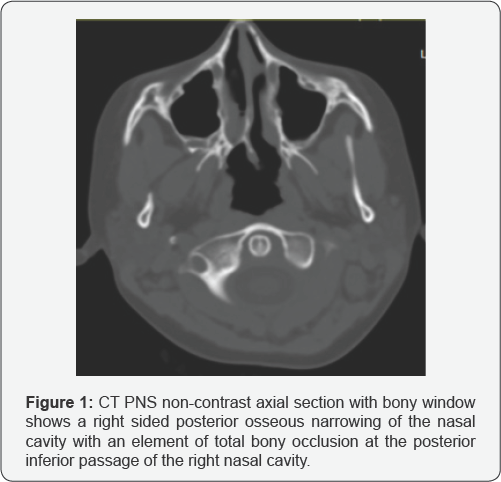
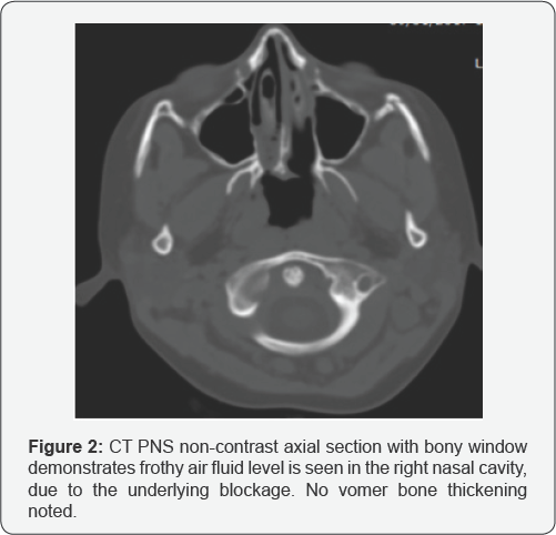
Imaging: CT scan of paranasal sinus was requested (Figures 1 & 2).


Findings of Non-contrast CT scan of PNS
a) Unilateral posterior osseous narrowing at the right posterior nasal cavity with an element of total bony occlusion at the posterior inferior passage of the right nasal cavity
b) Frothy air-fluid level noted opacifying the right nasal cavity.
c) No vomer bone thickening is noted
Diagnosis: Unilateral right sided choanal atresia
Discussion
Choanal atresia is a rare congenital disorder where there is narrowing of the posterior nasal cavity due to an obstruction at its end. This obstruction can be bony, membranous, or both. The obstruction can be unilateral, or may be rarely bilateral. Approximately 60% of the cases documented are unilateral [1].
Bilateral atresia can be diagnosed in the early neonatal days, as the child usually presents with severe respiratory distress. Unilateral choanal atresia is usually diagnosed later. The clinical diagnosis of choanal atresia is made when a catheter cannot be passed through the nose for some other reason. Some of the other maneuvers used for clinical diagnoses are lack of movement of a thin wisp of cotton under the nostrils while the mouth is closed, absence of fog on a mirror when it is placed under the nostrils, acoustic rhinometry and administering into the nose a colored solution that is visible in the pharynx.
The embryonic origin of choanal atresia is hypothesized to be due to a persistent bucco-pharyngeal or naso-buccal membrane [2]. Factors that can lead to an atretic choana are a medial outgrowth of the vertical or horizontal process of the palatal bone and persistent mesenchyme with misdirection of developmental flow [3]. Choanal atresia can be associated with some syndromes. For example it is a part of CHARGE syndrome, which stands for:
i. C- Coloboma
ii. H- Heart/cardiovascular anomalies
iii. A- Atresia of choana
iv. R- Retarded growth and development
v. G- Genital hypoplasia
vi. E- Ear anomalies
Some of the other associated syndromes are Crouzon syndrome, DiGeorge syndrome and Treacher Collins syndrome. Other associations include intestinal malrotation, craniosynostosis and congenital heart disease. Early pregnancy use of antithyroid drugs is also associated with the occurrence of choanal atresia [4]. CT in the axial plane is the most valuable tool in demonstrating choanal atresia. Here the entire nasal cavity can be visualized, and differentiation between bony and membranous components can be done. It can also be identified if the obstruction is partial or complete. The key findings are unilateral or bilateral posterior nasal narrowing with an obstruction with an airway less than 3 mm. An air-fluid level with frothy secretions may be observed as a result of the obstruction. There may be associated thickening of the vomer. Medial bowing of posterior maxillary sinus may also be observed. Septal deviation and stenosis can also be detected associated with this condition [5-7].
Management
a) Endoscopic perforation (for membranous types)
b) Full choanal reconstruction.
c) Surgery is the definitive treatment with two main approaches, namely transnasal or transpalatal.
The transnasal route is currently the preferred procedure and can be performed in a minimally invasive fashion with endoscopic instrumentation. It is a safe and rapid procedure even in very young children, with no complications and a high rate of success. The use of a navigation system for surgical planning and intraoperative guidance and powered instrumentation can improve treatment outcome. The transpalatal approach is more invasive and reserved for failed endoscopic cases.
References
- Szeremeta W, Parikh TD, Widelitz JS (2007) Congenital nasal malformations. Otolaryngol Clin North Am 40 (1): 97-112.
- Stankiewicz JA (1990) The endoscopic repair of choanal atresia. Otolaryngol Head Neck Surg 103(6): 931-937.
- Tewfik TL, Der Kaloustian VM (2011) Choanal atresia. Rhinogram demonstrating blockage of radiopaque dye at posterior choana. Choanal atresia, Workup. eMedicine. Drugs, Diseases and Procedures 4-11.
- Andersen SL, Olsen J, Wu CS (2013) Birth defects after early pregnancy use of antithyroid drugs: a Danish nationwide study. J Clin Endocrinol Metab 98 (11): 4373-4381.
- Craig DH, Simpson NM (1959) Posterior choanal atresia. J Laryngol Oto 75: 603-606.
- Maniglia AJ, Goodwin WJ (1981) Congenital choanal atresia. OtolaryngolClin North Am 14:167-173.
- ÜlfetVatansever, Rinodotdvan Duran, BetulAcunascedil, MuhsinKoten, Mustafa Kemal Adali (2005) Bilateral Choanal Atresia in Premature Monozygotic Twins. Journal of Perinatology 25: 800-802.


