



 OAJS Home



Frequency of Thyroid Malignancy in Patients Undergoing Thyroidectomy for Multinodular Goiter
Muhammad Zafar Mengal*, Yaseen Rafi and Khalid Masood Gondal
Department of surgery KEMU/Mayo Hospital, Lahore
Submission: March 24, 2017; Published: April 10, 2017
*Corresponding author: Muhammad Zafar Mengal, Department of surgery, Assistant Professor of Surgery King Edward Medical University, Lahore, Tel: +923334088934; Email:zafar.mengal@yahoo.com
How to cite this article: Muhammad Zafar Mengal*, Yaseen Rafi and Khalid Masood Gondal. Frequency of Thyroid Malignancy in Patients Undergoing Thyroidectomy for Multinodular Goiter. Open Access J Surg. 2024; 3(4): 555616. DOI: 10.19080/OAJS.2024.03.555616.
Abstract
Introduction: Thyroid disease commonly presents as multi-nodular goiter (MNG). Thyroid nodules have been reported to be found in4-7% of the population on neck palpation and in 30-50% of the population by ultrasonography (USG). Dietary iodine deficiency has been associated with thyroid malignancy and northern areas of Pakistan are known for endemic iodine deficiency goiter and hence the high frequency of goiter in local population.
Objective: To determine the frequency of thyroid malignancy in patients undergoing thyroidectomy for multinodular goiter Study setting: North Surgical Ward, Department of Surgery, Mayo Hospital, Lahore.
Duration of study: January 15, 2016 to July 15, 2016.
Study design: Prospective, Observational, Cross sectional study
Subjects & methods: Total of 132 patients was included in this study. Fine needle aspiration cytology was done in cases with a dominant nodule (nodules) which is growing hard, irregular nodule which was detected on clinical examination and on ultrasound. All data including age, gender, relevant investigations like ultrasonography, fine needle aspiration cytology, type and duration of surgery and final histopathology report were recorded on a standard form. The data were entered and analyzed by using SPSS version 21. Data were stratified for age, gender, size of goitre and duration of symptoms. Stratified groups were compared by using chi-square test. P-value <0.05 was considered as statistically significant.
Results: Overall incidence of Malignancy was 6.8%. Nine patients had malignancy, while rest of the patients was benign 123(93.2%).
Conclusion: Incidence of malignancy in multinodular goitre should not be overlooked as many of the patients with thyroid cancer can present with multinodular goitre.
Keywords: Multinodular goiter; Malignancy; Thyroidectomy
Abbreviations: MNG: Multi-Nodular Goiter; USG: Ultrasonography; LCA: Leukocyte Common Antigen; PAP: Peroxidase Anti-peroxidase Technique; TC: Thyroid Carcinoma; FNAC: Fine-Needle Aspiration Cytology
Introduction
Thyroid cancer is the most common of the endocrine malignancies and it represents <1% of all human tumors [1]. In most countries, a steady increase in the incidence of thyroid cancer (mainly papillary carcinomas) is observed in both sexes, [2] in all races and ethnicities [3]. Some findings suggest that the increase in thyroid cancer incidence may be related to increases in the use of thyroid ultrasound and FNAC [4]. Multinodular goitre (MNG) is a multinodular enlargement of the thyroid gland. MNG usually presents as an anterior neck mass, but other nodules may be detected incidentally during radiological evaluation of the neck. The reported incidence of missed malignancy in patients with MNG is between 0.7% and 2.2% if ultrasound-guided fine needle aspiration biopsy is used [5]. Multinodularity of the goiter should not be considered as low risk of malignancy and delay for surgical intervention.
Changes in the size of gland, the appearance of new and hard nodules or cervical lymphadenopathy may indicate malignant change and prompt indication for surgery [6]. In previous reviews MNG has long been regarded as benign however recent evidences have shown that incidence of malignancy in multi nodular goiter should not be underestimated [5-11]. In a local study, 9 out of 94 patients of multinodular goiter were found to have malignancy on histopathology, so overall incidence of malignancy in MNG was 9.5% with M: F ratio 1:2 with most common variant was papillary carcinoma [6]. In another local study, 105 patients with MNG underwent thyroidectomy, 8(7.6%) cases were malignant and frequency was papillary carcinoma (75%), follicular carcinoma (12.5%) and anaplastic carcinoma (12.5%) [8]. A relative higher incidence of malignancy (15%) was found in another local study with frequency of papillary carcinoma (66.7%), follicular carcinoma (26.7%), medullary carcinoma (6.7%) [9].
An Indian study showed frequency of 10% [10] and a Nepalese study showed 13% [11] but a much higher frequency i.e.23.1% was found in an international study [12]. It has been indicated that malignancy may be present in non dominant nodules and risk should not be under judged, however dominant nodule in patients with MNG was about 2.5 times more malignant than a non dominant nodule [13]. It is also recommended that FNAC for non dominant nodules could enhance the chance of finding malignancy in an MNG [13].
Objective
Objective of this study was to determine the frequency of thyroid malignancy and its types in patients undergoing thyroidectomy for multinodular goiter.
Operational Definitions
i. Goiter: It was a term used to describe any enlargement of the thyroid gland, which could be caused by iodine deficiency or a thyroid disorder.
ii. Multinodular Goiter: A multinodular goiter contains multiple distinct nodules within the goiter.
iii. Malignancy: It was measured on histopathology with presence of cells of irregular shape and size with calcification, hemorrhage or necrosis present in them.
Materials and Methods
This Prospective, Observational, Cross sectional study was carried out north Surgical Ward, Department of Surgery, Mayo Hospital, Lahore for six months from January to July 2016 I which 132 cases was calculated with 95% confidence level, 5% margin of error and taking expected percentage of malignancy i.e. 9.5% [7] in patients undergoing thyroidectomy for multinodular goiter. Sampling technique was non-probability, consecutive sample. Patients of age 20-60 years of either gender with medical record of clinically and radio logically proved multinodular goiter were included in study to undergo thyroidectomy and patients with solitary nodule of thyroid, Graves’ disease, Toxic MNG, history of irradiation to neck, pre operatively proven malignancy in MNG, metastasis (on clinical examination), recurrence of malignancy (on medical record) and pre operatively proven inadequate sample on FNAC were excluded from study. After approval from hospital ethical committee, 132 patients who fulfilled the inclusion criteria were enrolled in the study from Surgical Wards of Department of Surgery, Mayo Hospital, Lahore.
Informed consent of patients was obtained for this study. Basic demographic information of each patient (name, age, sex) was noted. After admission to hospital detailed history and thorough clinical examinations were carried out to reach the provisional diagnosis. Patient was investigated by performing blood tests, thyroid hormone levels and ultrasound neck to see number of nodules and calcification present in them. Fine needle aspiration cytology was done in cases with a dominant nodule (nodules) which is growing hard, irregular nodule which was detected on clinical examination and on ultrasound. Then patients underwent thyroidectomy with predictable safety of recurrent laryngeal nerve and parathyroid gland. All surgeries were performed under general anesthesia. After surgery patients were shifted to ward. The patients were closely monitored during the immediate postoperative period for complications like hemorrhage, respiratory obstruction, vocal cord paralysis, and surgical site infection. Surgical sample was sent to the histopathology department for assessment of type of lesion.
The specimens were evaluated on Haematoxylin and Eosin stained sections from paraffin embedded 10% buffered formalin fixed tissue blocks. Special stains like reticulin and Congo red was performed whenever required. Immunohistochemical staining with leukocyte common antigen (LCA), thyroglobulin, cytokeratin’s and vimentin were performed by using peroxidase fable to label histological types of thyroid cancer. Peroxidase antiperoxidase technique (PAP) technique was applied whenever indicated. Reports were assessed and thyroid malignancy was labeled (as per operational definition). Then in cases of thyroid malignancy, type of malignancy was noted. All pre and postoperative findings were recorded in detail in a pre designed Performa and results were evaluated.
Data Analysis Procedure
The collected data were entered and analysed through SPSS version 21. Quantitative variables like age, size of goitre and duration of symptoms were presented in the form of mean ± S.D. Qualitative variables like gender, malignancy and type of malignant tumour were presented in the form of frequency and percentage. Data were stratified for age, gender, size of goitre and duration of symptoms. Stratified groups were compared by using chi-square test. P-value <0.05 was considered as statistically significant.
Data Analysis Procedure
The collected data were entered and analysed through SPSS version 21. Quantitative variables like age, size of goitre and duration of symptoms were presented in the form of mean ± S.D. Qualitative variables like gender, malignancy and type of malignant tumour were presented in the form of frequency and percentage. Data were stratified for age, gender, size of goitre and duration of symptoms. Stratified groups were compared by using chi-square test. P-value <0.05 was considered as statistically significant.
Results
In this study, 132 cases of multi-nodular goitre were included. Of the chosen 132 MNG cases, 41(31.1%) were males and 91(68.9%) were females, with a striking female predominance. Among these patients, 42(31.8%) were between 20-30 age group, while 25(18.9%), 23(17.4%) and 42(31.8%) were between 31-40, 41-50 and 51-60 age groups respectively. Mean age of the patients was 40.44±13.25 with 20 and 60 as minimum and maximum ages. Most of the patients 87(65.9%) had >6 months of duration of symptoms, while 45(34.1%) had <6 months of duration of symptoms.
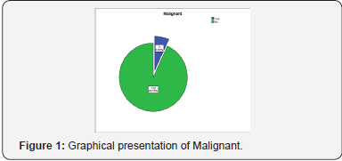
Mean size of the goiter was 1.85cm. 75(56.8%) patients had 0.80-1.60 cm size of goiter, while 48(36.4%) and 9(6.8%) had 1.70-2.30 cm and 2.40-2.90 cm size of goiter. Overall incidence of Malignancy was 6.8%. Nine patients had malignancy, while rest of the patients was benign 123(93.2%). In types of malignancy, 7(5.30%) patients were papillary malignant, while 2(1.52%) were follicular malignant.
By applying Chi-square test, it was concluded that, there was no association between malignancy and gender (p>1.000). Malignancy had equal effect on gender. It was concluded that, there was an association between malignancy and age groups (p<0.021). Malignancy was affected on elder patients. It was also concluded that, there was no association between malignancy and size of goiter and duration of symptoms as well (p>0.866, 0.960) (Figure 1).
Discussion
Multi-nodular goitre (MNG) is defined as the palpation of multiple distinct nodules in the enlarged thyroid gland. The aetiopathogenesis fail to detect the nodules when they are less than 1 cm in diameter [14,15]. In MNG, surgery is offered for cosmesis, the compressive symptoms, toxicity and for the suspicion of malignancy [16]. A long standing and hitherto unresolved issue is whether MNG is significantly associated with malignancy [17]. MNG had been traditionally thought to be at a low risk for malignancy as compared to a solitary nodule thyroid [16,18,19]. However, various studies have shown that the risk is quite high in MNG also. A study which was conducted by Benzarti et al. in Tunis found a 9.5% incidence of malignancy in MNG, [20,21] whereas Sarajevo reported an 8% incidence of malignancy in MNG in his study [20,22]. Prades et al. from France, however, reported quite a high incidence i.e. 12.2% [20,23].
The most common variety of malignancy which has been documented in the literature is papillary carcinoma [16,24]. The incidence of carcinoma in MNG in our study was 6.5% and the most common type of malignancy which was observed was papillary carcinoma 5.30%. This was consistent with the observations which were made by Benzarti et al. [20] in Tunis. A thyroid nodule should be viewed with suspicion if it is seen as a dominant nodule in the MNG, which is hard, irregular, fixed, and rapidly increasing, which is seen along with cervical lympadenopathy, recurrent laryngeal nerve palsy, extremes of age and the male sex. A patient with a history of neck irradiation or a family history of thyroid carcinoma (TC) should make the suspicion of MNG strong [20].
High-frequency, real-time ultrasonography and fine-needle aspiration cytology (FNAC) are the indispensible tools which are used in the pre-operative evaluation of MNG for malignant foci. The important sonographic findings which are suggestive of malignancy in the thyroid nodules are micro-calcifications (which are present in about 22% of the thyroid cancers), irregular margins of the nodules, a complex echogenicity and smaller nodules [24]. It has been postulated that the thyroid cancers would have manifested with more overt signs and symptoms of local invasion or metastasis by the time they had reached a significant size [24]. FNAC is a fast and inexpensive investigation which can be done to obtain cellular samples [24]. A series of reviews have reaffirmed its importance in the assessment of the thyroid nodules. However, a negative FNAC report does not exclude with certainty the possibility of a carcinoma, especially in MNG, where the error in sampling the right area is greater [16,19]. FNAC of a suspicious nodule under USG guidance is of great help.
Thyroid carcinomas account for 1% of all the malignancies and they are the most common endocrine tumours [20]. The incidence of TC varies considerably in different regions of the world. Globally, the incidence of TC has increased by up to fivefold during the past 60 years [18,25-29]. Thetumours are rare in children and their frequency increases with age. Overall, females have a higher incidence of TC [20]. Ionizing radiation, iodine deficiency and other factors have been attributed for the increase in TC, but these findings are inconsistent [18]. Hormonal factors, lactation suppresant drugs and fertility medications have been implicated for the high incidence of TC in females [26]. However, recent studies have reported no significant risk which has been associated with the use of hormone replacement therapy or fertility drugs [20,25,26]. It also has been proposed that the availability of better and more sensitive diagnostic tools may be responsible for the increasing incidence of TC [18,30].
Papillary micro-carcinoma is one subtype of papillary carcinoma, which was a frequent incidental finding in many autopsy studies [30]. The World Health Organization has defined it as a papillary thyroid carcinoma which measures ≤ 10 mm in the greatest dimension [30]. The literature provides us with conflicting information regarding the prognosis and the management of these lesions. Recent studies have suggested that the micro-carcinomas classically progress to a clinically evident disease if they are left untreated [27,30]. The treatment of papillary micro carcinoma should be similar to that of papillary thyroid cancer [28,30].
Conclusion
Incidence of malignancy in multinodular goitre should not be overlooked as many of the patients with thyroid cancer can present with multinodular goitre.<
References
- Pacini F, Castagna M, Brilli L, Pentheroudakis G (2012) Thyroid cancer: ESMO Clinical Practice Guidelines for diagnosis, treatment and follow-up. Ann Oncol 23(7): 110-119.
- La Vecchia C, Malyezzi M, Bosetti C, Garavello W, Beruccio P, et al. (2014) Thyroid cancer mortality and incidence: A global overview. Int J Cancer 136(9): 2187-2195.
- Magreni A, Bann D, Schubart J, Goldenberg D (2015) The Effects of Race and Ethnicity on Thyroid Cancer Incidence. JAMA Otolaryngol Head Neck Surg 141(4): 319-323.
- Zevallos J, Hartman C, Kramer J, Sturgis E, Chiao E (2014) Increased thyroid cancer incidence corresponds to increased use of thyroid ultrasound and fine-needle aspiration: A study of the Veterans Affairs health care system. Cancer 121(5): 741-746.
- Bombil I, Bentley A, Kruger D, Luvhengo T (2014) Incidental cancer in multinodular goitre post thyroidectomy. S Afr J Surg 52(1): 5-9.
- Rahman MM, Ali MI, Karim MA, Arafat MS, Hanif M, et al. (2015) Frequency of Malignancy in Multinodular Goitre. Bangladesh Journal of Otorhinolaryngology 20(2): 75-79.
- Bhatti ZA, Phulpoto JA, Shaikh NA (2013) Multinodular goiter; frequency of malignancy. Professional Med J 20(6): 1035-1041.
- Memon W, Khanzada TW, Samad A, Kumar B (2010) Incidence of thyroid carcinoma in multinodular goiters. Rawal Medical Journal 35(1): 65-67.
- Ullah I, Hafeez M, Ahmad N, Muahammad G, Gandapur S (2014) Incidence of thyroid malignancy in multinodular goiter. J Med Sci 22(4): 164-165.
- Hanumanthappa M (2012) The Incidence of Malignancy in Multi-nodular Goitre: A Prospective Study at a Tertiary Academic Centre. J Clin Diag Res 6(2): 267-270.
- Shrestha D, S Shrestha (2015) The incidence of thyroid carcinoma in Multinodular goiter: A retrospective study. Journal of College of Medical Sciences-Nepal 10(4): 18-21.
- Campbell MJ, Seib CD, Candell L, Gosnell EJ, Duh QY, et al. (2014) The Underestimated Risk of Cancer in Patients with Multinodular Goiter After a Benign Fine Needle Aspiration. World J Surg 39(3): 695-700.
- Paksoy N, Yazal L, Coark S (2011) Malignancy rate in nondominant nodules in patients with multinodular goiter. Experience with 1,606 cases evaluated by ultrasound-guided fine needle aspiration cytology. Cytojournal 8(1): 19.
- Pang HN, Chen CM (2007) The incidence of cancer in nodular goitres. Ann Acad Med Singapore 36(4): 241-243.
- Tan GH, Gharib H, Reading CC (1995) Solitary thyroid nodule. Comparison between palpation and ultrasonography. Arch Intern Med 155(22): 2418-2423.
- Pedamallu R, Pedamallu SB, Rama Rao K, Pedamallu CS (2008) Incidence of occult carcinoma in multi-nodular goitre which was diagnosed on the basis of the histopathological findings. The Internet Journal of Surgery 17(1).
- Abu-Eshy SA, Khan AR, Khan GM, Al-Humaidi MA, Al-Shehri MY, et al. (1995) Thyroid malignancy in multi-nodular goitre and in asolitary nodule. J.R. Coll. Surg. Edinb 40(5): 310-312.
- Memon W, Khanzada TW, Samad A, Kumar B (2010) Incidence of thyroidcarcinoma in multi-nodular goitres. Rawal Med J 35(1): 65-67.
- Gandolfi PP, Frisina A, Raffa M, Renda F, Rocchetti O, et al. (2004) The incidence of thyroid carcinoma in multi-nodular goitre: aretrospective analysis. Acta Biomed 75(2): 114-117.
- Najumul Haq R, Ali Khan B, Ahmed Chaudhry I (2009) Prevalence of malignancy in goiter-a review of 718 thyroidectomies. J Ayub MedColl Abbottabad 21(4).
- Benzarti S, Miled I, Bassoumi T, Ben Mrad B, Akkari K, et al. (2002) Thyroid surgery (356 cases): the risks and complications. Rev Laryngol Otol Rhinol (Board) 123(1): 33-37.
- Alagic-Smailbegovic J, Kapidzic A, Sutalo K, Resic M, Hadzic E (2005) Surgical treatment of thyroid gland disease. Med Arh 59: 241-243.
- Prades JM, Dumollard JM, Timoshenko A, Chelikh L, Michel F, et al. (2002) Multi-nodular goitre: surgical management and histopathologicalfindings. Eur Arch Otolaryngol 259(4): 217-221.
- Koh KB, Chang KW (1992) Carcinoma in multi-nodular goitre. Br J Surg 79(3): 266-267.
- Memon A, Darif M, Al Saleh K, Suresh A (2002) Epidemiology of thereproductive and hormonal factors in thyroid cancer: evidence from acase control study in the Middle East. Int J Cancer 97(1): 82-89.
- La Vecchia C, Ron E, Franceschi S, Dal Maso L, Mark SD, et al. (1999) A pooled analysis of the case-control studies of thyroidcancer; Oral contraceptives, menopausal replacement therapy and other female hormones. Cancer Causes Control 10(2): 157-166.
- Barbaro D, Simi U, Meucci G, Lapi P, Orsini P, et al. (2005) Thyroidpapillary cancers: Micro-carcinoma and carcinoma, incidental cancersand non-incidental cancers - are they different diseases? Clin Endocrinol (Oxf) 63(5): 577-581.
- Kucuk NO, Tari P, Tokmak E, Aras G (2007) Treatment for micro-carcinoma of the thyroid-a clinical experience. Clin Nucl Med 32(4): 279-281.
- Whelan SL, Parkin DM, Masuyer E (1990) The patterns of cancer in five continents. IARC Sci Publ 102: 152-53.
- Cerci C, Cerci SS, Eroglu E, Dede M, Kapucuoglu N, et al. (2007) Thyroid cancer in toxic and non-toxic multi-nodular goitre. J Postgrad Med 53(3): 157-160.

























