Duplication of Gallbladder: A Mini Review and Review of Literature
Prabhu Singh, Rahul, Saket Kumar and Abhijit Chandra*
Department of Surgical Gastroenterology, King George’s Medical University, India
Submission: March 11, 2017; Published: March 22, 2017
*Corresponding author: Prabhu singh, Department of Surgical Gastroenterology, King George’s Medical University, Lucknow, UP, India, Tel: +915222258660; Fax:+915222256116; Email:abhijitchandra@hotmail.com
How to cite this article: Prabhu Singh, Rahul, Saket Kumar and Abhijit Chandra*. Duplication of Gallbladder: A Case Report and Review of Literature. Open Access J Surg. 2024; 3(1): 555604. DOI: 10.19080/OAJS.2024.03.555604.
Abstract
Gallbladder duplication is a rare anomaly of number of the gallbladder which occurs in approximately 1 in 4000 birth. A 22-year-old man admitted in our department for evaluation of Extrahepatic Portal Venous Obstruction with massive splenomegaly and was found to have duplicated gallbladders. Preoperative diagnoses of such rare anomalies are of paramount important to avoid bile duct injury as well as vascular injury.
Keywords: Dupication of gallbladder; MRCP
Introduction
Case report
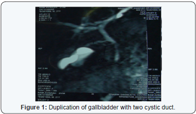
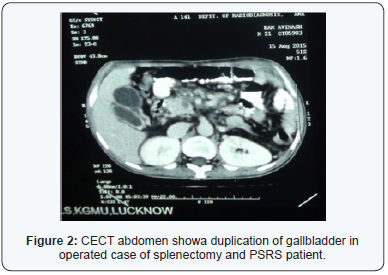
A 22-year-old non-alcoholic young male was admitted in Surgical Gastroenterology ward for Extrahepatic Portal Venous Obstruction with massive splenomegaly. During evaluation he was found to have duplicated gallbladder, detected by abdominal ultrasonography as well in Magnetic Resonance Cholangiopancreatography (MRCP). The patient was asymptomatic and had no significant past medical history including hospitalization. On physical examination, his abdomen was distended with massive splenomegaly, and the gallbladder was not palpable. Laboratory data were unremarkable. After comprehensive evaluation patient underwent proximal splenorenal shunt (PSRS) with splenectomy for his recurrent upper gastrointestinal bleeding and pancytopenia which were persistent after endoscopic management (recurrent bleeding from esophageal varices despite endoscopic variceal band ligation). Diagnosis of DG was made during postoperative period on follow-up Doppler ultrasound abdomen for patency of shunt. On retrospective evaluation, his MRCP shows duplication of gallbladder with separate necks and cystic ducts. Patient was asymptomatic for his duplication of gallbladder. His liver function test was normal.
The 2 cystic ducts separate entering the common bile duct (Figure 1). Contrast enhanced Computed Tomography (Figure 2), magnetic resonance imaging, and ultrasonography revealed no findings to suggest malignancy or benign biliary disease. The patient was asked to follow up using ultrasonography every year.
On follow up after 1,3,6 months, he remained asymptomatic with no more fever and pain and was able to do his routine work.


Discussion and Review of Literature
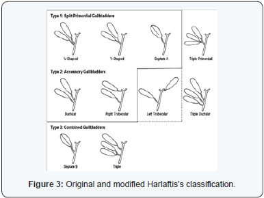
In 31 BC, the first case in human being was reported in a sacrificial victim of Emperor Augustus [1] Duplication of the gallbladder is a uncommon developmental anomaly of the extrahepatic biliary system with reported the incidence of 1 in 4000. In occurs as exuberant budding developing biliary tree during 5-6 weeks of embryogenesis. Harlaftis et al. [2] report, 207 duplicated and 8 triple gallbladders [2]. Two type of classification this congenital abnormality are described by Boyden’s and Harlaftis’s respectively [2,3]. But classification of surgical relevance is the Harlaftis classification (Figure 3). Basis of classification is the duplication of gallbladder in relation to cystic duct. Type one is duplication gallbladder with a single cystic duct also known as ‘split primordium group’ or ‘vesica fellea divisa’ .Type Two is duplication gallbladders with separate cystic duct draining into common bile duct, also known as ‘accessory group’ or ‘vesica fellea duplex’. Our case was type 2 duplication gallbladder.

There is no gender predilection, mostly diagnosed as incidental finding in adults, during cholecystectomy or at autopsy [4]. Recent improvements of technique, such as magnetic resonance cholangiopancreatography, have enabled diagnosis of this anomaly being as noninvasive and no radiation exposure [5,6]. Duplicated gallbladder may be incidentally (as in our case) or when symptoms associated with cholelithiasis and cholecystitis occur. Such clinical condition should be differentiated from type two choledochal cyst, abnormal fold or diverticulum of gallbladder, Phrygian cap, localized pericholecystic fluid collection, or focal adenomyomatosis, and intraperitoneal vascular bands across the gallbladder.
Presentation can be asymptomatic or biliary colic, acute or chronic cholecystitis. Less common pathologies in duplicated gallbladders include perforation, adenocarcinoma, and adenomyomatosis [3]. The management of duplicated gallbladder is similar to that of other gallbladder disease. Because there is no evidence of increased risk, surgery is not indicated for duplicated gallbladders when discovered incidentally without disease in both gallbladders. Patients with this pathology can be asymptomatic or symptomatic. If symptomatic, surgery to remove the gallbladders is indicated. Although rare, it is important to be aware of duplex gallbladders pre-operatively. As in our case it was missed preoperatively as well as intraoperatively. Preoperative diagnosis can avoid the duplication gallbladder being missed intra-operatively with these patients re-presenting with biliary symptoms at a later date.
However, if one or both gallbladders cause symptoms, cholecystectomy should be done for both gallbladders. Laparoscopic cholecystectomy can be done as per indication as a standard of care. Relevance of congenital anomalies of extrahepatic biliary system is because it case cause bile duct injury during open or laparoscopic choelcystectomy. Preoperative diagnosis is of paramount importance to avoid such disaster. Intraoperative cholengiography can be considered in unclear scenario.
Conclusion
Duplication of gallbladder is a uncommon anomaly of gallbladder number. If symptomatic, it requires preoperative diagnosis for appropriate management. Asymptomatic case can be observed.
References
- Udelsman R, Sugarbaker PH (1985) Congenital duplication of the gallbladder associated with an anomalous right hepatic artery. Am J Surg 149(6): 812-815.
- Boyden EA (1926) The accessory gall-bladder: an embryological and comparative study of aberrant biliary vesicles occurring in ma and the domestic mammals. Am J Anat 38(2): 177-231.
- Harlaftis N, Gray SW, Skandalakis JE (1977) Multiple gallbladders. Surg Gynecol Obstet 145(6): 928-934.
- Skandalakis JE, Gray SW, Ricketts R, Skandalakis LJ, Dodson T (1993) The extrahepatic ducts and the gallbladder. In: Embryology for Surgeons: The Embryological Basis for the Treatment of Congenital Anomalies. (2nd edn), Williams & Wilkins, Baltimore, USA, 297-303.
- Kim RD, Zendejas I, Velopulos C, Fujita S, Magliocca JF, et al. (2009) Duplicated gallbladder arising from the left hepatic duct: report of a case. Surg Today 39(6): 536-539.
- Singh B, Ramsaroop L, Moodley J, Satyapal KS (2006) Duplicated gallbladder: an unusual case report. Surg Radiol Anat 28(6): 654-657.


