




 OAJGGM Home



Abstract
Facial aging is a multifactorial process characterized by significant changes in the superficial and deep fat compartments of the face. This review summarizes the current understanding of volumetric and topographic changes in facial fat related to aging, analyzing the influence of sex, age, harmful habits, lifestyle, medication use, and chronic diseases. Evidence shows that facial fat compartments do not age uniformly: while deep compartments undergo progressive atrophy and volume loss (up to 18.4% over a decade), superficial compartments exhibit variable patterns that include selective hypertrophy in certain regions, such as the double chin, and ptosis in others. Sexual dimorphism influences soft tissue thickness, with women exhibiting greater thickness in the zygomatic and infraorbital regions, although this difference diminishes with age. Factors such as body mass index, smoking, certain medications, and chronic diseases can accelerate or alter these patterns of fat aging. Understanding these variations is essential for developing personalized facial rejuvenation strategies based on anatomical evidence.
Keywords:Facial aging; Fat compartments; Superficial fat; Deep fat; Senescence; Facial volume
Introduction
Aging of the human face is a complex biological process that goes beyond the formation of skin wrinkles [1]. For decades, attention focused on dermal changes and the effects of gravity; however, recent research has revolutionized our understanding by identifying that the face is composed of fat compartments both superficial and deep that undergo volumetric and positional changes that manifest with age but do not always present in the same way in every individual, as they are influenced by multiple intrinsic and extrinsic factors [1,2].
Facial fat is not a homogeneous mass but is organized into independent units delimited by fascial septa and retaining ligaments [3-5]. The superficial compartments (such as the nasogenian fat, medial malar fat, and double chin fat) are located above the Superficial Musculoaponeurotic System (SMAS), while the deep compartments (such as the deep medial malar fat and suborbital fat) are located below this layer and are firmly anchored to the periosteum [3-5]. This compartmentalization explains why fat aging is not uniform and exhibits selective patterns of atrophy, hypertrophy, and ptosis [2,3].
Aging is associated with skeletal changes (bone resorption), ligamentous laxity, and changes in skin quality, but it is the changes in fat deposits that largely determine the appearance of an aging face: hollowing of the malar region, deepening of the nasolabial folds, the appearance of a double chin, and the prominence of dark circles under the eyes [1,2,6,7]. These changes do not occur in isolation but are influenced by multiple factors, such as genetics, race, the individual’s sex, chronological age, lifestyle habits (smoking, sun exposure, nutrition, and sleep duration), the use of certain medications, and the presence of chronic diseases such as diabetes or lipodystrophies [8-11].
There are numerous factors to consider when evaluating facial aging; ranging from the treating physician’s anatomical knowledge to the factors that influence the changes that occur over the years. For a multimodal facial rejuvenation treatment, it is necessary to understand the anatomy of the superficial and deep fat compartments and to analyze variations in their volume and topography based on sex, age, as well as the influence of harmful habits, lifestyle, medication use, and history of chronic diseases thereby establishing correlations between fat aging patterns and their implications for facial rejuvenation [6,12,13].
Throughout history, concepts and theories regarding facial aging have evolved, and based on this, facial rejuvenation treatment protocols have been updated to achieve a more harmonious and comprehensive result. This served as the motivation for this study, which aims to determine the structural and volumetric changes in the superficial and deep facial fat pads associated with aging, while analyzing the factors that influence these changes [1,2,6].
Method
A literature search was conducted in the PubMed, Scopus, Scielo, and Web of Science databases for articles in English and Spanish using three search strategies that combined descriptors with Boolean operators, limiting the “AND” operator to distinct terms and the “OR” operator to synonyms:
Old age (or) aging (and) facial fat compartments (or) facial fat
pads
Facial volume (and) old age (and) rejuvenation
Facial fat compartments (and) determining factors (or) age
(or) lifestyle (or) associated diseases (or) medication use
Results
Of the 230 sources identified, 33 articles published between 2020 and 2026 were selected, prioritizing systematic reviews, original articles, longitudinal cohort studies, cadaveric studies, and imaging analyses (CT, MRI, ultrasound) that quantitatively assess facial fat compartments in adult populations; studies linking fat aging to demographic, clinical, or environmental factors. We excluded 197 articles focused exclusively on rejuvenation techniques without baseline anatomical analysis; case series with insufficient sample sizes (n<10); conference abstracts, case reports, and articles not available in full text.
We analyzed the information found based on determining factors such as sex, age, race, substance use, lifestyle, medication use, and chronic diseases, all of which are related to volumetric and topographic changes in facial fat with aging.
Discussion
Anatomy of the Facial Fat Compartments
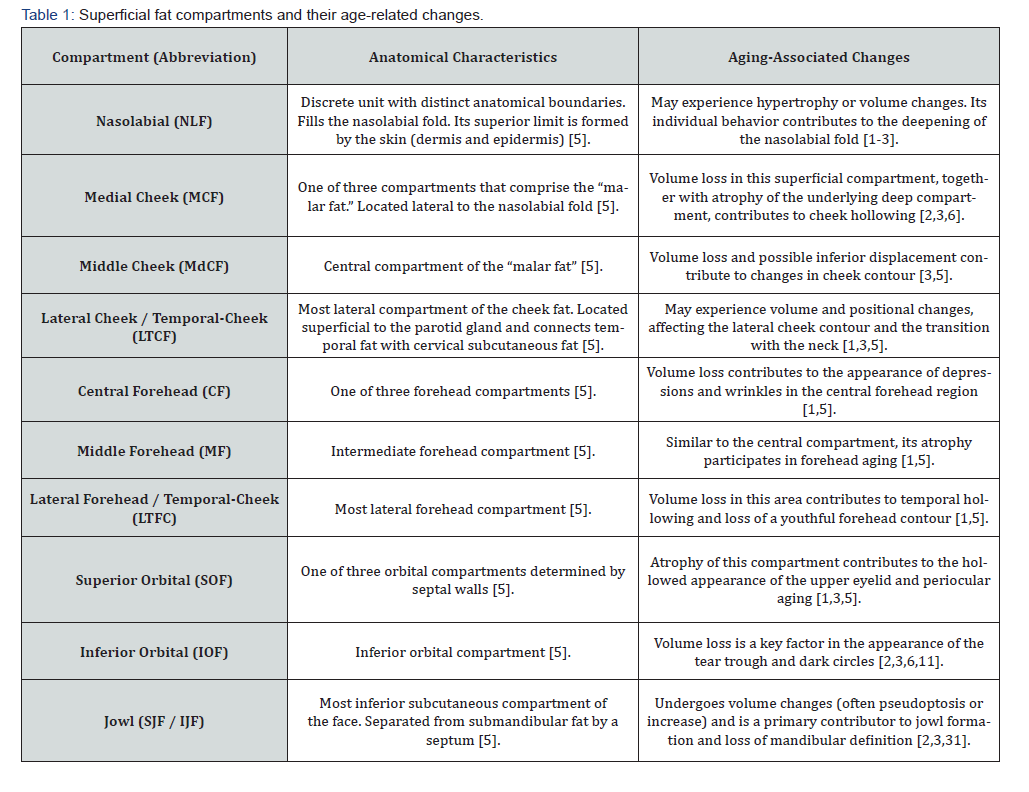
The human face exhibits a laminar organization into several histological layers depending on the aesthetic subunit, in which the following are always present: skin, subcutaneous fat (which houses the superficial compartments), SMAS, deep fat (deep compartments), and periosteum [1,4,5] (Tables 1 & 2).
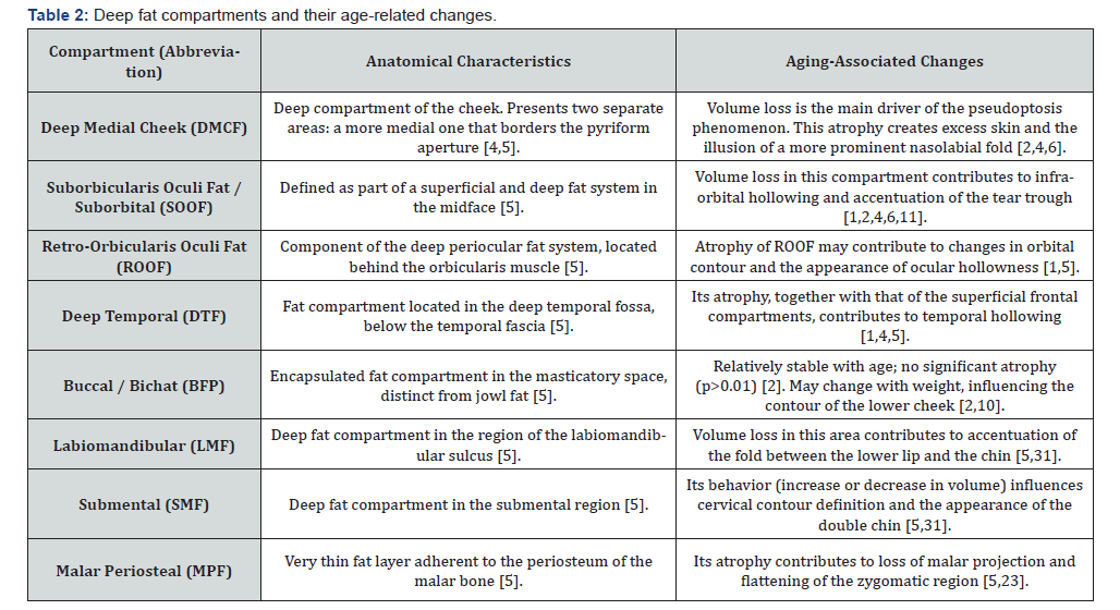
A key characteristic is that the deep compartments are firmly anchored to the underlying bone and provide structural support, whereas the superficial compartments are more mobile and are influenced by muscle contraction [4,5]. This anatomical difference determines their divergent aging patterns [2,3].
Age-Related Volumetric and Topographic Changes
Facial adipose tissue is one of the areas most affected by the passage of time. The deep subcutaneous tissue gives the face its youthful position, contour, and dimensions and is therefore critical for mitigating the morphological changes that occur over time. It is responsible for the concave and convex lines that define a youthful face [2,6]. Rohrich and Pessa [5] describe the superficial fat compartments of the face. These move with facial expressions and shift as we age. Each compartment can age independently, and this affects the others in a cascade of events. With age, a loss of volume occurs in deep fat pads, causing superficial compartments to shift and form furrows, which contributes to the malpositioning of soft tissues [2,6]. Furrows form and become more pronounced in transition areas between superficial fat pads, which no longer have deep support because it has diminished [6,12].
Not all fat compartments of the face change in the same way; while some atrophy, others retain their volume and cause asymmetry, as is the case with the nasogenian fat compartment, where superficial and deep fat pads converge [2,3]. The study of these accumulations of fat cells is important because it allows for the selective replenishment of those that have been depleted [2,6]. A cadaveric study involving 63 specimens (38 women, 25 men, mean age 71 years) revealed that the mean size of adipocytes in the nasogenian (superficial) fat is significantly larger than that in the deep medial malar fat (p<0.0001), suggesting that the superficial and deep compartments possess distinct metabolic and structural properties [4]. This difference may explain why superficial fat tends to undergo hypertrophy in certain regions while deep fat tends to atrophy [2,4].
Facial adipose tissue holds the secret to a youthful face. Each person will lose it according to their own genetics, gender, and lifestyle. Every individual has a cellular program that determines when these cells will begin to dissipate, leading to a decrease in facial volume. Some people lose cells more slowly than others. This is why some people may appear younger than their peers of the same age. Lifestyle can damage fat deposits. Marathon runners and triathletes, who push themselves physically, have very little adipose tissue [14,15]. People who prefer to sleep on the same side with their face pressed down against the mattress may reduce fat cells in that area because very little oxygen or nutrients flow to the tissue [16].
Fat loss causes the facial contours to sag or droop, no matter how well you’ve cared for your skin. Ultimately, changes in facial adipose tissue can influence changes in other tissues such as the skin, muscles, and bones and vice versa [1,2,6,7].
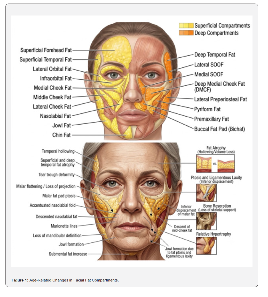
Evidence shows that the aging of facial fat is characterized by non-uniform, compartment-specific changes [2,3] (Figure 1). Regarding overall volume loss, a longitudinal CT study conducted on 19 individuals over a mean period of 11.4 years demonstrated a significant decrease in total facial fat volume (from 46.48 cc to 40.81 cc, p<0.01). Superficial fat decreased from 26.11 cc to 23.14 cc (p<0.01), representing an 11.3% loss of initial volume, while deep fat decreased from 11.00 cc to 8.99 cc (p<0.01), with an 18.4% loss [2]. This finding confirms that atrophy predominantly affects the deep compartments, which supports the pseudoptosis theory; the loss of deep support contributes to the sagging of the overlying superficial compartments [2,6].
This is why the new approach for aging faces is based on recognizing the importance of facial adiposity. Patients with better facial volume always appear younger, even without having undergone any procedures [6,12].
Regarding specific regional patterns, a strategically important area for the loss of fat and tissue—especially in women—is the temporal region, which is often neglected and overlooked but plays a key role in preventing facial aging [1,5]. As we age, these areas sink, causing the forehead to appear narrower—a hallmark of the “pyramid of aging.” Caring for this often-overlooked area of the face has a significant impact on facial rejuvenation [6,7,13].
The malar and periorbital fat compartments are the first to be affected, which accentuates dark circles and the nasolabial folds [2,3,6]. The orbital fat compartment is not immune to the loss of soft tissue volume that accompanies aging. As the orbital fat compartments begin to diminish, a person appears more tired and older. Later, the eyes become sunken and cast shadows, making them appear darker and more hollow. In extreme cases, atrophy of this fat compartment is so severe that the orbital rim becomes visible, accompanied by the appearance of the tear trough and bone resorption, which exacerbates the process [6,7,11].
The face loses fat in some areas but may accumulate it in others. In the case of cheek formation, when the perioral fat located around the dimples of the mouth begins to decrease along with the preauricular fat located at the angles of the face, just in front of the ears, it creates a forward shift [2,3].
The Influence of Sex on Changes in Facial Fat
Sexual dimorphism is a determining factor in the morphology and aging pattern of facial fat compartments. Anatomical studies have shown that women have greater soft tissue thickness in the zygomatic and infraorbital regions compared to men, while men exhibit greater thickness in most regions of the facial midline [17]. This sex difference is also evident at the cellular level: the mean size of adipocytes is significantly larger in women than in men (p<0.0001), although this dimorphism is attenuated in the fat deposits of overweight individuals, suggesting a complex interaction between sex and body mass index [4]. With advancing age, sex differences in soft tissue thickness tend to decrease progressively; linear regression models show that both age and elevated BMI mitigate the significant sex dimorphism observed in younger populations [17]. Women experience more pronounced changes in deep fat regions, requiring more aggressive volumetric restoration strategies in the midface during rejuvenation [7], whereas men, who have greater thickness in the superficial compartments, may benefit from focal approaches to correct ptosis and irregular contours [4,17]. This sex-related variability in fat aging underscores the importance of developing personalized therapeutic strategies that consider not only the patient’s chronological age but also their sex and the specific characteristics of their fat compartments [6,7,17].
Influence of Habits and Lifestyles
Lifestyle has a decisive influence on changes in facial fat compartments during aging. Evidence shows that these factors act through well-defined mechanisms that can accelerate, mitigate, or even exacerbate volume loss and changes in facial fat distribution [8,9,18,19]. The following section analyzes the main modifiable determinants.
Tobacco use accelerates skin aging and contributes to the loss of elasticity in the skin and facial ligaments [18,19]. Although specific evidence on the direct impact of smoking on facial fat is limited, it is known that tobacco-induced oxidative stress promotes the degradation of collagen and elastin, which facilitates the ptosis of the superficial fat compartments [18]. Impaired microvasculature reduces blood supply to adipocytes, accelerating the atrophy of the deep fat compartments [19]. Furthermore, chronic smoking is associated with a greater deepening of the nasolabial folds and a premature loss of mandibular angle definition [18].
Body weight significantly influences the morphology of facial fat, with differential effects depending on BMI and the rate of weight change [4,8]. Low BMI (< 20 kg/m²): Accelerates facial atrophy. A low BMI accelerates facial aging by mimicking and exacerbating the volume loss typical of old age. The cadaveric study by Wan et al. demonstrated that the ratio of deep to superficial fat (a key indicator of supportive volume) is significantly lower in individuals with a BMI <20 (0.15) compared to those with a normal (0.31) or high (0.34) BMI [4]. This suggests that extreme thinness primarily affects the deep compartments, which are responsible for the face’s structural support, leading to premature sagging [4,8]. Following massive weight loss (due to bariatric surgery or medications such as GLP-1 agonists), the most significant loss of volume occurs in the mid-cheek region, nasolabial folds deepen, and neck sagging becomes more pronounced a phenotype associated with a perception of greater apparent age [8,20].
Normal BMI (18.5–24.9 kg/m²): Relative balance. In individuals with a normal BMI, changes in facial fat are primarily due to intrinsic aging, with gradual atrophy of the deep compartments and variable hypertrophy of the superficial ones [2,3]. However, rapid weight loss—even starting from a normal BMI—produces significant changes that resemble accelerated facial aging [8,20].
Extreme weight loss (as seen in eating disorders) results in a gaunt appearance characterized by accentuated nasolabial folds and sunken cheeks, reflecting the loss of both fat compartments [8,9,21]. Fat distribution in anorexia nervosa shows a severe reduction in subcutaneous fat mass, resulting in an emaciated face with prominent cheekbones and periorbital hollowing [21]. A study using an advanced genetic design (Mendelian randomization) confirmed a causal relationship between genetic predisposition to obesity and accelerated facial aging, suggesting that the underlying mechanisms go beyond the simple “filling or emptying” process [8].
Ultraviolet (UV) radiation is a primary extrinsic factor in the aging of facial fat. It not only damages the superficial skin but also affects the connective tissue that supports the fat compartments [18]. Solar radiation induces the production of inflammatory cytokines such as IL-11, IL-1α, IL-6, and TNF-α in the skin; these molecules inhibit the differentiation of preadipocytes into mature adipocytes, blocking the formation of new fat tissue and contributing to the atrophy of the superficial compartments [16,22]. Although actinic damage primarily affects the epidermis and dermis, solar elastosis and the degradation of retaining ligaments can accelerate the ptosis of the superficial compartments [16,18].
The use of broad-spectrum sunscreens prevents this radiationinduced inflammatory process, suggesting that adequate photoprotection may slow the loss of facial fat associated with photoaging [22]. In addition, diet plays a modulatory role: a higher intake of monounsaturated fatty acids from olive oil has been associated with a lower risk of severe photoaging, suggesting a beneficial effect of the Mediterranean diet on the preservation of facial fat [22,23].
The position in which we sleep exerts repetitive and prolonged pressure on the face, which can have long-term structural consequences [24]. Sleeping on one’s side—especially always on the same side—compresses the face for 6 to 9 hours a day. This constant pressure can “deflate” the malar and buccal fat pads, as well as compress the skin and contribute to the appearance of wrinkles and facial asymmetries [24]. Experts in dermatology and plastic surgery note that repetitive compression can gradually soften the fat pads in the cheeks, creating an appearance of volume loss and accentuating nasolabial folds, especially on the side on which one sleeps [24]. A preventive strategy is to try sleeping on your back or using special pillows that minimize pressure on the face [24].
The influence of physical exercise on facial fat during aging is a complex, two-sided issue, where the effects depend critically on the type, intensity, and metabolic context of the exercise [14,25].
Accelerating effect (fat loss): Intense, prolonged cardiovascular exercise (such as running or cycling) burns calories and reduces the percentage of total body fat. This generalized lipolysis also affects facial fat, which can result in a thinner, sunken face with an aged appearance (similar to the “runner’s face” or “gym face” effect) [14,25]. It has been observed that elite athletes with low body fat percentages have a more “gaunt” facial appearance due to the loss of superficial fat, which accentuates facial shadows and creases [14]. During intense exercise, the body diverts blood flow away from the skin and superficial fat toward the active muscles; this relative hypoxia affecting adipocytes may contribute to their long-term reduction [25]. In addition, strenuous exercise generates free radicals that, combined with sun exposure and aging, damage collagen and elastin, contributing to sagging and ptosis of the already weakened fat compartments [14,25].
Protective Effect (Muscular Support): There is an opposing approach: exercising the facial muscles to combat volume loss. Aging involves atrophy of muscles and fat; the theory is that exercising the facial muscles causes them to hypertrophy, plumping the skin from within and improving the contour, in a manner analogous to “adding volume” beneath the surface [26]. A pilot study published in JAMA Dermatology (2018) is one of the key references in this field. In 16 women aged 40–65 who performed facial exercises for 30 minutes daily over 20 weeks, a significant increase in the fullness of the upper and lower cheeks was observed. Blinded evaluators estimated that the participants looked, on average, three years younger at the end of the study [26]. The study concludes that these exercises can improve the appearance of aging, possibly by strengthening the underlying muscles and improving facial contour [26].
A synthesis of the evidence suggests that the effect of exercise on facial fat depends on the balance between caloric expenditure (which reduces fat) and muscle tone (which improves support). Strenuous and prolonged cardiovascular exercise may contribute to the atrophy of fat compartments by reducing total body fat and blood flow, while specific facial exercises offer a potential way to counteract the loss of support, although further studies with control groups are needed [14,25,26].
Prolonged stress and lack of sleep affect the body through the hormone cortisol and by disrupting cellular repair processes [27,28]. Chronically elevated cortisol (associated with stress) breaks down collagen, the protein that keeps the skin firm and elastic, which can lead to thinner, sagging skin that is prone to wrinkles [27]. Restful sleep is crucial for cell renewal and collagen synthesis, while sleep deprivation exacerbates the effects of cortisol and can contribute to facial swelling due to fluid retention [28]. Although direct evidence on the impact of stress and sleep on facial fat compartments is limited, it is known that elevated cortisol promotes the redistribution of body fat toward visceral fat deposits, which could indirectly alter the distribution of facial fat [27].
Diet and fluid intake form the foundation for healthy skin and body fat [23]. A diet high in processed foods and sugars can promote inflammation, while extreme calorie restriction accelerates fat loss [8,23]. Adequate hydration keeps the skin smoother and can help reduce puffiness and the appearance of sagging [23]. There is an association between a diet with a moderate caloric surplus and rich in healthy fats (such as the monounsaturated fatty acids found in olive oil, avocados, and nuts) and improvements in facial fullness and skin quality [22,23].
The influence of modifiable factors opens the door to preventive strategies for fat-related aging. Body weight control appears to be crucial: being overweight may mitigate atrophy of the deep compartments (although it may also contribute to double chin hypertrophy) [4,8], while severe weight loss accelerates facial emaciation [8,21]. Sun protection, smoking cessation, and proper nutrition (with sufficient intake of vitamins and antioxidants) can preserve skin and ligament quality, slowing the ptosis of the superficial compartments [18,19,22,23].
Influence of Race
Ethnic and racial differences are a determining factor in the aging of facial fat compartments, modulating both baseline morphology and the patterns of atrophy, hypertrophy, and ptosis that develop with aging [17,29]. Although the progression of aging may be similar across population groups, the patterns and rate of change vary significantly among different races and ethnicities [29]. In individuals of Asian descent, the combination of thicker skin, greater amounts of superficial fat, and denser fibrous connections between the SMAS and the parotidomaseteric fascia reduces the prevalence of superficial wrinkles and severe soft-tissue ptosis, although thickening of the superficial fat and increased moderate ptosis are distinctive features of aging in this population [29]. In contrast, white/Caucasian women tend to exhibit earlier signs of facial aging compared to Hispanic, Asian, and African American women, with a higher prevalence of linear wrinkles on the forehead and in the periorbital region [17,29] Populations of African descent and African Americans, characterized by thicker skin, heavier malar fat pads, and potentially weaker skeletal support, exhibit aging marked by the inferomedial descent of the malar fat pads, fat accumulation in the midface region, and accentuation of the nasolabial folds [17,29]. Systematic studies have demonstrated that ethnic groups exhibit distinctive patterns in the thickness of facial soft tissues, with women showing greater thickness in the deep fat regions and men in the superficial regions—findings that support the need for personalized and culturally sensitive rejuvenation strategies [17,29]. Understanding these anatomical variations and their impact on fat-related aging is essential for the development of evidence-based therapeutic approaches tailored to the specific characteristics of each population group [6,17,29].
Effects of Medication Use
Drugs can induce profound and often specific changes in the morphology of facial fat—phenomena that sometimes overlap with or are confused with physiological aging but that result from specific pathophysiological mechanisms [15,30]. One of the most widely studied examples is facial lipoatrophy associated with antiretroviral therapy for HIV, a lipodystrophy syndrome that affects approximately 50% of patients on antiretroviral therapy (ART) and is characterized by a disproportionate loss of subcutaneous fat in the face and extremities, which may coexist with central fat accumulation in the trunk and abdomen [15]. Protease inhibitors, introduced as standard therapy in 1996, were the first to be associated with this effect, although subsequent studies have identified thymidine analog reverse transcriptase inhibitors, particularly stavudine and zidovudine, as the drugs with the highest risk of inducing lipoatrophy [15]. A comparative study showed that the incidence of facial atrophy was 48% in patients treated with stavudine compared with 22% in those receiving zidovudine (p=0.011), and it has been observed that replacing these drugs with more modern alternatives such as abacavir or tenofovir offers substantial protection against lipoatrophy [15]. Computed tomography scans of affected patients confirm the nearly complete loss of subcutaneous fat in the malar and temporal regions, a change that, beyond its aesthetic impact, has been linked to depression, low self-esteem, and a significant impairment in quality of life [15].
In recent years, the widespread use of GLP-1 receptor agonists, such as semaglutide, for weight loss has brought to light a new phenomenon, popularly known as “Ozempic face,” which illustrates how pharmacologically induced weight loss can exacerbate the typical changes associated with facial aging [20]. Unlike HIV-associated lipoatrophy, which appears to have a direct effect on adipose tissue, the effect of GLP-1 agonists on facial fat is primarily a consequence of generalized lipolysis secondary to rapid and significant weight loss [20]. However, the loss of volume is not uniform: a study that quantified volumetric changes in patients treated with semaglutide documented an average decrease of 41.8% in the volume of superficial temporal fat and a 69.9% reduction in superficial cheek fat, predominantly affecting the superficial compartments of the midface region [20]. This loss of volume in key areas such as the cheeks, temples, chin, and periorbital region leads to a sunken or emaciated facial appearance that accentuates wrinkles and skin laxity; it has been estimated that patients who experience massive weight loss may appear up to five years older than their peers without a history of extensive weight loss [8,20].
On the other hand, systemic corticosteroids represent a completely different pharmacological mechanism of facial fat alteration, mediated by a central redistribution of body fat secondary to excess cortisol [19]. Prolonged use of these drugs, such as prednisone, is a well-known cause of iatrogenic Cushing’s syndrome, whose most characteristic facial feature is the “moon face”—a rounded facial swelling and fullness that contrasts sharply with the atrophy observed in old age [30]. This effect, which is often accompanied by fat accumulation at the nape of the neck (buffalo hump), is usually gradually reversible when the corticosteroid dose is reduced or adjusted [30].
More recently, cancer immunotherapy has introduced a new and rare adverse effect on facial adipose tissue. Immune checkpoint inhibitors (ICIs), such as nivolumab and pembrolizumab, have been associated in isolated cases with the development of acquired generalized lipodystrophy, which includes a total and permanent loss of both facial and non-facial fat [11]. Cases have been documented in which loss of facial volume was the patient’s primary concern, significantly aging their appearance. Symptoms typically begin, on average, about 7.44 months after the start of treatment with ICIs [11]. Although fewer than ten cases have been reported, the severity of the condition and the difficulty in finding reconstructive options due to the lack of suitable autologous fat donor sites make this a significant clinical challenge, for which alternatives such as dermal grafts or autologous fat grafting have been explored [11].
Taken together, the evidence shows that multiple drug classes—from antiretrovirals and GLP-1 agonists to corticosteroids and immune checkpoint inhibitors—can induce substantial and specific changes in facial fat compartments, whether by promoting their selective atrophy, central redistribution, or generalized loss secondary to lipolysis [15,20,30]. These effects, which often overlap with the signs of chronological aging, underscore the importance of a detailed medication history in the clinical evaluation of the patient and point to the need for personalized therapeutic strategies that take into account the underlying etiology of facial volume changes [6,15,20].
Impact of Chronic Diseases
Chronic diseases are a determining factor in the acceleration and alteration of aging patterns in the facial fat compartments, acting through pathophysiological mechanisms that go beyond the process of physiological senescence [9,11]. Acquired facial lipoatrophy, characterized by the loss of subcutaneous adipose tissue that causes flattening or indentation of the facial contour, can manifest as a complication of various systemic diseases [11]. Among the most common are connective tissue disorders associated with panniculitis, such as deep lupus erythematosus or scleroderma, which can induce localized and asymmetric loss of facial fat [11]. Lipodystrophies, both hereditary and acquired, represent another group of disorders with a profound impact on the facial fat architecture; Barraquer-Simons syndrome, a rare, acquired lipodystrophy that predominantly affects women, presents with a symmetrical and progressive loss of subcutaneous fat in the face, neck, and shoulder girdle, giving patients an aged and cachectic appearance that negatively affects their self-image and social status [11]. In the context of HIV infection, highly active antiretroviral therapy (HAART) is a well-documented cause of facial lipoatrophy, affecting approximately 55% of patients, with 47% experiencing facial fat loss [15]. Diabetes mellitus, for its part, exerts a more subtle but equally significant influence on facial fat aging; patients with type 2 diabetes exhibit accelerated skin aging mediated by the formation of advanced glycation end products (AGEs) and chronic oxidative stress, which promote the degradation of collagen and elastin, compromising the structural integrity of the retaining ligaments that support the fat compartments [9]. Likewise, chronic kidney disease (CKD) is associated with premature multisystemic aging, driven by mechanisms such as increased allostatic load and the activation of pro-aging pathways; in advanced stages, alterations in bone and mineral metabolism secondary to hyperparathyroidism can lead to facial skeletal deformities that alter the bony base upon which the fat compartments rest [7]. Chronic inflammatory diseases, such as rheumatoid arthritis, also contribute to changes in facial fat, both due to persistent systemic inflammation and the prolonged use of corticosteroids, which induce a characteristic redistribution of body fat with accumulation in the facial region (“moon face”) [11,30]. Taken together, the evidence shows that chronic diseases modulate the aging of facial fat compartments through pathways that include inflammation, oxidative stress, metabolic disorders, and the iatrogenic effects of pharmacological treatments, leading to phenotypes of premature aging that require an integrative diagnostic and therapeutic approach that considers both the underlying pathology and its consequences on facial architecture [9,11,20].
The reviewed evidence shows that the superficial and deep fat compartments of the face do not age uniformly: while the deep compartments undergo progressive and significant atrophy—with losses of up to 18.4% over a decade—the superficial compartments exhibit variable patterns that include selective hypertrophy in the double chin and ptosis in the malar regions [2,3,31]. This morphological heterogeneity, mediated by differences in bony anchorage, mobility, and the metabolic properties of adipocytes [4,5], constitutes the anatomical basis of the senile phenotype characterized by sagging of the midface, deepening of the nasolabial folds, formation of the double chin, and loss of definition of the mandibular angle [1,2,6].
Sexual dimorphism emerges as a significant modulator of these changes: women exhibit greater thickness in the deep fat regions and experience more pronounced changes in the midface, while men show greater thickness in the superficial compartments, suggesting the need for sex-specific therapeutic strategies [4,7,17]. Toxic habits and lifestyle factors—particularly smoking, body mass index, sun exposure, and intense physical exercise—act as accelerators or mitigators of fat-related aging through mechanisms that include oxidative stress, chronic inflammation, generalized lipolysis, and collagen degradation [8,18,19,16,14]. Likewise, certain medications—antiretrovirals, GLP-1 receptor agonists, and corticosteroids—and chronic diseases such as diabetes, lipodystrophies, and autoimmune diseases induce premature aging phenotypes that may be confused with physiological senescence but that result from specific pathophysiological mechanisms [9,11,15,20,30]. Ethnic and racial differences, although less studied, influence soft tissue thickness and ptosis patterns, underscoring the importance of personalized and culturally sensitive approaches [17,29].
The clinical implications of these findings are substantial. The concept of pseudoptosis—the loss of deep support that causes the descent of superficial compartments—has revolutionized aesthetic medicine by establishing that volumetric restoration must begin with the deep compartments before addressing the superficial ones [2,6,12,13]. Filler techniques targeting specific compartments, rather than indiscriminate wrinkle filling, have proven to be more effective and long-lasting in correcting midface sagging and improving facial contours in a natural way [6,20,21,26]. This approach, based on anatomical evidence, requires a detailed understanding of interindividual variability, which is influenced by the patient’s sex, age, BMI, race, and medication history [8,12,13,29].
Conclusion
The findings of this review confirm that the aging of facial fat is a highly compartmentalized and non-uniform process, determined by the interaction of multiple intrinsic and extrinsic factors [1-3]. This understanding has shifted the traditional paradigm of facial aging from a passive phenomenon of “deflation and sagging” toward an active and region-specific model [2,6,12].
The aging of facial fat is a multifactorial and compartmentspecific process, modulated by a complex network of biological, behavioral, pharmacological, and environmental factors [1,2,8,9]. Understanding this variability is no minor academic exercise: it is the foundation upon which truly personalized facial rejuvenation strategies must be built—strategies that respect each patient’s anatomy and move away from standardized protocols to embrace a comprehensive, evidence-based approach centered on the individuality of each face [6,12,13]. Integrating this knowledge into clinical practice will not only improve aesthetic outcomes but also preserve facial function and harmony, restoring to patients not only a more youthful appearance but also a facial identity that reflects their history and well-being [1,2,6,32,33].
References
- Nam KI, Shin S, Song J (2025) Anatomical Importance of Fat Compartments in Facial Aging: A Review. Anat Biol Anthropol. 38(3): 145-152.
- Boehm LM, Morgan A, Hettinger P, Matloub HS (2021) Facial Aging: A Quantitative Analysis of Midface Volume Changes over 11 Years. Plast Reconstr Surg 147(2): 319-327.
- Guduk SS, Cenkeri HC, Cicek ED, Kus S (2022) Evaluation of aging changes of the superficial fat compartments of the midface over time: A computed tomography study. J Cosmet Dermatol 21(4): 1430-1435.
- Wan D, Amirlak B, Giessler P, Rasko Y, Rohrich RJ, et al. (2014) The differing adipocyte morphologies of deep versus superficial midfacial fat compartments: A cadaveric study. Plast Reconstr Surg 133(5): 615-622.
- Rohrich RJ, Pessa JE (2007) The fat compartments of the face: anatomy and clinical implications for cosmetic surgery. Plast Reconstr Surg 119(7): 2219-2227.
- Ramanadham SR, Rohrich RJ (2015) Newer Understanding of Specific Anatomic Targets in the Aging Face as Applied to Injectables: Superficial and Deep Facial Fat Compartments--An Evolving Target for Site-Specific Facial Augmentation Plast Reconstr Surg 136(5 Suppl): 49-55.
- Mendelson B, Wong CH (2012) Changes in the facial skeleton with aging: implications and clinical applications in facial rejuvenation. Aesthetic Plast Surg 36(4): 753-760.
- Liu XJ, Sultan MT, Li GS (2024) Obesity, Glycemic Traits, Lifestyle Factors, and Risk of Facial Aging: A Mendelian Randomization Study in 423,999 Participants. Aesthetic Plast Surg 48(5): 1005-1015.
- Lian J, Zhang Z, Yang W, Pan L, Pan X, et al. (2026) Exploring the Causal Relationship and Potential Mediating Mechanisms Between Obesity and Facial Aging: A Two-Sample, Multivariable and Mediation Mendelian Randomization Study Clin Cosmet Investig Dermatol 19: 1-11.
- Akşamoğlu M, Muluk NB, Şahan MH (2024) Volumetric changes in buccal fat pad with aging and body mass index. J Comput Assist Tomogr 48(1): 156-164.
- Garg A (2011) Lipodystrophies: Genetic and Acquired Body Fat Disorders. J Clin Endocrinol Metab 96(11): 3313-3325.
- Lambros V (2007) Observations on periorbital and midface aging. Plast Reconstr Surg 120(5): 1367-1376.
- Cotofana S, Gotkin RH, Morozov SP (2018) The relationship between bone remodeling and the volume of the facial fat compartments: A computed tomography study. J Craniofac Surg 29(7): 1844-1849.
- Wilmshurst S (2021) Body composition and facial morphology in endurance athletes. J Sports Sci 39(14): 1580-1588.
- Carr A, Cooper DA (2000) Adverse effects of antiretroviral therapy. Lancet 356(9239): 1423-1430.
- Fisher GJ, Kang S, Varani J, Csorgo ZB, Wan Y, et al. (2002) Mechanisms of photoaging and chronological skin aging. Arch Dermatol 138(11): 1462-1470.
- Wang Y (2022) Sex differences in facial soft tissue thickness: A systematic review and meta-analysis. Forensic Sci Int 337: 111372.
- Morita A (2007) Tobacco smoke causes premature skin aging. J Dermatol Sci 48(3): 167-174.
- Freiman A, Bird G, Metelitsa AI, Barankin B, Lauzon GJ (2004) Cutaneous effects of smoking. J Cutan Med Surg 8(6): 415-423.
- Kitzinger HB, Karle B, Hacker S (2022) The Impact of Massive Weight Loss on Facial Aging: A Systematic Review. Aesthetic Plast Surg 46(3): 1189-1198.
- Rigaud D (2022) Facial fat distribution in anorexia nervosa: A prospective study. Int J Eat Disord 55(8): 1025-1034.
- Krutmann J, Schikowski T, Morita A, Berneburg M (2021) Environmentally-induced (extrinsic) skin aging: Exposomal factors and underlying mechanisms. J Invest Dermatol 141(4): 1096-1103.
- Ivić NP, Mirić L, Čarija A (2022) Skin aging: natural and protective factors. Acta Dermatovenerol Croat 30(2): 102-111.
- Zimbler MS, Kokoska MS, Thomas JR (2021) Sleep position and facial aging: A review of the evidence. Facial Plast Surg Clin North Am 29(1): 15-22.
- Plotkin DL, Roberts MD, Haun CT, Schoenfeld BJ (2021) The effects of endurance exercise on subcutaneous adipose tissue: A review. Sports Med 51(11): 2317-2332.
- Alam M, Walter AJ, Geisler A, Roongpisuthipong W, Sikorski G, et al. (2018) Association of facial exercise with the appearance of aging. JAMA Dermatol 154(3): 365-367.
- Chen Y, Lyga J (2014) Brain-skin connection: stress, inflammation and skin aging. Inflamm Allergy Drug Targets 13(3): 177-190.
- Sundelin T, Lekander M, Kecklund G (2015) Sleep loss alters facial appearance and increases perceptions of age, fatigue, and sadness. Psychoneuroendocrinology 56: 121-128.
- Lee JM (2023) Racial and ethnic differences in facial aging patterns: A systematic review. J Clin Med 12(15): 5012.
- Price NJ, Bertagna X, Grossman AB, Nieman LK (2006) Cushing's syndrome. Lancet 367(9522): 1605-1617.
- Kim JH (2023) Age-related changes of the superficial and deep fat compartments in the lower face: A computed tomography study. J Craniofac Surg 34(2): 145-150.
- Gkantidis N (2021) Genetic and environmental influences on facial soft tissue thickness: A twin study. Am J Phys Anthropol 174(3): 523-532.
- DeLorenzi C (2023) Newer approaches to facial volumization with hyaluronic acid fillers. Aesthet Surg J 43(Suppl 1): 15-26.

























