




 OAJGGM Home



Abstract
Total nasal defects, whether caused by trauma or pathology, require reconstruction of skin, mucosa, cartilage, and bone. Typically, the use of a radial forearm free flap is employed for the inner nasal lining. This report presents the case of a 69-year-old male victim of a gunshot wound to the submental region, with an avulsive exit wound encompassing almost the entirety of the nasal aperture. This patient’s post-injury course represents the first account of such a defect being reconstructed primarily using one donor site, the scalp, utilizing a peri cranial flap for intranasal lining, a calva rial bone graft with auricular cartilage for the nasal substructure, and a paramedian forehead flap for the external skin. This technique can be especially useful if free tissue transfer is not indicated in a patient, or if future radial forearm free flap(s) is/are planned for other uses.
Keywords:Total Nasal Defect; Nasal Reconstruction; Total Nasal Reconstruction; Subtotal Nasal Reconstruction; Pericranial Flap; Nasal Defect
Abbreviations: SIGSW: Self-Inflicted Shotgun Gunshot Wound; OMS: Oral and Maxillofacial Surgery; UABMC: University of Alabama at Birmingham Medical Center; ORIF: Open Reduction Internal Fixation; RFFF: Radial Forearm Free Flap
Introduction
The use of locoregional flaps for nasal reconstruction dates as far back as 600 BC when Sushruta described what is now known as the paramedian forehead flap. This flap has gone through numerous iterations over the years until Millard and Menick made key modifications that gave rise to the modern paramedian forehead flap used for complex nasal reconstruction. Complex defects often involve various reconstructive techniques to restore external coverage, substructural support, and the internal nasal lining.
Total nasal defects, whether caused by trauma or pathology, require restoration of skin, mucosa, cartilage, and bone. Typically, the use of grafts, locoregional flaps, and free flaps from different donor sites have been used for reconstruction of total nasal defects. This report is the first in the English language detailing a case of total external nasal reconstruction almost entirely using a single donor site. A paramedian forehead flap was used for external skin coverage, underlying calvarial bone for hard tissue support, and the intervening pericranial tissue for the internal nasal lining. The only tissue used outside of the primary donor site was auricular cartilage harvested for reconstruction of the nasal ala. This technique has the potential for decreased operative time, hospital stay, and overall donor site morbidity while still achieving an acceptable aesthetic outcome.
Case Report
A 69-year-old male suffered a self-inflicted shotgun gunshot wound (SIGSW) to the lower face, midface, and nose. He was taken to a local hospital where he underwent surgical debridement, primary closure of the wounds, and a tracheostomy. He was transferred three days post-operatively for further management by the Department of Oral and Maxillofacial Surgery (OMS) at the University of Alabama at Birmingham Medical Center (UABMC), a tertiary level one trauma center.
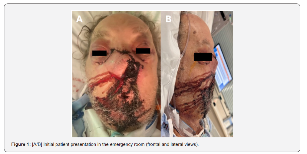
Upon presentation, the patient had severe microstomia with an oral commissure spanning less than one-centimeter due to subtotal vermillion avulsion and primary repair, subtotal nasal avulsion, a mandibular angle-to-angle continuity defect, and multiple midfacial factures (Figure 1).
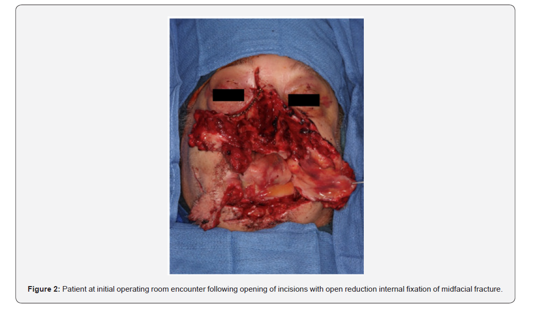
To eliminate the potential for excessive tissue scarring and a deformed facial structure largely incompatible with meaningful life, it was determined the patient would benefit from acute surgical intervention. The patient was taken to the operating room for a direct laryngoscopy, wound exploration, debridement of non-vital tissue, and open reduction internal fixation (ORIF) of the midfacial fractures (Figure 2). In separate procedures, a fibula free flap and later a radial forearm free flap were used to restore the avulsive mandibular defect and reconstruct the lower lip, respectively.
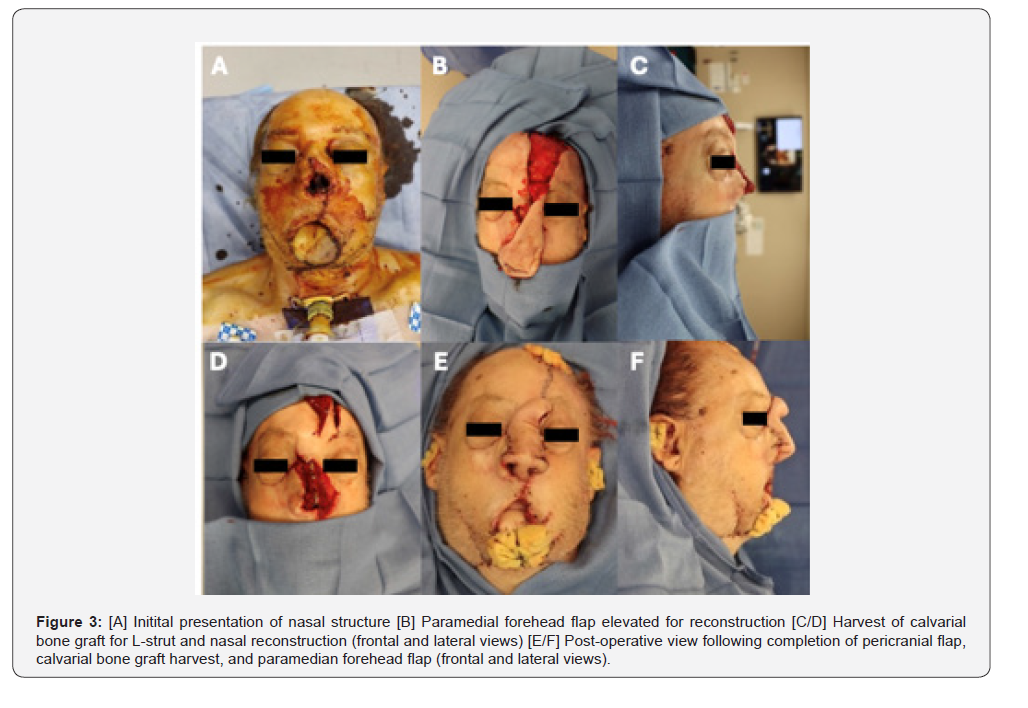
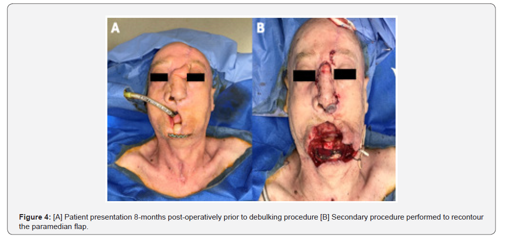
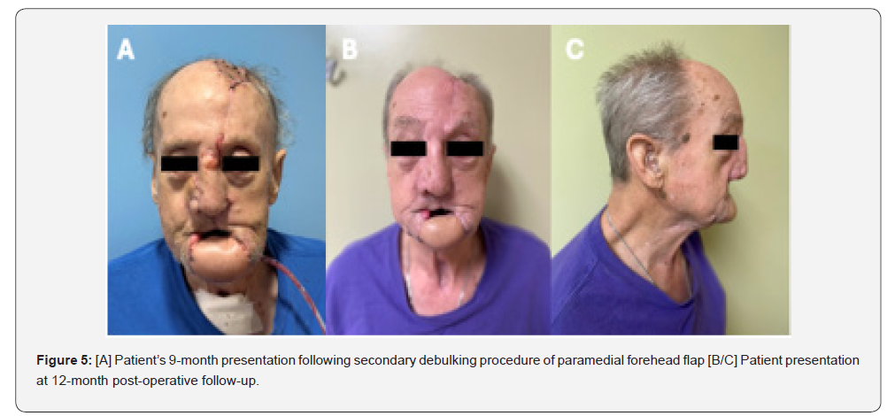
For the nasal component, reconstruction required addressing the internal lining, osseous and cartilaginous substructure, and external skin. Reconstruction of the nasal lining is typically performed with a radial forearm free flap. The use of local tissue was favored to avoid an additional free tissue transfer. First, a paramedian flap was harvested in a supraperiosteal fashion. Next, the scalp was widely undermined to further expose the pericranium. A pericranial flap was incised and reflected from the calvarial bone. Strips of calvarial bone were harvested in segments to fashion an L-strut framework to make both dorsal and columellar struts. The pericranium was rotated inferiorly for the inner lining, and the osseous struts were fixated in place using titanium midfacial miniplates. Auricular cartilage grafts were then fixated in place to reconstruct the bilateral ala. The pericranium was tent-poled to the surrounding bone and auricular grafts. Local tissue flaps were undermined and used to close the donor site defect. Finally, the paramedian flap was placed over the three-dimensional construct (Figure 3). The patient successfully underwent debulking of the paramedian flap with severing of the pedicle several months later, delayed significantly from the standard three-week window due to social issues (Figure 4). To accelerate healing of the donor site, some granulating tissue was debrided and a split thickness skin graft applied during the same operation as severing of the paramedian pedicle. At subsequent follow-up appointments, the patient was healing well with no current complaints or complications (Figure 5).
Discussion
Self-inflicted gunshot wounds (SIGSWs) to the face are the leading cause of gun-related deaths with injuries that are widely variable for those who survive [1]. Factors such as the strength of the tissue at the injury site and ballistic properties play a role in the severity of the facial defect [1]. Nasal reconstruction for fullthickness defects is a difficult task for surgeons due to the need to match the shape, color, and skin texture while restoring both functionality and aesthetics.
Transfer of tissue by means of either free or rotational flaps to the traumatized areas was expedited in this patient to provide early structural support for the soft tissues and prevent wound contracture. Our team employed the FACES protocol with the goal of earlier intervention to re-establish facial dimensions in an expedited fashion, decrease the need for more numerous revisions, and risk the patient being lost to follow-up [2-5]..
The typical workflow for total or subtotal nasal reconstruction often incorporates a radial forearm free flap used for reconstruction of the inner mucosal layer of the nasal cavity, followed by calvarial and cartilaginous grafting for the nasal substructure, finally followed by the paramedian flap for reconstruction of facial skin. Our report is the first published in the English language that almost entirely reconstructs the nose using one donor site (plus auricular cartilage grafts for nasal ala reconstruction), with the pericranial flap replacing a radial forearm free flap (RFFF). Both the RFFF and paramedian flaps have a success rate of 95- 99% and 94.9% respectively with the RFFF generally being described as the gold standard for this purpose in head and neck reconstruction [6-9]. Moreover, bone and fibrocartilage grafting serve as excellent options for helping to establish the structural integrity of the nose. Bone grafts for total nasal reconstruction are commonly harvested from the calvarium due to their slow rate of resorption. The calvarial bone is anatomically distant from critical nerves and vessels, decreasing the chance of morbidity from the grafting procedure [9,10]. Common complications of calvarial grafts include surface deformities in the donor or recipient site, graft fracture during harvest, seromas, and hematomas [11,12]. Of note, our patient did have a noticeable tissue deficit in the primary donor site due to the removal of three layers of tissue, coupled with postoperative scarring. Auricular fibrocartilage is commonly used to recreate the lateral cartilages, nasal tips, and provide support for the alar rims. Moreover, the auricular fibrocartilage graft is generally pliable and loses rigidity due load on the graft overtime.
Our approach entailed the utilization of the pericranial flap, calvarial and auricular cartilage, and the paramedian flap to decrease further donor site morbidly at distant donor site, as the patient was anticipated to require a RFFF for lower lip reconstruction. The pericranial flap repair of anterior skull base defects have a 90% complication-free and 95% overall success rate [13]. This can be considered an alternative method for reconstructing the internal nasal lining in patients with complex nasal defects where free tissue transfer is not desired, contraindicated, or it is not a viable surgical option performable by the reconstructive team. This patient’s postoperative course was complicated by a partially missing left pyriform rim, which led to some lateral collapse of the nose in the postoperative period. This was considered a minor concern, as the patient was overall pleased with the result.
Conclusion
Nasal reconstruction due to GSWs possess a unique challenge for surgeons to acquire both function and aesthetics. Though there are a wide range of tissue flaps and grafting techniques that can be used, clinicians should consider donor site morbidity, patient functional status, defect size, and options available to the surgical team when determining which reconstructive methods to utilize. Our technique was unique in its ability to avoid the use of free tissue transfer for this type of reconstruction.
References
- Peled M, Leiser Y, Emodi O, Krausz A (2012) Treatment protocol for high velocity/high energy gunshot injuries to the face. Craniomaxillofac Trauma Reconstr 5(1): 31-40.
- Gruss JS, Antonyshyn O, Phillips JH (1991) Early definitive bone and soft-tissue reconstruction of major gunshot wounds of the face. Plast Reconstr Surg 87(3): 436-450.
- Gruss JS, Van WL, Phillips JH, Antonyshyn O (1990) The importance of the zygomatic arch in complex midfacial fracture repair and correction of posttraumatic orbitozygomatic deformities. Plast Reconstr Surg 85(6): 878-890.
- Vasconez HC, Shockley ME, Luce EA (1996) High-energy gunshot wounds to the face. Ann Plast Surg 36(1): 18-25.
- Vayvada H, Menderes A, Yilmaz M (2005) Management of close- range, high-energy shotgun and rifle wounds to the face. J Craniofac Surg 16(5): 794-804.
- Wong CH, Wei FC (2010) Microsurgical free flap in head and neck reconstruction. Head Neck 32(9): 1236-1245.
- Hurvitz KA, Kobayashi M, Evans GR (2006) Current options in head and neck reconstruction. Plast Reconstru Surg 118(5): 122e-133e.
- Santamaria E, Granados M, Barrera FJL (2000) Radial forearm free tissue transfer for head and neck reconstruction: Versatility and reliability of a single donor site. Microsurgery 20(4): 195-201.
- Moscoso JF, Urken ML (1994) Radial forearm flaps. Otolaryngol Clin North Am 27(6): 1119-1140.
- Wei FC, Jain V, Celik N, Chen HC, Chuang DC, et al. (2002) Have we found an ideal soft-tissue flap? An experience with 672 anterolateral thigh flaps. Plast Reconstr Surg 109(7): 2219-2230.
- Demetriades D, Chahwan S, Gomez H (1998) Initial evaluation and management of gunshot wounds to the face. J Trauma 45(1): 39-41.
- Hollerman JJ, Fackler ML, Coldwell DM, Ben MY (1990) Gunshot wounds. 1: bullets, ballistics, and mechanisms of injury. AJR Am J Roentgenol 155(4): 685-690.
- Price JC, Loury M, Carson B, Johns ME (1988) The pericranial flap for reconstruction of anterior skull base defects. Laryngoscope 98(11): 1159-1164.

























