




 OAJGGM Home



Abstract
Ulcerations of the oral mucosa can be acute and self-limiting or chronic with impaired healing. Chronic, non-healing ulcers require prompt intervention to identify factors hindering proper healing and, more importantly, to determine whether the lesion is reactive or neoplastic.
Successful treatment outcomes rely on early detection and diagnosis, particularly in cases involving malignant neoplastic lesions. Age-related changes in oral mucosal tissue can also affect the healing process. In elderly patients, mucosal atrophy, reduced connective tissue elasticity, and the use of medications that contribute to mucosal dryness are factors that may impede ulcer healing. Consequently, food intake can be disrupted, leading to nutritional deficiencies that further compromise overall health. Additionally, age-related changes in immune function may delay healing. Therefore, detection, diagnosis, and early intervention are critical for preserving quality of life as patients age.
Keywords:Anterior-ventral tongue; Mucosa; Eosinophilic granulomas; Biopsy
Introduction
The lesion described in this case exhibits distinct hallmark of histologic features and have been reported on various intraoral sites in patients ranging from infants to older adults. In breastfed infants under two years of age, this lesion is consistently found on the anterior-ventral tongue, with natal or neonatal teeth implicated as the source of trauma. Interestingly, in this adult patient, who is also diagnosed with HIV, the ulcer also developed on the anterior-ventral tongue - the same site commonly affected in infants.
Notably, this lesion is not typically associated with comorbidities such as HIV/AIDS. Clinical evaluation of a large, nonhealing ulcer of the tongue, supported by histologic and cytologic findings resembling malignancy, led to the final diagnosis. Based on the clinical description and microscopic findings, the final diagnosis for this case was eosinophilic granuloma of the tongue.
Background
Eosinophilic granulomas (EG) are considered a benign disease entity with an etiology and pathogenesis that remain poorly understood. Trauma to the affected mucosa is commonly suspected as the cause. Eosinophilic granulomas are also solitary and benign lesions, which can often mimic oral malignancies such as squamous cell carcinoma. Rapid healing after biopsy or excision has been reported. The comparable entity in infants and neonates is called Rega-Fede disease [1-4].
This report highlights the occurrence of this poorly understood lesion in an HIV-infected individual who is also categorized as a member of the elder population who was currently taking medications to treat HIV. The management of HIV/AIDS normally includes the use of multiple antiretroviral drugs, antiretroviral therapy (ART), as a strategy to control HIV infection. There are several classes of antiretroviral agents that act on different stages of the replication cycle of HIV.
Case Report
A 62-year-old African male presented with a chief complaint of yellowish-white plaques on his tongue. He reported experiencing tenderness and pain for ten days, along with difficulty eating, but was uncertain about the lesion’s duration. Additional information obtained from the patient’s primary care team revealed that the growth on the tongue had first appeared approximately ten months earlier.
A review of the patient’s medical history revealed an HIV infection of 3–4 years’ duration, with an unknown mode of transmission. The patient stated he was unaware of how he contracted HIV. His current medications included Azithromycin, Bactrim, and Truvada. Azithromycin and Bactrim are antibiotics used to treat a variety of bacterial infections, while Truvada is used to manage HIV infection by inhibiting the virus’s ability to use the host’s CD4 cells to replicate [5,6].
An oral examination revealed a fixed yellow mass on the anterior-ventral one-third of the patient’s tongue (Figure 1). The classic ulcer, typically characterized by a break in the tongue mucosa, was not clearly visible in this case due to a thick fibrinous covering on the anterior portion of the tongue. The dorsal surface exhibited focal areas of whitish-yellow plaques (Figure 2).
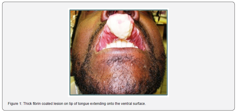
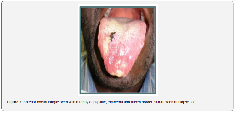
An incisional biopsy was performed on one of the affected sites on the dorsal surface. The specimen was fixed in formalin and sent to pathology for histologic preparation and evaluation.
Microscopic Description
Low-power microscopic examination revealed a segment of ulcerated mucosa with underlying edema adjacent to an area of hyperplastic epithelium (Figure 3). The underlying fibrous connective tissue contained numerous vascular spaces accompanied by a marked inflammatory infiltrate.
A medium-power view demonstrated infiltration of inflammatory cells into the submucosal muscle layer. High-power examination revealed a thickened lamina propria containing chronic inflammatory cells, including numerous lymphocytes, eosinophils, and some neutrophils. Additionally, large, atypical cells with pale-staining nuclei were observed. Occasional large cells with hyperchromatic nuclei, were also present.
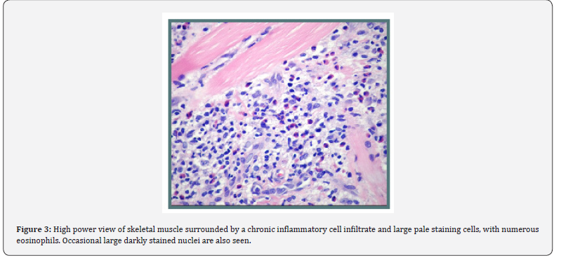
Based on the clinical description and microscopic findings, the final diagnosis for this case was “Eosinophilic granuloma of the tongue”.
Follow-Up
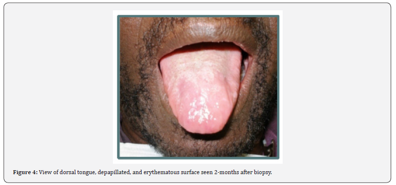
After the biopsy and subsequent suture removal, the patient was lost to follow-up for two months. Upon returning to the dental office, an oral examination revealed a depapillated and erythematous area of the tongue at the previously affected site. Despite mucosal atrophy on the anterior tongue, signs of good healing were evident (Figure 4) [7]. The yellowish-white plaques observed at the initial presentation were no longer present. The patient described self-treatment, which included scraping the lesion and rinsing with hydrogen peroxide.
Discussion
An eosinophilic granuloma (EG) is a benign disease entity with an etiology [8] and pathogenesis that remain poorly understood. This lesion healed after an incisional biopsy [8] Trauma to the affected mucosa is commonly suspected as the cause [7]. In a review of 132 cases of oral traumatic granulomas, [9] Joseph and Bairava Sundaram found that the tongue was affected in more than 60% of cases [9]. Their review, however, did not address patients infected with HIV. In contrast, [10] reported a case of an ulcer on the left posterior lateral tongue with similar microscopic findings in a patient with AIDS [10].
The lesion in this report occurred on the anterior-ventral tongue, a site more commonly affected in infants less than 2 years old. Natal and prenatal teeth are usually the source of low-grade trauma to the tongue mucosa during suckling. In infants, this lesion is diagnosed as Riga-Fede’s disease - a condition described clinically and histologically by Antoni Riga and Francesca Fede, two Italian physicians of the late nineteenth century (1.2).
Sharp areas on orthodontic appliances, ill-fitting or poorly maintained oral prostheses, or sharp-edged restorations may cause trauma and irritation to oral mucosa, leading to ulcers like the one presented in this case. However, as in most cases of EG previously reported [7,8], this patient did not recall or report any history of trauma associated with the lesion. Sharp edges or cusps on natural teeth can also be implicated [11,12] but this patient neither wore dentures nor had anterior teeth in poor condition. His anterior teeth exhibited acceptable morphology and alignment.
Other intraoral sites where eosinophilic granuloma (EG) may occur include the gingiva, floor of the mouth, buccal mucosa, and retromolar pads. Lesions can range in size from a few millimeters with an innocuous appearance to very large and alarming, as seen in this case. The raised indurated border and peri-lesional erythema raised concerns about potential malignancy [13]. Similarly, the histologic findings were equally concerning. Based on the clinical presentation and the patient’s medical history, the differential diagnosis included a major aphthous ulcer, a secondarily infected non-healing ulcer, and squamous cell carcinoma.
Microscopic Examination and Special Stains
Microscopic examination included a Periodic acid-Schiff (PAS) special stain to assess the presence of fungal infection. Ulcerations of the oral mucosa are susceptible to secondary infection by oral microbes, including viruses, bacteria, and fungi [14]. Candidiasis is the most common fungal infection in the oral cavity, especially in immunocompromised individuals. When Candida species secondarily infect oral ulcerations, mucosal integrity is compromised, leading to prolonged healing [15]. However, the ulcerated lesion in this case tested PAS-negative for candidiasis. No other special stains were ordered.
The Enigmatic Nature of Eosinophilic Granuloma
The term “enigmatic” has been used by various researchers to describe the behavior of EG. It’s questionable origin, pathogenesis, and spontaneous resolution following biopsy are poorly understood. Another poorly understood but consistent feature of EG is its positive reaction to the CD30 immunohistochemical stain. CD30 is commonly requested to evaluate atypical changes related to lymphoproliferative disorders (LPDs), a spectrum of lymphocyte abnormalities often associated with compromised immune systems [16,17].
CD30 is a transmembrane protein of the tumor necrosis factor family, and a tumor-cell surface marker expressed in various lymphomas, including anaplastic large-cell lymphoma, primary cutaneous lymphoma, and Hodgkin’s lymphoma [16,17]. It is also expressed by activated B-cells, T-cells, natural killer cells, eosinophils, and mast cells. Additionally, CD30 expression has been associated with non-lymphoid malignancies such as embryonic carcinoma, melanoma, and pancreatic cancer [18].
Histologic Considerations and Differential Diagnosis
The histology of EG includes chronic inflammatory cells
with prominent eosinophils and large, pale-staining irregular
cells. These findings resemble features of Kimura’s disease [18],
Hodgkin’s lymphoma, and Langerhans cell histiocytosis (LCH).
a) Kimura’s Disease is a benign chronic inflammatory
disorder characterized by increased proliferation of chronic
inflammatory cells in lymphoid tissue, particularly in the head
and neck [18].
b) Hodgkin’s Lymphoma is a malignant disorder of the
immune and lymphatic systems [19].
c) Langerhans Cell Histiocytosis (LCH) has a less clearly
defined classification, with some researchers considering
it “cancer-like” and others viewing it as an “autoimmune
phenomenon.” The National Cancer Institute classifies LCH as a
cancer [12,19,20] (PDQ).
Despite their clinical differences, these conditions share histologic features involving eosinophilic infiltration.
Role of Eosinophils in Immunity and Tissue Repair
Eosinophils, once thought to primarily defend against parasitic infections, are now recognized as contributors to both innate and adaptive immunity. These white blood cells, containing large cytoplasmic granules, respond to pathogens by degranulating and releasing cytotoxic proteins [21] Travers). However, this process can also lead to host tissue damage, as seen in conditions such as contact dermatitis, severe asthma, drug hypersensitivity, and eosinophilic esophagitis. Theoretically. In this case, delayed healing of the tongue lesion may have resulted from cytotoxins released by degranulating eosinophils.
In adaptive immunity, eosinophils regulate immune responses through cytokines and chemokines, mediating cellular activity, antibody synthesis, and antigen presentation for destruction by T-cells. They also play roles in tissue repair, remodeling, irritable bowel syndrome, and graft-versus-host disease. Their infiltration into solid malignant epithelial tumors highlights their involvement in immune system modulation [21,22].
Wound Healing and Tissue Remodeling
This large eosinophilic granuloma (EG) developed at the site
of a non-healing ulcer, suggesting an interruption in the processes
of wound healing and tissue remodeling. Wound healing typically
occurs in three phases23 Wang:
a) Hemostasis/Inflammation Phase – Neutrophils and
macrophages cleanse the wound by removing pathogens and
cellular debris.
b) Proliferative Phase – Granulation tissue forms, preparing
the site for epithelialization.
c) Remodeling Phase – Tissue is reorganized, and cells
involved in repair undergo apoptosis.
Disruption in any of these phases can impair healing [23]. For example, poor macrophage cleansing or defective granulation tissue formation may delay repair. Additionally, dysregulated apoptosis may prematurely remove cells required for epithelial remodeling, resulting in prolonged inflammation or unstable tissue growth. Given eosinophils’ regulatory role in apoptosis and cellular proliferation, their involvement in this lesion may have contributed to disrupted wound healing and tissue remodeling. Intriguingly, the biopsy may have acted as a “reset,” triggering a correction in the healing and remodeling processes.
Conclusion
The ulcerated lesion in this case suggests mucosal injury with delayed healing and spontaneous resolution. This report explored the lesion’s pathophysiology, including its CD30 positivity and association with lymphoproliferative disorders, as well as its histologic similarities to other inflammatory and malignant conditions. The prominent eosinophilic infiltration highlights the complexity of EG, making it challenging to identify its precise cause.
This case showed that the eosinophilic granulomatous lesion was not typically associated with comorbidities such as HIV/ AIDS. Clinical evaluation of a large, non-healing ulcer of the tongue, supported by histologic and cytologic findings resembling malignancy, led to the final diagnosis eosinophilic granuloma of the tongue. Resolution of the lesion was observed after incisional biopsy. Further research into eosinophil activity and its impact on wound healing may help elucidate the mechanisms underlying EG development and resolution.
References
- Riga A (1881) Of an early childhood illness. Probably untreated, with pathological movements. Naples.
- Fede F (1890) Of sublingual production or Riga disease. Proceedings of the Italian Congress of Pediatrics 1890. Napoli 1891, 251.
- Lakkam BD, Astekar M, Shakir A, Adeeba S (2021) Traumatic ulcerative granuloma with stromal eosinophilia: a puzzle. J Oral and Maxillofacial Pathol 25: 42-45.
- Butler JN, Kobayashi TT (2017) Traumatic ulcerative granuloma with stromal eosinophilia: a malignant-appearing benign lesion. Cutaneous Med for the Practitioner 100(2): 28-31.
- Eggleton JS, Nagali S (2020) Highly Active Antiretroviral Therapy (HAART) In: StatPearls.
- Tran BX, Fleming M, Do HP, Nguyen LH, Latkin CA (2018) Quality of life improvement, social stigma and antiretroviral treatment adherence: implications for long-term HIV/AIDS care. AIDS Care 30(12): 1524-1531.
- Brown R, Farquharson A, Peppers CG, Lawrence L, Mills GD (2020) A case of cheilitis granulomatosa/orofacial granulomatosis. Clinical, cosmetic and investigational dentistry 12: 219-224.
- Tobias RSF, Oliveira ARS, Carvalho SP, Vêncio EF, Silva MAG (2025) Traumatic ulcerative granuloma with stromal eosinophilia on the tongue: a case report. Oral Surgery, Oral Medicine, Oral Pathology and Oral Radiology 139(5): e29.
- Joseph BK, Sundaram BD (2010) Oral traumatic granuloma: report of a case and review of literature. Dental Traumatology 26(1): 94-97.
- Cunha VS, Zanol JDR, Sprinz E (2007) Riga-Fede-Like Disease in an AIDS Patient. Journal of the International Association of Physicians in AIDS Care 6: 273-274.
- Siu A, Landan K, Ramos DM (2015) Differential Diagnosis and Management of Oral Ulcers. Seminars in Cutaneous Medicine and Surgery 34(4): 171-177.
- PDQ Langerhans Cell Histiocytosis Treatment (2020) PDQ® Pediatric Treatment Editorial Board. Bethesda, MD: National Cancer Institute.
- Neto CIJ, Gondak R (2024) Traumatic ulcerative granuloma with stromal. Dentistry 16(3): e377.
- Muzyka BC (2005) Oral Fungal Infections. Dental Clinics of North America 49: 49-65.
- Deepa AG, Nair BJ, Sivakumar TT, Joseph AP (2014) Uncommon opportunistic fungal infections of oral cavity: A review. J Oral Maxillofac Pathol 18(2): 235-243.
- Gottesman SR (2016) CD 30: Receptor, Marker, Target; Pathology and Laboratory Med 8: 27-36.
- Vaillant JAA, Stang CM (2021) Lymphoproliferative Disorders. In: StatPearls. Treasure Island (FL): StatPearls.
- Guimaraes CS, Levy MN, Sapadin A, Vidal C (2009) Kimura’s Disease. Case Reports in Medicine p. 4.
- Darling MR, Cuddy KK, Rizkalla K (2012) Hodgkin Lymphoma of the Oral Mucosa. Head and Neck Pathol 6(4): 507-510.
- Shea CR (2020) Langerhans Cell Histiocytosis. Medscape.
- Travers J, Rothenberg ME (20151) Eosinophils in mucosal immune responses. Mucosal Immunol, Division of Allergy and Immunology, Cincinnati Children’s Hospital Medical Center, Cincinnati, OH, USA 8(3): 464-475.
- Diny NL, Rose NR, Cihakova D (2017) Eosinophils in Autoimmune Diseases. Frontiers in Immunol.
- Wang P, Huang B, Yeh C, Chen Y, Horng HC (2018) Wound Healing. Journal of the Chinese Medical Association 81(2): 94-10.

























