




 JOJUN Home



Abstract
Background: Radical cystectomy (RC) remains the cornerstone in the management of muscle-invasive and high-risk non-muscle-invasive bladder cancer. Despite the advances in perioperative care and surgical technology, RC continues to carry high morbidity and mortality rates.
Objective: This review examines current evidence regarding indications, surgical approaches, oncologic and functional outcomes, adjunctive therapies, and emerging directions in RC.
Methods: A narrative review of the literature was conducted, analyzing studies on open, laparoscopic, and robotic-assisted radical cystectomy, with particular emphasis on urinary diversion techniques, neoadjuvant and adjuvant therapies, and quality-of-life outcomes.
Results: Open radical cystectomy remains the gold standard; however, minimally invasive and robotic-assisted techniques demonstrate comparable oncologic efficacy with reduced blood loss, faster recovery, and lower perioperative morbidity. Urinary diversion options—including ileal conduit, Indiana pouch, and orthotopic neobladder-affect postoperative function and quality of life differently. Neoadjuvant cisplatin-based chemotherapy improves survival, while immunotherapies and gene-targeted approaches show promising efficacy for cisplatin-unfit or BCG-refractory patients. Lymph node involvement remains a critical prognostic factor, and organ-sparing techniques enhance functional outcomes in selected patients.
Conclusion: Radical cystectomy continues to evolve with the integration of minimally invasive techniques, enhanced perioperative protocols, and immunotherapeutic advances. Future research should focus on optimizing patient selection, refining robotic approaches, and exploring molecular and genomic predictors to personalize therapy and improve postoperative quality of life.
Keywords:Radical Cystectomy; Bladder Cancer; Robotic Surgery; Urinary Diversion; Neoadjuvant Chemotherapy; Immunotherapy; Quality of Life
Abbreviations: RC: Radical Cystectomy; Mibcmuscle-Invasive Bladder Cancer; NMIBC: High-Risk Non-Muscle Invasive Bladder Cancer; LRC: Laparoscopic Radical Cystectomy; ORC: Open Radical Cystectomy; UCB: Urothelial Carcinoma Of The Bladder; TURBT: Trimodal Therapy, Which Combines Transurethral Resection Of Bladder Tumor; CIS: Carcinoma In Situ; NBI: Narrow-Band Imaging; MRI: Magnetic Resonance Imaging; FISH: Fluorescence In Situ Hybridization; NMP22: Nuclear Matrix Protein 22; PLND: Pelvic Lymph Node Dissection; RARC: Robotically Assisted Radical Cystectomy; TURBT: Transurethral Resection Of Bladder Tumors; LRC: Laparoscopic Radical Cystectomy; SIRS: Systemic Inflammatory Response Syndrome; MIBC: Muscle-Invasive Bladder Cancer; RARC: Robotic Assisted Laparoscopic Radical Cystectomy; ONB: Orthotopic Neobladder; PLND: Pelvic Lymph Node Dissection; TURBT: Transurethral Resection Of Bladder Tumor; Mucb: Metastatic Urothelial Carcinoma Of The Bladder; ROS: Reactive Oxygen Species; MMC: Mitomycin C; EMDA: Electromotive Drug
Introduction
Urinary cancers can be divided into urinary bladder cancer, which constitutes 90% of the urothelial carcinoma, and upper tract urothelial carcinoma [1]. The optimal treatment of urinary bladder cancer is radical cystectomy (RC), which can be conducted for muscle-invasive bladder cancer (MIBC), high-risk non-muscle invasive bladder cancer (NMIBC), and metastatic urinary bladder cancer [2]. In 2020, around 573,000 novel cases of urinary bladder cancer were recorded on a global level, leading this type of cancer to be considered the 10th most diagnosed cancer, with 200,000 deaths recorded in the same year [1]. The surgical technique differs between males and females. The usual anterior pelvic exenteration involves the excision of the urinary bladder and other reproductive organs such as the uterus, fallopian tubes, ovaries, and anterior vaginal wall, in case of females [3]. For male patients, the bladder, prostate, seminal vesicles, distal ureters, and regional lymph nodes are excised “en bloc” [4].
To be able to overcome many limitations, the preservation of genital or pelvic organs for both sexes enhances the postoperative outcomes and the reproductive function [4]. NMIBC, also known as the superficial type, begins with an abnormal increase in the number of urothelial cells lining, known as urothelial hyperplasia. It can be divided into Ta, T1, and carcinoma in situ (CIS) [5]. On the other hand, MIBC is known to have an elevated risk of death due to distant metastases and starts with the development from dysplasia to flat CIS in addition to high-grade non-invasive lesions. It includes stage T2 and beyond [5]. MIBC accounts for 30% of the cases and can go up to 40 % if taking into consideration the cases that develop from NMIBC, which is the most common type in patients affected with BC [2]. This review discusses the indications of RC and explores its different surgical approaches, from open radical cystectomy (ORC) to minimally invasive approaches, which include laparoscopic radical cystectomy (LRC), robotic-assisted radical cystectomy (RARC), and urinary diversion techniques. Additionally, the review states the treatment outcomes and goes through special considerations and controversies.
Indications of Radical Cystectomy
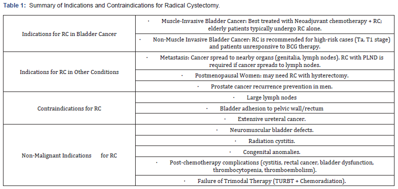
Urothelial Carcinoma of the bladder (UCB) in both MIBC and NMIBC is found to be one of the major reasons for RC [6]. In MIBC, the best treatment is integrating neoadjuvant chemotherapy with RC, given the patient’s preference to preserve their continence [7]. On the other hand, several studies show that the ideal treatment for bladder cancer in elderly people is RC only, excluding aged individuals from their selection criteria to go through combined approach [8]. Regarding NMIBC patients, RC is suitably performed for those with greater risks: stage Ta or T1 [6,9]. Moreover, RC with urinary diversion is suggested to those who have NMIBC and do not respond to Bacillus Calmette-Guerin (BCG) therapy, which is known to delay or stop the spread of bladder cancer [10].
RC also plays a role when cancer is found in areas next to the bladder, mostly the genital parts of both genders. Although there is a small probability that UCB metastasizes to the ovaries, it is believed that women, especially those in their postmenopausal years, should perform hysterectomy along with the RC [11]. Concerning the male genitalia, RC is done to prevent the recurrence of prostate cancer, which was proven by a study conducted by Gomez et al., where 20 patients underwent RC with prostatesparing. Of these 20 patients, 12 were staged previously as pT2 (pathological stage), where the tumor is limited to the prostate gland, 7 were staged in the past as pT1, where the tumor is found in a minimal percentage in the prostate gland, and 1 patient was staged as pT3, where the tumor has grown outside the prostate gland but not broadly. Given these conditions, prostate cancer has developed in only 4 patients without recurrence [11].
Once the cancer metastasizes from the bladder to the surrounding lymph nodes, RC associated with pelvic lymph node dissection (PLND) should be done to limit its spread [11,12]. Nonetheless, patients cannot go through RC when the lymph nodes are large, the bladder is firmly attached to the pelvic wall or the rectum, or the cancer expands extensively through the ureters. In these cases, only chemotherapy is applicable [12].
Occasionally, patients do RC for non-cancerous problems when the bladder is critically damaged or induces pain, or for postchemotherapy complications. Accordingly, the non-malignant indications to perform RC are neuromuscular defects of the bladder when it’s difficult to control the bladder, radiation cystitis when the bladder is inflamed due to chemotherapy sessions, and congenital errors when children are born with problems leading to cognitive impairment and physical disabilities [13].
For instance, patients who choose to keep the bladder and go through chemotherapy face a higher risk of cystitis, rectal cancer, proctitis, and bladder defect due to the temporary and extended toxicity that is generated by chemotherapy. In addition, they are at risk of thrombocytopenia, thromboembolism, and gastrointestinal problems [7]. Furthermore, patients with bladder malfunction undergo RC after failing to respond to Trimodal Therapy, which combines transurethral resection of bladder tumor (TURBT) and chemoradiation [10]. Nevertheless, as people start aging, partial cystectomy, which is less invasive, is done instead of RC as urinary complications such as bladder dysfunction and other medical problems arise [8].
Suspicious patients undergo bladder biopsy using whitelight cystoscopy to have a clear vision of the bladder. In the case of patients with bladder cancer, red velvety areas are observed, which are a hallmark of carcinoma in situ (CIS) that grow and proliferate on the bladder walls [12,14].
The use of photodynamic cystoscopy, also known as Fluorescent/blue-light cryptoscopy, aids in the diagnosis [12,14]. In this type of cryptoscopy, a substance such as hexaminolevulinic acid or 5-aminolevulinic acid is introduced in the bladder using a catheter and absorbed by the malignant cells. Once absorbed, blue light is used during this procedure, and the tumor cells appear red. Sometimes, type I errors occur in many cases, especially if the bladder is inflamed, patients are under BCG therapy, or patients have performed lately TURBT [12]. Another endoscopic approach to detect bladder cancer is Narrow-Band imaging (NBI) [12,14]. In this technique, two specific wavelengths of light are used to render the blood vessels darker for a better evaluation of the tissue. Doctors use other diagnostic tools to detect CIS and then perform RC [14].
Supplementary diagnostic tests can be performed, such as complete blood count, liver function tests, and alkaline phosphatase tests [14]. Imaging techniques, including Magnetic Resonance Imaging (MRI), Computed tomography, Chest X-ray, and scanning methods for the pelvis and abdomen, can also be used to detect bladder cancer [14]. Moreover, genetic diagnostic tools can be adopted, such as Fluorescence in situ hybridization (FISH), which is a molecular technique allowing the detection of specific DNA sequences, inspecting the chromosomal number, and assessing chromosomal rearrangement in the cell. This technique uses fluorescent probes to quickly assess abnormal chromosomes, specifically 3,7,9, and 17, since they are the most prone to abnormalities in bladder malignancies. Additionally, tumor markers, specifically nuclear matrix protein 22 (NMP22), are released by bladder malignancies and can therefore be detected [14] (Table 1).
Surgical Approach to Radical Cystectomy
RC is a complex process consisting of a three-part “en bloc” operation that includes (1) bladder removal; (2) pelvic lymphadenectomy; and (3) urinary diversion [15-17]. In males, excision of the prostate and seminal vesicles follows, while in females, removal of the uterus, adnexa, and anterior vaginal wall is required [15]. ORC with pelvic lymph node dissection (PLND) represents the gold standard procedure in the treatment of muscle-invasive bladder cancer and high-risk non-muscle invasive bladder cancer [18]. Nonetheless, despite the recent remarkable advancements in surgical techniques and expertise, radical cystectomy remains a highly morbid procedure with complications occurring in up to two-thirds of patients within 90 days of the surgery [19].
Patient age, operative risks secondary to medical comorbidities, or post-operative functional concerns can require an alternative procedure to a radical cystectomy, such as minimally invasive cystectomy: laparoscopically, robotically assisted radical cystectomy (RARC), or robotic-assisted laparoscopic radical cystectomy [20]. A systematic review reported on the statistics of the surgical approach in radical cystectomies, findings that 35.7% of the cases were open surgery, 32.9% were performed laparoscopically, and 25.2% were robotic-assisted surgeries [21]. The review also highlights the prevalence of ileal conduit as the most common diversion method, accounting for nearly 82% of diversions in patients requiring such intervention [21]. RC differs from simple cystectomy in the aspect that simple cystectomy is referred to as supratrigonal or prostate sparing and often leaves behind the trigone urothelium along with the pelvic lymph nodes [16].
Simple cystectomies are performed in patients with unrelenting symptoms, nonetheless not for oncologic control. Urinary diversion is a surgical intervention performed after a cystectomy, aiming to redirect the stream of urine [22]. One described method, the ileal conduit, has been in practice since the 1930s and was described and made popular in a landmark article by Bricker in 1950 [22]. Diversions are divided into continent and incontinent, with several types and procedures discussed and elaborated in the literature. Extensive literature has been published on the use of external beam radiation therapy for the treatment of MIBC, with the most common therapy being a Transurethral Resection of Bladder Tumors (TURBT) [20]. Response rates ranged from 60 to 80% with a 5-year survival rate of patients [20]. While a radical TURBT allows bladder preservation in cases of urothelial bladder cancer, and involves the complete transmural resection of visible tumors [23], although extremely rare, the risk of extravesical seeding of tumor cells persists. Such a technique remains eligible in only 11-35% of patients with MIBC [23]. In the case of an unresectable disease, RC should not be performed, especially with large unresectable lymph nodes [24]. In such cases, a computed tomography-guided biopsy is recommended, and chemotherapy is provided if positive.
Laparoscopic Radical Cystectomy (LRC)
The LRC technique is a new procedure approaching standardization after duplicating the well-established ORC surgery [25]. A retrospective study found that LRC was associated with reduced blood loss, decreased post-operative ileus or pain, and shorter hospital stay compared to ORC [25]. No significant differences were reported regarding operative time and postoperative complication rates. Furthermore, the available data suggest that a urinary diversion performed laparoscopically provided a statistically significant decrease in operating time, blood loss, and transfusion rate, and a faster return of patients to oral intake and ambulation [25]. Studies report that minimally invasive surgery can reduce the severity of systemic inflammatory response syndrome (SIRS) compared to open surgery, with a noted advantage in the elderly population [26]. Conversely, while the laparoscopic approach seems appealing, especially in the elderly population, no studies addressed the outcomes of a partial cystectomy specifically in this age group [20].
Such a phenomenon can be explained by the strict indications and pre-operative requisites, making only 5-10% of patients with muscle-invasive bladder cancer (MIBC) eligible for this procedure [20]. The procedure begins with the ureters dissected from the bladder and transected distally. For males, the dissection begins in the cul-de-sac between the rectum posteriorly and the prostate, seminal vesicles, and bladder anteriorly. It is carried out distally up to the mid-prostate until the anterior part of the bladder is mobilized and the dorsal vein is secured [25]. The urethra is closed before the transaction to avoid the spillage of urine with tumor cells. Following transection, the specimen is placed in a bag and removed. For females, two procedures can be performed: a uterus-sparing and vaginal sparing LRC, or a conventional anterior exenteration [25]. Lymph node dissection is performed bilaterally towards the aortic bifurcation after the completion of RC.
Robotic Assisted Laparoscopic Radical Cystectomy (RARC)
Although patient care has had significant advancements, ORC still presents multiple perioperative complications and a high mortality rate as compared to RARC [16]. RARC is an extracorporeal procedure during which a 5-8 cm incision is performed to retrieve the bladder specimen and perform the bowel anastomoses, and if necessary, neobladder construction. In RARC, urinary diversion can be performed via an extracorporeal or an intracorporeal approach. The use of RARC is characterized by improved surgical precision and dexterity while reducing the surgeon’s fatigue. However, in a comparison of surgical time, robotic procedures were found to take longer operative time than open procedures, with the mean being 390 minutes for RARC compared to 300 minutes for ORC [27]. Furthermore, RARC remains a complex procedure with a proposed learning curve of 30 cases to reach extirpative proficiency [27].
As a procedure, RARC is performed via a five or six-port transperitoneal approach [15]. The patient is first placed in the steep Trendelenburg position with the legs in low lithotomy position, the robotic camera being placed 4-5 cm above the umbilicus [28], three robotic ports, and two assistant ports at the level of the umbilicus or above. The use of the da Vinci Xi Robotic System is usually used to perform robot-assisted Laparoscopic Radial Cystectomy. Using a Veress needle, the pneumoperitoneum is incised about 5 cm above the umbilicus. The abdomen is insufflated to 15 mmHg, and an 8 mm camera port is inserted in the midline. The incision of the peritoneum frees the bladder from the rectum [15]. Following dissection of the bladder, lymphadenectomy is performed, then urinary diversion, usually via a small laparotomy, although it can be entirely performed robotically. A detailed step-by-step procedure can be found in (Figure 1).
Operating Room Configuration and Patient Preparation: Prior to anesthesia induction, patients are provided 5000 units of heparin for deep vein thrombosis prophylaxis. A Foley catheter and an orogastric tube are placed, with appropriate intravenous antibiotics administered for the coverage of enteric organisms [29].
Urinary Diversions
Diversion is a necessary procedure performed following a cystectomy, with the aim of restoring urine flow and voiding. In the case of incontinent urinary diversions, a cutaneous pouch is used for continuous urine drainage, such as an ileal conduit. While for continent diversion, the patient voids through the native urethra or self-catheterizes through a surgical stoma [22]. In that case, a urine reservoir can be attempted via a cutaneous stoma for catheterization, in addition to a neobladder construction with anastomosis to the native urethra [22]. Comparing continent and incontinent urinary diversions, surgical time is lengthened in the case of continence. Criteria for the type of technique include age, physical condition, intestinal, hepatic, and renal functions, tumor stage and location, previous radiation therapy, and life expectancy.
The literature mostly reports on the use of bowels in urinary diversions acting as a conduit or a reservoir for urine storage. Basically, the bowel segment is respected, and an anastomosis links it to the genitourinary tract [22]. Given the refluxing characteristic of the conduit, an anti-reflux anastomosis can be performed; however, it is associated with an increased chance of uretero-ileal anastomotic stricture. One example of incontinent urinary diversions is the ileal conduit, which consists of isolating and mobilizing a segment of the distal ileum, about 15-20 cm, and using it to construct and implant the ureters separately into its proximal ends. The continuity of the gastrointestinal tract is restored with an ileal-ileal anastomosis, making sure to preserve the terminal part of the ileum necessary for vitamin B12 and bile salts absorption.

A less commonly practiced procedure, sigmoid conduit,
remains in use for patients with a history of abnormal distal
column, inflammatory bowel disease, or pelvic radiation [22].
The sigmoid conduit consists of isolating and mobilizing a 12-15
cm segment of the sigmoid colon to be used for a urine conduit
and drainage through a cutaneous ostomy. The gastrointestinal
continuity is restored through a colo-colic anastomosis. As
opposed to the ileal conduit, the thick musculature of the colon
allows reflux prevention mediated by submucosal tunnels.
Indiana Pouch: The Indiana pouch technique is among
the most common continent cutaneous reservoir diversions
performed. Through an incision made along the taenia coli, the
procedure involves mobilization and resection of 20-25 cm of
the cecum and ascending colon to the hepatic flexure, including a
segment of the terminal ileum (15-18 cm from the ileocecal valve)
[22]. The continuity of the gastrointestinal tract is constructed
through an ileocolic anastomosis. The constructed stoma may
be in the right lower quadrant or brought to the umbilicus. The
ureters are then anastomosed to the colonic segment separately
and tunneled into the posterior taenia to prevent reflux [22]. To
maintain continence, several surgical techniques can be used.
For instance, the appendix can be used as a catheterizable stoma
(Mitrofanoff procedure). Alternatively, a tapered ileum can
provide the same functions (Monti procedure) [22].
Orthotopic Neobladder (ONB): In 1959, the first successful
bladder replacement was initially performed, known as the
Camey procedure. This technique requires longer surgical
time with longer bowel segments. Unlike alternative methods,
a neobladder preserves the ability to void through the native
urethra, waiving the need for a stoma or any external devices. It
can be constructed from the ileum, colon, or both, with the use of
the small bowel as a reservoir being the most preferred technique
nowadays. Several types of ONB have been created, such as:
Camey I and II (U-shaped), modified Camey II (Z-shaped), Kockpouch,
T-Pouch, Studer clutch bag, Hautmann (W-shaped), Abol-
Enein and Ghoneim modification of the W pouch, and the Vescica
Ileale Padovana (VIP, circularly shaped) [30]. The two most
common surgical techniques to create ONBs include the Studer
and Hautmann pouches.
Studer Pouch: An ileal pouch is created by respecting a 60
cm segment of the ileum, 20-25 cm proximal to the ileocecal valve
[22]. The antimesenteric borders are folded in half and then are
oversewn together. The intact proximal ileal limb functions as a
reflux prevention mechanism.
Hautmann Pouch: Another ileal pouch created by respecting
a 60 cm segment of the ileum and detubularizing and rearranging
it into a W shape. An anastomosis links the ureters to the
neobladder, then the urethra is anastomosed to a buttonhole
incision in one of the limbs. The outside edges of the W are closed
in a side-to-side manner.
Pelvic Lymph Node Dissection (PLND)
Dissection is performed from the site of the proximal ureter and common iliac artery crossover to the lateral circumflex iliac vein, inguinal ligament, and node of Cloquet and from the genitofemoral nerve crossing the psoas muscle to the bladder [17]. If grossly palpable nodes are found, the margins should be extended till the paraaortic lymph nodes. In men, preserving potency and fertility can be achieved with a prostate sparring technique that has a high selectivity from the patient perspective [17]. Leaving the neurovascular bundle undisturbed decreases the risk of erectile dysfunction, while the preservation of the distal sphincter complex improves continence rates. In contrast, for females, sparring and preservation of pelvic organs contributed to the prevention of vaginal dryness and dyspareunia. In the case of the absence of oncological invasion, such organs can be spared.
Neoadjuvant and Adjuvant Therapies for Bladder Cancer
Treatment Strategies for Muscles Invasive & Metastatic Bladder Cancer (MIBC & MUCB)
The rate of recurrences is high in patients with MBIC even after RC, and the neoadjuvant chemotherapy provides only a 5% increase in the overall survival rate and may be pointless in some patients [31]. On the contrary, adjuvant chemotherapy is more effective because of its ability to eliminate any remaining or undetected cancer cells that may still be present even after radical cystectomy, hence, reducing the risk of recurrences [32]. For patients with metastatic or locally advanced bladder cancer, a cisplatin-containing regimen is the standard chemotherapy approach due to its ability to target rapidly dividing cancer cells and control the progression of the disease. According to the SWOG 8710 trial, a combination of neoadjuvant chemotherapy (cisplatin-based) followed by radical cystectomy and pelvic lymph node dissection is considered the standard treatment for muscleinvasive bladder cancer.
Numerous clinical studies suggest either adjuvant or neoadjuvant chemotherapy for locally advanced or metastatic bladder cancer based on the combination of either the conventional MVAC regimen (methotrexate, vinblastine, doxorubicin, and cisplatin) or gemcitabine/cisplatin. For non-metastatic MBIC (T2T4N0M0), the optimal treatment is neoadjuvant cisplatinbased chemotherapy before undergoing RC. Trimodal therapy that includes maximal transurethral resection of bladder tumor (TURBT) and chemoradiation is considered an alternative for RC for patients deemed suitable for this approach [32]. In cases where the tumor does not respond to TMT or recurs after initial treatment, physicians refer to RC as a backup or followup treatment. Notably, around half of the patients with nonmetastatic MIBC in the US do not receive any treatment intended to eliminate the cancer or instead of curing the disease, they only receive treatment aimed at managing symptoms or prolonging survival [32].
A systematic review conducted by Williams and his colleagues (2019) emphasizes the importance of ensuring more patients receive adequate treatment either through TMT or RC. Whereas, in patients with metastatic urothelial carcinoma of the bladder (mUCB), especially patients with locally advanced disease (T4b or N1-3), are primarily treated with chemotherapy. RC may then be performed following chemotherapy to remove the remaining cancerous cells [32]. Patients with metastatic urothelial carcinoma of the bladder (mUCB), especially patients with locally advanced disease (T4b or N1-3), are primarily treated with chemotherapy. RC may then be performed following chemotherapy to remove the remaining cancerous cells [32].
Advances in Intravesical Therapy for Non-Muscle Invasive Bladder Cancer Patients
The intravesical administration of immunomodulators such as BCG, either alone or combined with interferon (IFN-α), is considered an effective approach for patients with NMIBC, either in the first-line treatment (refractory cases) or secondline (relapsed cases). This treatment placed with tumor cells stimulates the secretion of many cytokines, including IL-1, IL- 2, IL-6, and most importantly, tumor necrosis factors (TNF-α). Also, it contributes to the production of reactive oxygen species (ROS), which play a crucial role in eliminating cancerous cells by causing extensive damage to their DNA [33]. According to Di Stasi et al. (2006) and Di Lorenzo et al. (2010), it is recommended to administer BCG injection before mitomycin MMC because this intravesical chemotherapy increases the bladder mucosal permeability, thus enhancing the delivery of the drug to the targeted tissues. This combination of therapy slows the rate of progression in patients with Ta (Noninvasive papillary carcinoma) and T1(tumor invading the lamina propria) illness as compared to patients with Tis (carcinoma in situ) disease, which highlights the need for an alternative strategy [33].
For high-risk NMIBC patients unable to tolerate BCG injection, intravesical therapy through chemotherapeutic drugs is the best choice to potentially reduce stage progression and relapses. These chemotherapeutic drugs, including mitomycin C (MMC), thiotepa (N, N’, N”-triethylene thiophosphoramide), gemcitabine, cisplatin, 5-fluorouracil, doxorubicin, carboplatin, and epirubicin, are either used individually or in combination. According to Sylvester et al. (2004), mitomycin C (MMC), epirubicin, and pirarubicin are more effective than thiotepa in reducing the bladder cancer relapse. Furthermore, to achieve a higher response rate in NMIBC patients at high risk, MMC must be combined with an electromotive drug (EMDA) that facilitates the transportation of the chemotherapeutic drug into the tumor cells [33].
A phase II trial, KEYNITES-057 cohort study, reveals that pembrolizumab monotherapy is an FDA-approved optimal treatment for patients at high risk and unresponsive to BCG [31]. Another emerging immunotherapy that also targets high-grade, BCG-unresponsive NMIBC patients is intravesical rAd-INFa/syn3 (Adstiladrin), which uses a non-replicating adenovirus to deliver a gene for recombinant interferon-α2b (IFNα2b) directly into the bladder [31]. Several other options are available to achieve better treatment outcomes, such as heating the bladder wall to 42 ± 2 °C using microwave equipment coupled with cooled MMC, which ameliorates drug absorption, distribution, and metabolism [33]. Adding to this, these drugs inhibit tumor development through the production of oxidative stress, numerous chromosomal anomalies, and profound DNA strand breakage.
To improve the overall survival rate in patients at high risk of NMIBC, many physicians recommend beginning the treatment with radical cystectomy. However, this approach is not considered the standard treatment for this type of cancer, leading to an ongoing debate about the optimal approach for treating patients at high risk of NMIBC with several options, including adjuvant therapies and RC. While in patients with subsequent recurrences, physicians begin the treatment with salvage cystectomy and then continue with chemotherapy [33]. In MIBC patients, the presence of distant metastasis leads to many treatment failures, which highlights the need for more active systemic therapies like a combination of cytotoxic chemotherapy with ICIs. This dual stimulates the immune system to recognize and kill cancerous cells.
Combination of Neoadjuvant Immunotherapy with Chemotherapy and Radiation
In MIBC patients, the presence of distant metastasis leads to many treatment failures, which highlights the need for more active systemic therapies like a combination of cytotoxic chemotherapy with immune checkpoint inhibitors (ICIs). This dual stimulates the immune system to recognize and kill cancerous cells. In a phase II trial conducted by the BLASST-1 study, a combination of gemcitabine and cisplatin (G+C) with nivolumab (an ICI) in patients with MIBC at stages cT2-T4aN<1 reports a significant pathologic complete response (49%) and pathologic response rate (66%), suggesting a reduction of the tumor. Subsequently, cystectomy is performed 6-8 weeks following therapy without any unexpected complications [31].
Despite the high risk of mortality, there are limited treatment options for patients with MIBC who are unfit for the cisplatin treatment. In this situation, pembrolizumab, another immune checkpoint inhibitor, is considered a good option due to its effectiveness in the advanced stages of cancer and its low health risks. The phase II PURE trial shows that pembrolizumab given before surgery is beneficial for MIBC patients. It achieves significant tumor response rates, especially in patients with high PD-L1 expression. Likewise, the ABACCUS trials investigate atezolizumab before surgery in urothelial carcinoma patients unable to receive cisplatin and demonstrate a high complete pathological response (approximately 31%). After the treatment with post-pembrolizumab, the tumor burden mutation is significantly reduced in patients as compared to TMB scores prior to therapy.
Several ongoing trials are investigating the use of immune checkpoint inhibitors (ICIs) in combination with radiation therapy for treating muscle-invasive bladder cancer (MIBC). The PLUMMB trial, a phase I study, aims to assess the safety and tolerability of weekly radiation therapy paired with pembrolizumab in patients with either metastatic or locally advanced urothelial carcinoma of the bladder [31]. In the first cohort of the PLUMMB trial, pembrolizumab is administered at a dose of 100 mg every three weeks alongside weekly radiation therapy (36 Gy in six fractions). However, patients who required radiation for bladder cancer reported significant toxicities, including Grade 3 urinary toxicities and Grade 4 rectal perforation. This indicates the importance of the dose modification. Additionally, the phase Ib/II DUART trial investigates the combination of radiation therapy with adjuvant durvalumab for one year in patients with locally advanced bladder cancer (T3-4, N0-2, M0) ineligible or unfit for surgery. This trial demonstrates a high percentage of complete response rate (71%) and a disease control rate of 95%.
Importance of Biomarkers in the Treatment of MIBC
As personalized medicine advances in the curative setting, identifying reliable biomarkers to predict treatment response has become a dire need. Data from perioperative immunotherapy trials show that some biomarkers, such as PD-L1 expression and tumor mutational burden (TMB), which are relevant in the metastatic settings, may also play a pivotal role in muscle-invasive bladder cancer (MIBC) [31]. The PURE and NABUCCO trials reveal that neoadjuvant single-agent immune checkpoint inhibitors (ICIs) are more effective in patients whose tumors have high PDL1 expression (CPS ≥10%).
In contrast, the ABACUS trial does not indicate any similar correlation with using atezolizumab as a neoadjuvant. Increased Biomarkers like TMB, increased activity of T cells in the tumor microenvironment, and some immune gene signatures also have a positive correlation with favorable outcomes to neoadjuvant ICIs. These findings raise questions about responses to ICIs in patients with PD-L1–negative tumors and whether biomarkers’ effectiveness depends on the clinical context alone. Combining multiple biomarkers can improve patient selection for these therapies. Tumor tissue obtained during TURBT for immune studies will be critical for understanding immunotherapy responses, aiding in patient selection and bladder-preserving strategies in the future.
Treatment Outcomes in Radical Cystectomy
Overall Survival and Disease-Free Survival Key Metrics
Patients who undergo RC following a main response to chemotherapy (but with residual disease) have similar survival rates to patients with a total response to chemotherapy (with no residual disease) [34]. In a study, the 2-year progression-free survival was demonstrated to be similar in RARC and ORC, as 72% (95% CI, 63.6 to 78.2) [34]. In addition, some studies found that survival at 5 years was almost 60% and at 10 years it decreased to 40%, which is expected in these patients who may have numerous comorbidities [35].
Impact of Lymph Node Involvement
According to the literature, lymph Node metastasis is present in more than 20% of radical cystectomy patients, and it is an important prognostic factor that predicts the survival of these patients. In fact, a 43% 10-year recurrence-free survival is associated with a lymph node density of 20% or less compared to 17% with a density more than 20% [36]. Therefore, the number of positive nodes and their percentage are independent predictors of survival [37]. Additionally, integrating systemic chemotherapy before or after radical cystectomy increases chances of survival, and a thorough node dissection is an essential part of this strategy [37].
Some studies show that PLND improves survival and that it is implemented as an essential routine element of RC for Urothelial Carcinoma of the Bladder (UCB). However, some debate is still present about the optimal extent of PLND for maximal oncologic control. This is due to the perioperative complications and costs that may increase with an extended PLND, although it could potentially help eradicate this metastatic disease [34]. Additionally, in comparisons between extended and standard PLND, an increase in the 5-year recurrence-free survival was shown in the extended PLND cohort as compared to the limited PLND (35% versus 7%) [34].
Surgical Technique Differences
In comparison studies made between ORC and RARC in terms of oncological outcomes and complication rates, no significant difference was found. However, RARC is associated with reduced blood loss and faster diet recovery, but longer operative time [38,39]. In another comparison between ORC and RARC, regarding the outcomes in continent diversions, similar rates of continence were recorded, regardless of an open or minimally invasive approach [40]. Additionally, a review of 99 patients following RARC, a 5-year cancer-specific survival of 68% was recorded, which is similar to the 71% rate seen in ORC patients [41].
After RC, Positive surgical margins PSM are used as a predictor for metastatic progression. These were also found to be similar in ORC and RARC. The overall PSM rate was 4.2% with a 5-year disease-specific survival rate of 32% for patients with PSM versus 72% for negative margins [40]. However, in another study, a 15% rate of metastasis was seen in the robotic cohort, compared to higher (17-35%) rates in the open patients. Also, cancer-specific 5-year survival was approximately 70% in ORC patients, while over a 1-to-2-year follow-up, a 95% follow-up rate was seen in RARC [35].
Complications and Morbidity
RC, whether open or robotic, has complication incidence being reported to be in the range of 30-70% after surgery [34]. RC-related complications are divided into early and late complications; early are those that occur within 30 days of the surgery, and late complications occur between 30-90 days [34].
Short-Term Complications
Studies show that RC patients have immediate postoperative issues, which are seen in as many as 60% of patients, and from which 13% are major complications [42]. These can include the gastrointestinal, infectious, and wound-related categories [34]. These include surgical site infections, UTI, paralytic ileus, urinary leaks, dehydration, and bowel obstruction complications. Economic implications of these complications are highlighted by some studies that show that these short-term issues contribute to increased hospital costs for both surgical techniques [34,43].
Sometimes, UBC and UTUC can be diagnosed together, which necessitates a combined RC with radical nephroureterectomy (RNU). Compared to radical cystectomy alone, this procedure has higher complexities and complication rates [38]. This procedure has many complications that include infection-related adverse events, acute kidney failure, ureter-intestinal anastomosis leak, port hernia, retroperitoneal hemorrhage, and prolonged paralytic ileus [38].
Long-Term Complications
Some common late complications that affect RC patients are related to gastrointestinal, infectious, and genitourinary categories. These can be ventral and parastomal hernias, ureteroenteric strictures, and calculus formation. These complications may be related to the use of the bowel segment and the diversion performed [34]. Moreover, some studies compared RARC and ORC using the 5-grade modified Clavien system and found that RARC patients had a complication rate at 90 days of 62% versus 66% for the open group [40].
Mortality Rates
RC’s associated perioperative morbidity and mortality are relatively high [39], despite progress in surgical techniques and perioperative care (1.2%–3.2% at 30 days and 2.3%–8% at 90 days) [42]. Moreover, it appears that in high-volume academic centers, mortality is lower, although the patient population at these centers has more comorbidities [34]. Moreover, urinary diversion (UD) is the reason behind the most significant morbidity following radical cystectomy [39].
The mean survival period of patients in palliative care, in whom urinary diversion is undertaken, varies between three and nine months and depends on the metastatic load [44]. In RARC, UD is performed in two ways: either with an extracorporeal or with an intracorporeal approach. However, it was shown that in patients who have poor cardio-respiratory status, implementing an intracorporeal approach may reduce surgical trauma and cardiorespiratory complications, and it is performed in expert centers [39].
Quality of Life
RC-related complications and readmission rates following the procedure are high, which may decrease the quality of life (QoL) in patients [45]. QoL is influenced by several factors, including the type of urinary diversion, sexual function, and psychosocial adjustments.
Impact of Urinary Diversion
Following RC, the UD procedure is related to complications and long-term QoL. The method used in RC depends on the patient’s and surgeon’s preferences [46], there are the orthotopic neobladder ONB and the ileal conduit IC methods [45]. The patients who undergo the IC method live with the disadvantage of having continuous abdominal stoma, which affects body image and psychosocial well-being. But they do not have urinary retention or incontinence problems [45].
However, the ONB group of patients has the advantage of voiding through the urethra but experiences urinary retention and incontinence and has a lower QoL, but this is argued upon by other studies [45]. Furthermore, few studies evaluated QoL in female patients and showed that the most affecting element of QoL is incontinence, particularly at night, which has a negative impact on many social life areas [46]. In addition to physical aspects, RC has significant psychosocial implications. Patients with urinary diversions frequently report concerns related to body image, social stigmatization, and depression.
Sexual Function
Sexual function is a critical component of QoL, particularly for younger patients undergoing RC. A study that examined female patients based on the Female Sexual Function Index (FSFI) found that 37.5% of these patients experienced decreased sexual desire, and 45% of them were unable to achieve orgasm anymore, and that almost half of them were only able to have vaginal intercourse successfully [45]. However, FSFI does not capture sexual dysfunction in sexually inactive patients. Additionally, studies found that even with nerve-sparing approaches, postoperative sexual activity will not be the same as the baseline [47]. In other studies, ONB reconstructions had a big effect on body image and sexual life after the procedure [47].
Furthermore, only 3% patients who underwent the IC method in UD could maintain penile erection compared to 23% of ONB patients, but both have the same level of sexual desire [45]. However, no meaningful differences in sexual function between patients who have undergone different UD methods have been proven, but it is shown that recovery in men any significant sexual function in men following RC is low [45]. Overall, while advancements in surgical techniques and postoperative care have enhanced QoL for many patients, long-term support remains critical to addressing the multifaceted impact of RC.
Special Considerations in Radical Cystectomy
Regarding the complications of RC, the data showed that half of the cases above 70 years showed complications, in addition to 26% of that category presented loss of renal function compared to 7% of cases under 70 years old in five years post operation [2]. Also, the studies reported that 90% daytime and 85 % of nighttime continence in male patients who are not using pads or diapers, and 5 % self-catheterization after 36 months of follow-up [4]. Now for the sexual function, 14-100% is the recovery range in males for erectile function that received nerve-sparing technique with or without saving the prostate, while for the females, the sexual activity was reduced in the female cases that used nonneurovascular preservation [4].
For the post-surgery continence rate in females, it was noted for daytime and nighttime respectively that 57% and 42.9% of females are pad and diaper-free after performing the vaginalsparing technique and neobladder, in addition to 30.6% requiring self-catheterization [4]. The minimally invasive robotic surgery showed improved outcomes before and after the operation in clinical practice, and the robotic-assisted radical cystectomy diminished critical complications for 30 and 90 days [48]. In other words, this type of surgery can replace the open procedure with having comparable, if not superior, safety [49]. For the survival rates, it was mentioned by the International Robotic Cystectomy based on a 5-year scale that 67%, 75%, and 50% respectively, for recurrence-free, cancer-specific, and overall survival rates, and it was noted that those results were comparable to ORC [49].
Regarding the quality of life, it was mentioned by some studies that emotional well-being for both RARC and ORC was significantly higher at 3 and 6 months after surgery than baseline, in addition to presenting that 63 days is the time needed for affected individuals to target 90% in the overall Convalescence and Recovery Evaluation (CARE) difference index [49]. For many surgeries especially the ones at the level of the abdomen, the minimal invasive procedure are preferred due to the benefits in recovery and complications in addition to this the robotic assisted surgery improves precision and skills for the surgery and also in the reduction of blood loss and in the cost effective and also increase in the length of surgery with less fatigue for the surgeon [2].
Future Research Considerations
Genomic and Molecular Profiling
The emergence of acquired or inherent resistance to the cytotoxic effects of the drugs, along with the presence of efficient DNA repair mechanisms in the tumor cells, plays a pivotal role in reducing the effectiveness of therapeutic agents in treating bladder cancer. When DNA repair proteins are overexpressed, they may interfere with the normal function of p53, suppressing its ability to halt cell division, reconstruct the damaged DNA, or trigger cell programmed death in case of severe impairment. As a result, therapeutic outcomes that rely entirely on eliminating cancerous cells are undermined [33]. Many advancements in gene therapy and RNA-based treatments are crucial in enhancing the therapeutic strategies of bladder cancer. According to Abuzeid et al. (2009), a combination of cisplatin and mutant DNA repair gene therapy contributes to the regression of the tumor along with extensive DNA damage and telomere degradation. Another treatment strategy, applied in vitro, refers to the usage of small activating duplex RNA (dsP21-322) delivered directly into the bladder (intravesically) through lipid nanoparticles.
These RNA fragments activate the p21 gene involved in regulating cell growth and programmed cell death. As a result, this technique is beneficial in contributing to tumor regression in 40% of the mice [33]. In case of cisplatin-resistant cancerous cells (cisplatin-resistant NTUB1/P human bladder carcinoma cells), a combination of bifunctional alkylating agents and arsenic trioxide (ATO) prevents the activity of protein kinase B (AKT), which inactivates the function of the DNA repair protein. As a result, it reduces the growth cisplatin cisplatin-resistant cancerous cells [33]. Another study by Singh et al. (1996) demonstrates that J82/MMC-2 bladder cancer cells become more resistant to mitomycin C (MMC), a chemotherapeutic medication. This is explained by increased DNA repair caused by DNA polymerase beta overexpression. In a different clinical study, Bellmunt et al. (2007) show that when patients with advanced metastatic bladder cancer receive chemotherapy regimens like gemcitabine/ cisplatin or gemcitabine/cisplatin/paclitaxel, low levels of the protein excision repair cross-complementing 1 (ERCC1) are linked to higher survival rates [33].
Advances in the understanding of the biology of bladder cancer have led to the development of various drugs that are either under evaluation or expected to be available soon, preoperatively. These include modifications in fibroblast growth factor receptor, human epidermal growth factor receptor-2, DNA damage response, angiogenesis, epigenetics, and immune cell infiltration. Combining targeted therapies with immunotherapy is also being explored [31]. For instance, inhibiting vascular endothelial growth factor can influence the tumor microenvironment by reversing immunemediated suppression caused by myeloid-derived suppressor cells and regulatory T cells. Additionally, histone deacetylase inhibitors can enhance tumor immunogenicity by upregulating the expression of MHC complex I and II and other co-stimulatory molecules [31].
Emerging Techniques
Palliative radical cystectomy is not a standard treatment for patients with muscle-invasive urothelial carcinoma of the bladder (mUCB) to control bleeding and other local symptoms due to its high risk of morbidity and mortality. However, the future introduction of immune checkpoint inhibitors may be helpful in improving patients’ outcomes and extending overall survival rates [32]. Even with the progress in technology, open RC manifests high rates of surgical complications and mortality. Therefore, minimally invasive surgery is considered a better option because it contributes to a faster recovery and fewer complications without affecting cancer-free survival rates. Moreover, robotassisted surgery enhances surgeon precision and reduces fatigue. This explains why many ongoing studies aim to determine if RARC can optimize the outcomes of this high-risk operation [32].
In 2015, a randomized controlled trial conducted by Bochner and colleagues comparing open RC and RARC revealed that RARC has less blood loss despite taking longer to perform. Another randomized controlled trial (RAZOR) by Parekh and colleagues’ shows similarity between RARC and ORC in terms of 2-year progression-free survival rates, thus indicating that RARC is not worse than ORC in preventing cancer recurrences or progression. Additionally, a Cochrane review evaluates five randomized trials involving 541 patients with muscle-invasive bladder cancer (MIBC) who undergo either open radical cystectomy (ORC) or robotic-assisted radical cystectomy (RARC).
The study finds minimal differences between the two approaches in terms of cancer recurrence and major complications. When compared with other similar reviews, it emphasizes a shared conclusion that extracorporeal urinary diversion contributes to post-operative complications. The review also addresses the potential benefits of intracorporeal diversion and advancements in robotic technology, such as using indocyanine green to reduce uretero-enteric structures, in improving patient outcomes [32]. This emphasizes the ongoing prospect of robotic assistance in improving surgical procedures, reducing the likelihood of complications, and enhancing postoperative results for high-risk MIBC.
Conclusion
RC remains the cornerstone treatment for muscle-invasive and selected high-risk NMIBC, offering durable oncologic control when performed with appropriate patient selection and perioperative optimization. However, its high complication rates, postoperative functional decline, and impact on quality of life highlight the need for continued refinement in surgical and perioperative strategies. Minimally invasive and robotic-assisted approaches demonstrate comparable oncologic outcomes to open surgery while improving perioperative recovery and patient comfort. Nonetheless, their cost-effectiveness, learning curve, and long-term functional results require further validation through multicenter randomized trials. Moreover, the integration of neoadjuvant and adjuvant systemic therapies, including immune checkpoint inhibitors and gene-targeted treatments, represents a paradigm shift toward personalized, multimodal care.
Future studies should prioritize identifying robust molecular and imaging biomarkers to predict treatment response, refine patient stratification, and guide individualized decision-making. There is also a pressing need to develop standardized measures for postoperative quality of life, sexual function, and psychosocial well-being, especially among women and elderly populations. Ultimately, advancing radical cystectomy lies not only in surgical innovation but in a multidisciplinary, patient-centered approach that combines precision surgery, tailored systemic therapy, and survivorship-focused rehabilitation to achieve optimal long-term outcomes.
Authors’ Contributions
Conception, organization, and writing of the first draft: JP.J., E.C., C.ElK., T.S., B.B., Writing of the first draft and editing: JP.J., E.C., C.ElK., T.S., B.B.. Review/editing: JP.J., K.K.K. Conception and design, and review/editing of the manuscript: JP.J.. All authors have read and agreed to the published version of the manuscript.
Financial support and sponsorship
This review article is based on publicly available literature; therefore, no financial support was required for its preparation.
Key Chemicals Implicated in RCC Development
Occupational exposure to such compounds as Trichloroethylene, Asbestos and Cadmium has been found to have an increased association with the development of Renal Cell Carcinoma. [29-31]. In a multicenter study of over 1700 patients with Renal Cell Carcinoma and 2300 controls, It was observed that there was an increased risk of cancer in asbestos exposed individuals (RR 1.3,95% CI 1.1-1.8), cadmium (RR 2, 95% CI 1-3.9) and gasoline (RR 1.6 CI 1.2-2.2) [29].
Trichloroethylene (TCE)
Trichloroethylene; a petroleum byproduct used as a metal degreasing solvent and cleaner, also used in the production of other metals. It was also used in the past as a grain fumigant and anesthetic agent. Studies have shown that exposure to Trichloroethylene has been linked with pathogenic variants of the von Hippel-Lindau (VHL) tumor suppressor gene. Toxic metabolites derived from TCE are reported to induce chronic renal tubular lesions and may exert genotoxic effects on the proximal convoluted tubule. The relationship between pathogenic variants of the VHL tumor suppressor gene and exposure to TCE was evaluated in 44 patients with RCC and known exposure to the toxin, 107 with RCC but no known exposure, and 97 controls [32]. A specific mutational hot spot in the VHL gene was found in 39 percent of individuals who were exposed to the toxin (TCE) [32].
Cadmium
Cadmium is also implicated in the development of RCC. It is found in metals as Copper, Zinc, and Steel, and can be found in cement, phosphate fertilizers, batteries, and some paints and plastics. Individuals who work in the smelting, welding, and mining industries are also at high risk of cadmium exposure. Cadmium workers who smoke particularly have a high incidence of RCC [33]. Inhalational cadmium toxicity tends to be a contributing factor in renal injury [34,35]. Its diagnosis is based solely on clinical presentation and a history of cadmium exposure. Cadmium toxicity is confirmed by evaluating the blood level of cadmium as well as urine levels of cadmium. An estimate of the ratio of urine cadmium to creatinine levels suggests kidney injury from cadmium exposure [36,37]. The earliest sign of cadmium nephrotoxicity is tubular proteinuria. It can be present as glycosuria, aminoaciduria, hypercalciuria, polyuria, reduced glomerular filtration, and mild metabolic acidosis. Rarely, a patient may present with end-stage kidney disease. Risk factors for cadmium toxicity are iron deficiency, increasing age, and diabetes mellitus [33-37].
Cadmium induces the production of reactive oxygen species, leading to oxidative damage to cellular components such as DNA, lipids, and proteins. This contributes to the development of cancer. It also breaks strands of DNA and causes mutations, thereby activating oncogenes and suppressing tumour suppressor genes. Cadmium also accumulates in the proximal tubules of kidney cells and directly damages them [33-37].
Asbestos
Asbestos is a fibrous silicate mineral, carcinogenic to humans, used in construction, fireproofing, and insulation. Epidemiological evidence suggests it is one of the causes of renal cell carcinoma. This is complex because the physicochemical characteristics of asbestos fibers, their size, surface reactivity, chemical composition, exposure time, and dose vary among patients. Other factors such as genetics, alcohol, smoking, a sedentary lifestyle, and environmental factors such as solvents (mainly trichloroethylene), pesticides, dust, and medications also influence this type of cancer. Renal fibrosis is observed in patients exposed to asbestos, where these fibers can be transported through the blood to kidney cells, where they can cause inflammatory processes and, therefore, genetic alterations. Asbestos fibers have also been observed in urine [38,39].
Industries that process plastics, dyes, synthetic fibers, and lubricants use benzene, an organic hydrocarbon that, like gasoline, is a pollutant and is considered a Group 1 carcinogen by the International Agency for Research on Cancer. Prolonged exposure can cause kidney, urinary, and bladder cancers. However, the carcinogenesis of benzene is debatable, as is asbestos. Benzene is metabolized in the liver to benzene oxide and several reactive metabolites, which can reach the bone marrow and other organs such as the kidney. They are excreted in the urine, damaging the urinary tract and causing carcinogenicity through two main mechanisms: covalent binding to DNA (genotoxicity) and the generation of reactive oxygen species through oxidative damage [40,41].
Other Chemicals
Regarding the chemical carcinogenesis of kidney cells, it is essential to mention environmental factors such as pesticides and herbicides. Thirty-eight pesticides associated with kidney cancer have been studied, with an increased risk in people exposed to the herbicide 2,4,5-T and with years of exposure to three herbicides (atrazine, cyanazine, and paraquat) and two insecticides (chlorpyrifos and chlordane) [42,43]. Renal cell carcinoma is the most common form of kidney cancer. Its incidence has been increasing in the United States. Previous studies have found associations between renal cell carcinoma and pesticide use in agriculture [42].
Mechanistic Insights into Chemical Carcinogenesis in ccRCC
Clear cell renal cell carcinoma (ccRCC) develops through a complex interplay of genetic, epigenetic, inflammatory, and metabolic changes, often triggered by environmental carcinogens like cadmium, arsenic, nitrosamines, and polycyclic aromatic hydrocarbons. These substances drive ccRCC by causing DNA damage, disrupting epigenetic regulation, inducing chronic inflammation and fibrosis, and interfering with the hypoxia-inducible pathway, particularly via the Von Hippel–Lindau (VHL) tumor suppressor gene [43-47]. The process typically begins with direct DNA damage, where environmental carcinogens form adducts, oxidative lesions, and strand breaks, primarily in proximal renal tubular cells. Chemicals like arsenic and cadmium further hinder DNA repair mechanisms by inhibiting key enzymes, increasing mutations that inactivate tumor suppressor genes (e.g., VHL, SETD2) and activate oncogenes, promoting clonal expansion [48,57].
Beyond direct DNA damage, chemical exposures profoundly influence epigenetic changes. DNA methylation, a major alteration, silences crucial tumor suppressor genes like VHL, RASSF1A, CDKN2A, and TIMP3 by adding methyl groups to their promoters. VHL promoter methylation, seen in up to 70% of sporadic ccRCC cases, functionally mimics VHL mutations, disrupting hypoxia signaling [46,53]. Global hypomethylation can destabilize the genome, while specific hypermethylation further silences anti-proliferative genes. Carcinogens also alter microRNA (miRNA) expression, for instance, by reducing DICER expression, which impairs miRNA processing. This leads to the downregulation of tumor-suppressive miRNAs (e.g., miR-9, miR-124-3) and upregulation of oncogenic miRNAs (e.g., miR-21, miR-17-5p, miR-210), collectively promoting cell proliferation, survival, immune evasion, and angiogenesis [45,56]. Notably, miR-2355-5p, responsive to HIF-2α, targets multiple tumor suppressors [44].
Chronic inflammation and fibrosis exacerbate these changes, fostering a pro-tumor microenvironment. Environmental factors elicit sustained immune responses, releasing inflammatory cytokines (IL-6, TNF-α) and chemokines that attract immune cells, generating reactive oxygen species (ROS) that inflict further DNA damage and epigenetic alterations. Pro-fibrotic cytokines, like TGF-β, drive epithelial-to-mesenchymal transition (EMT) and extracellular matrix deposition, enhancing tissue remodeling, angiogenesis, and invasive potential, ultimately contributing to metastasis [51].
Central to ccRCC development is the disruption of the hypoxia-inducible pathway due to VHL gene inactivation. Normally, pVHL ubiquitinates HIF-α subunits (HIF-1α, HIF-2α) for degradation. Loss or silencing of VHL, through mutation, methylation, or miRNA repression, stabilizes HIF proteins even under normal oxygen. This "pseudohypoxic" state drives transcription of genes promoting angiogenesis (VEGF, PDGF), glycolysis (GLUT1, LDHA), and cell survival (EPO, IGF-2) [47,49]. HIF-2α is particularly critical, promoting oncogenic transcription, upregulating oncogenic miRNAs (miR-210, miR-2355-5p), and altering enhancer landscapes to amplify gene expression [55]. HIFs also stimulate histone demethylases, supporting an active oncogenic program.
Mutations in chromatin-modifying enzymes (PBRM1, SETD2, BAP1, KDM5C) are also significant, often co-occurring with VHL mutations. Loss-of-function in these genes disrupts transcriptional regulation, leading to persistent abnormal gene expression. For example, SETD2 loss impairs DNA mismatch repair, while BAP1 loss is linked to dedifferentiation and poor prognosis [54].
A key downstream effect of VHL and HIF dysregulation is metabolic reprogramming. HIF activation induces the Warburg effect (glycolysis even with oxygen), upregulating glucose transporters (GLUT1) and glycolytic enzymes (hexokinase 2, LDHA). This allows tumor cells to thrive in low-oxygen conditions while suppressing mitochondrial oxidative phosphorylation, reducing ROS production, limiting apoptosis, and acidifying the microenvironment, promoting invasion and immune evasion [50,52].
These interconnected pathways DNA damage, epigenetic dysregulation, inflammation, hypoxia signaling, and metabolic reprogramming form a self-reinforcing loop. For instance, ROS-induced DNA damage can epigenetically silence DNA repair genes, which in turn stabilize HIF activity, promoting inflammatory cytokines and fibrosis. This inflammatory microenvironment perpetuates hypoxia and oxidative stress, with miRNAs acting as crucial intermediaries. This results in a highly adaptable tumor capable of resisting apoptosis, promoting angiogenesis, remodeling its environment, and metastasizing [44-57].
Chemical carcinogenesis in ccRCC is a multifaceted process driven by environmental toxins, initiating DNA mutations, epigenetic silencing, inflammatory signaling, and hypoxia-driven transcription. Understanding this intricate network clarifies how environmental exposures lead to ccRCC and reveals diverse opportunities for prevention, early detection, and personalized therapy [44-57].
Clinical Features and Prognostic Implications
Clear cell renal cell carcinoma (ccRCC) is the most common histologic subtype of renal malignancy, accounting for over 75% of all kidney cancers. While genetic predispositions and lifestyle factors such as smoking, obesity, and hypertension are well-established risk contributors, increasing attention has been given to the role of environmental and occupational exposures particularly to carcinogenic chemicals such as trichloroethylene (TCE), benzene, cadmium, and petroleum-based solvents. TCE, a volatile chlorinated solvent used in metal degreasing and dry cleaning, has been classified as a Group 1 human carcinogen by the International Agency for Research on Cancer (IARC), with substantial epidemiologic evidence linking long-term exposure to an elevated risk of ccRCC [58,59].
Evidence suggests that ccRCC associated with chemical exposure may demonstrate distinct clinical behaviors. While tumor stage at diagnosis is generally similar between exposed and non-exposed patients, some studies have reported increased tumor aggressiveness in the former group. Case-control studies involving TCE-exposed workers have shown a higher frequency of Fuhrman grade 3 and 4 tumors, more frequent sarcomatoid differentiation, and increased vascular invasion all of which are associated with worse prognosis [60,61]. Moreover, transcriptomic profiling studies have demonstrated that exposure-related tumors are more likely to belong to the ccB molecular subtype, which is characterized by increased angiogenic, proliferative, and inflammatory signatures and correlates with inferior survival outcomes compared to the more indolent ccA subtype [62]. These findings suggest that while tumor burden may not initially appear greater, the biological behavior of chemically induced tumors tends to be more aggressive, with shorter progression-free and overall survival [63].
Treatment responsiveness in chemically associated ccRCC remains an area of active research. Although clinical trials have not yet stratified ccRCC patients by chemical exposure history, molecular features of these tumors suggest potential differences in therapy sensitivity. Tumors with high HIF pathway activation and VEGF expression may be more responsive to VEGFR tyrosine kinase inhibitors such as sunitinib or axitinib [64]. However, the co-occurrence of mutations in tumor suppressor genes like BAP1 frequently observed in exposure-associated tumors has been linked to poor outcomes and resistance to both targeted and immunotherapeutic agents [65]. The absence of prospective, exposure-stratified therapeutic trials represents a significant gap in the current literature. Molecularly, exposure-related ccRCC is characterized by a distinctive mutational landscape. The VHL gene, a tumor suppressor inactivated in over 90% of ccRCC cases, frequently shows mutation patterns in exposed individuals that are consistent with chemical-induced genotoxicity, including G-to-A transitions and base substitutions linked to oxidative damage [66]. Beyond VHL, genes involved in chromatin remodeling and epigenetic regulation such as PBRM1, SETD2, and BAP1 are often mutated. Among these, BAP1 mutations are particularly notable, as they correlate with high-grade, aggressive tumors and reduced cancer-specific survival [67,68]. Epigenetically, exposure-related ccRCC demonstrates global DNA hypomethylation and promoter hypermethylation of tumor suppressor genes, contributing to widespread enhancer remodeling and oncogenic transcriptional reprogramming [69]. Inflammatory pathways including NF-κB and C/EBP are often activated, promoting immune evasion and metastasis [70].
Recent research has also highlighted oxidative stress-related pathways as key players in chemical carcinogenesis. The NRF2–KEAP1 signaling axis, typically repressed in ccRCC, is paradoxically activated in a subset of chemically induced tumors, potentially reflecting an adaptive mechanism to chronic oxidative stress from environmental toxins [71]. While NRF2 activation can promote tumor survival, it may also contribute to therapy resistance, particularly against anti-angiogenic agents and immune checkpoint inhibitors. These insights suggest that chemical exposure history is not only a risk factor for tumor initiation but also a determinant of tumor biology, therapeutic vulnerability, and prognosis [71].
In conclusion, ccRCC arising from chemical carcinogenesis, especially due to agents like trichloroethylene, presents with unique clinical and molecular features. While tumor stage at diagnosis may be similar to sporadic cases, these tumors tend to be biologically more aggressive, exhibit distinct mutational signatures, and activate transcriptional programs associated with inflammation and oxidative stress.
Prevention, Screening, and Public Health Strategies
Regulatory responses to chemical carcinogenesis in ccRCC have focused primarily on establishing occupational exposure limits and implementing protective workplace standards. Studies have shown that males considered as 'substantially exposed to organic solvents' showed a significant excess risk for RCC leading to stricter regulations on solvent exposure in industrial settings [72,73]. International agencies have developed exposure limits for known nephrotoxic substances, including heavy metals like cadmium, lead, and arsenic, which have been associated with increased renal cancer risk. The International Agency for Research on Cancer (IARC) has classified crystalline silica as a Group 1 human carcinogen, prompting workplace safety regulations to limit dust exposure through engineering controls, personal protective equipment, and regular monitoring. However, regulatory gaps remain for emerging chemicals and combined exposures, where occupational exposures to specific types of dusts, specifically glass fibre, mineral wool fibre, and brick dust have shown increased RCC risk, yet comprehensive workplace standards for these materials are still evolving [73].
Early detection of chemical-induced ccRCC faces substantial obstacles due to the long latency period between exposure and cancer development, often spanning decades. The relationship between renal cancer risk and these occupational dust exposures was examined allowing for a 20-year lag between exposure and diagnosis, highlighting the extended timeframe that complicates screening efforts [74]. Biomonitoring for chemical exposure presents technical challenges, as many carcinogenic substances are metabolized quickly, making it difficult to establish causative links between past exposures and current cancer risk. The lack of specific biomarkers for chemical-induced renal carcinogenesis means that screening programs must rely on imaging techniques like contrast-enhanced CT scans, which are expensive and may not detect early-stage disease [75]. Additionally, the heterogeneous nature of chemical exposures in occupational settings makes it challenging to develop standardized screening protocols that account for multiple simultaneous exposures and their synergistic effects.
Effective occupational health surveillance for chemical carcinogenesis requires comprehensive exposure assessment and long-term worker follow-up programs. Expert-based exposure assessment teams reviewed all occupational questionnaires, rating frequency and intensity of occupational exposure to 72 specific agents, demonstrating the complexity of surveillance systems needed to track chemical exposures accurately [76,77]. Surveillance programs must account for the fact that substantial exposure to metals and solvents may be nephrocarcinogenic, with evidence for gender-specific susceptibility, requiring tailored monitoring approaches for different worker populations. Modern surveillance systems integrate occupational history databases, exposure matrices, and health outcome tracking to identify at-risk workers and implement preventive measures [78]. However, challenges remain in standardizing exposure assessment methods across different industries and countries, particularly in developing regions where industrial hygiene practices may be less stringent. The effectiveness of surveillance is further complicated by worker mobility between industries and the need for lifelong health monitoring to capture the delayed effects of chemical carcinogenesis [77].
Conclusion
Clear cell renal cell carcinoma (ccRCC) arising from chemical carcinogenesis, particularly due to agents like trichloroethylene, cadmium, and various industrial and environmental toxins, presents with unique clinical and molecular features that distinguish it from sporadic cases. While tumor stage at diagnosis may be similar, these chemically induced tumors tend to be biologically more aggressive, exhibit distinct mutational signatures—especially in VHL, PBRM1, SETD2, and BAP1 and activate specific transcriptional programs associated with inflammation, oxidative stress, and metabolic reprogramming. The pervasive influence of environmental and occupational chemical exposures on ccRCC underscores the critical need for a deeper understanding of the underlying molecular mechanisms, which encompass direct DNA damage, widespread epigenetic dysregulation, the fostering of pro-tumor inflammatory microenvironments, and profound alterations in the VHL-HIF pathway. Despite advancements in understanding these complex pathogenic pathways, significant challenges persist in prevention and early detection due to the prolonged latency periods and the absence of specific biomarkers for chemically induced renal carcinogenesis. Current regulatory responses, while beneficial in establishing occupational exposure limits, must evolve to address emerging chemicals, complex mixed exposures, and global disparities in safety practices. Future efforts must focus on developing targeted screening tools, refining exposure assessment methods, and implementing robust, long-term occupational health surveillance programs. Clinically, recognizing the distinct molecular profiles of exposure-related ccRCC is paramount, as these features may dictate differential responses to targeted therapies, immunotherapies, and metabolic inhibitors. By integrating a comprehensive understanding of chemical carcinogenesis with advanced diagnostic and therapeutic strategies, we can move closer to more effective prevention, earlier detection, and personalized management for patients affected by environmentally induced ccRCC.
References
- Zein M, Nasrallah AA, Abou Heidar NF, Najdi J, Hneiny L, et al. (2023) Concurrent radical cystectomy and nephroureterectomy indications and outcomes: a systematic review and comparative analysis. Ther Adv Urol 15: 17562872231171757.
- Aminoltejari K, Black PC (2020) Radical cystectomy: a review of techniques, developments and controversies. Transl Androl Urol 9(6): 3073-3081.
- Laukhtina E, Von Deimling M, Pradere B, Yanagisawa T, Rajwa P, et al. (2024) Urinary function in female patients after traditional, organ‐sparing and nerve‐sparing radical cystectomy for bladder cancer: a systematic review and pooled analysis. BJU Int 133(3): 246-258.
- Quesada-Olarte J, Álvarez-Maestro M, Gómez-Rivas J, Toribio-Vázquez C, Aguilera Bazán A, et al. (2020) Organ-sparing cystectomy techniques: Functional and oncological outcomes, review and current recommendations. Arch Esp Urol 73(10): 961-970.
- Garg M (2014) Prognostic and therapeutic applications of the molecular events in clinical management of urothelial carcinoma of bladder. J Exp Ther Oncol 10(4): 301-316.
- Aminoltejari K, Black PC (2020) Radical cystectomy: a review of techniques, developments and controversies. Transl Androl Urol 9(6): 3073-3081.
- Lyons MD, Smith AB (2016) Surgical bladder-preserving techniques in the management of muscle-invasive bladder cancer. Urol Oncol Semin Orig Investig 34(6): 262-270.
- Weizer AZ, Palella GV, Montgomery JS (2010) Managing muscle-invasive bladder cancer in the elderly. Expert Rev Anticancer Ther 10(6): 903-915.
- Zein M, Nasrallah AA, Abou Heidar NF, Najdi J, Hneiny L, et al. (2023) Concurrent radical cystectomy and nephroureterectomy indications and outcomes: a systematic review and comparative analysis. Ther Adv Urol 15: 17562872231171757.
- Nadal R, Apolo AB, Girardi DM, Hahn NM, Bellmunt J (2023) Systemic therapy issues: Immunotherapy in nonmetastatic urothelial cancer. Urol Oncol Semin Orig Investig 41(1): 27-34.
- Quesada-Olarte J, Álvarez-Maestro M, Gómez-Rivas J, Toribio-Vázquez C, Aguilera Bazán A, et al. (2020) Organ-sparing cystectomy techniques: Functional and oncological outcomes, review and current recommendations. Arch Esp Urol. 73(10): 961-970.
- Venkatramani V, Parekh DJ (2021) Surgery for Bladder and Upper Tract Urothelial Cancer. Hematol Oncol Clin North Am 35(3): 543-566.
- Moomjian LN, Carucci LR, Guruli G, Klausner AP (2016) Follow the Stream: Imaging of Urinary Diversions. RadioGraphics 36(3): 688-709.
- Garg M (2014) Prognostic and therapeutic applications of the molecular events in clinical management of urothelial carcinoma of bladder. J Exp Ther Oncol 10(4): 301-316.
- Steinberg PL, Ghavamian R (2012) Robotic-assisted radical cystectomy: current technique and outcomes. Expert Rev Anticancer Ther 12(7): 913-917.
- Aminoltejari K, Black PC (2020) Radical cystectomy: a review of techniques, developments and controversies. Transl Androl Urol 9(6): 3073-3081.
- Quesada-Olarte J, Álvarez-Maestro M, Gómez-Rivas J, Toribio-Vázquez C, Aguilera Bazán A, et al. (2020) Organ-sparing cystectomy techniques: Functional and oncological outcomes, review and current recommendations. Arch Esp Urol 73(10): 961-970.
- Albisinni S, Veccia A, Aoun F, Diamand R, Esperto F, et al. (2019) A systematic review and meta-analysis comparing the outcomes of open and robotic assisted radical cystectomy. Minerva Urol Nefrol 71(6): 553-568.
- Ariafar A, Salehipour M, Zeyghami S, Rezaei M (2022) The effect of retroperitonealization of ureteroileal anastomosis on perioperative complications of radical cystectomy with ileal conduit urinary diversion. Arch Ital Urol E Androl 94(2): 150-154.
- Weizer AZ, Palella GV, Montgomery JS (2010) Managing muscle-invasive bladder cancer in the elderly. Expert Rev Anticancer Ther 10(6): 903-915.
- Zein M, Nasrallah AA, Abou Heidar NF, Najdi J, Hneiny L, et al. (2023) Concurrent radical cystectomy and nephroureterectomy indications and outcomes: a systematic review and comparative analysis. Ther Adv Urol 15: 17562872231171757.
- Moomjian LN, Carucci LR, Guruli G, Klausner AP (2016) Follow the Stream: Imaging of Urinary Diversions. RadioGraphics 36(3): 688-709.
- Khalil MI, Alliston JT, Bauer-Erickson JJ, Davis R, Bissada NK, et al. (2019) Organ-sparing procedures in GU cancer: part 3-organ-sparing procedures in urothelial cancer of upper tract, bladder and urethra. Int Urol Nephrol 51(11): 1903-1911.
- Venkatramani V, Parekh DJ (2019) Surgery for Bladder and Upper Tract Urothelial Cancer. Hematol Oncol Clin North Am 35(3): 543-566.
- Ríos González E, López-Tello García JJ, Martínez-Piñeiro Lorenzo L (2009) Laparoscopic radical cystectomy. Clin Transl Oncol 11(12): 799-804.
- Jensen BT, Lauridsen SV, Jensen JB (2020) Optimal Delivery of Follow-Up Care After Radical Cystectomy for Bladder Cancer. Res Rep Urol 12: 471-486.
- Patel R, Szymaniak J, Radadia K, Faiena I, Lasser M (2015) Controversies in Robotics: Open Versus Robotic Radical Cystectomy. Clin Genitourin Cancer 13(5): 421-427.
- Moschovas MC, Seetharam Bhat KR, Jenson C, Patel VR, Ogaya-Pinies G (2021) Robtic-assisted radical cystectomy: Literature review. Asian J Urol 8(1): 14-19.
- Sandberg JM, Hemal AK (2016) Robot-assisted laparoscopic radical cystectomy with complete intracorporeal urinary diversion. Asian J Urol (3): 156-166.
- Fasanella D, Marchioni M, Domanico L, Franzini C, Inferrera A, et al. (2022) Neobladder “Function”: Tips and Tricks for Surgery and Postoperative Management. Life 12(8): 1193.
- Nadal R, Apolo AB, Girardi DM, Hahn NM, Bellmunt J (2023) Systemic therapy issues: Immunotherapy in nonmetastatic urothelial cancer. Urol Oncol Semin Orig Investig 41(1): 27-34.
- Aminoltejari K, Black PC (2020) Radical cystectomy: a review of techniques, developments and controversies. Transl Androl Urol 9(6): 3073-3081.
- Garg M (2014) Prognostic and therapeutic applications of the molecular events in clinical management of urothelial carcinoma of bladder. J Exp Ther Oncol 10(4): 301-316.
- Aminoltejari K, Black PC (2020) Radical cystectomy: a review of techniques, developments and controversies. Transl Androl Urol 9(6): 3073-3081.
- Steinberg PL, Ghavamian R (2012) Robotic-assisted radical cystectomy: current technique and outcomes. Expert Rev Anticancer Ther 12(7): 913-917.
- Ghodoussipour S, Daneshmand S (2019) Current controversies on the role of lymphadenectomy for bladder cancer. Urol Oncol Semin Orig Investig 37(3): 193-200.
- Lerner SP (2009) The Role and Extent of Pelvic Lymphadenectomy in the Management of Patients with Invasive Urothelial Carcinoma. Curr Treat Options Oncol 10(3-4): 267-274.
- Zein M, Nasrallah AA, Abou Heidar NF, Najdi J, Hneiny L, et al. (2023) Concurrent radical cystectomy and nephroureterectomy indications and outcomes: a systematic review and comparative analysis. Ther Adv Urol 15: 17562872231171757.
- Albisinni S, Veccia A, Aoun F, Diamand R, Esperto F, et al. (2019) A systematic review and meta-analysis comparing the outcomes of open and robotic assisted radical cystectomy. Minerva Urol Nefrol 71(6): 553-568.
- Patel R, Szymaniak J, Radadia K, Faiena I, Lasser M (2015) Controversies in Robotics: Open Versus Robotic Radical Cystectomy. Clin Genitourin Cancer 13(5): 421-427.
- Davis RB, Farber NJ, Tabakin AL, Kim IY, Elsamra SE (2016) Open versus robotic cystectomy: Comparison of outcomes. Investig Clin Urol 57(Suppl 1): S36.
- Venkatramani V, Parekh DJ (2021) Surgery for Bladder and Upper Tract Urothelial Cancer. Hematol Oncol Clin North Am 35(3): 543-566.
- Tzelves L, Skolarikos A, Mourmouris P, Lazarou L, Kostakopoulos N, et al. (2019) Does the Use of a Robot Decrease the Complication Rate Adherent to Radical Cystectomy? A Systematic Review and Meta-Analysis of Studies Comparing Open with Robotic Counterparts. J Endourol 33(12): 971-984.
- Stein R, Hohenfellner M, Pahernik S, Roth S, Thüroff JW, Rübben H (2012) Urinary Diversion --approaches and consequences. Dtsch Ärztebl Int 109(38): 617-622.
- Choi H, Park JY, Bae JH, Tae BS (2020) Health-related quality of life after radical cystectomy. Transl Androl Urol 9(6): 2997-3006.
- Fasanella D, Marchioni M, Domanico L, Franzini C, Inferrera A, et al. (2022) Neobladder “Function”: Tips and Tricks for Surgery and Postoperative Management. Life 12(8): 1193.
- Laukhtina E, Von Deimling M, Pradere B, Yanagisawa T, Rajwa P, et al. (2024) Urinary function in female patients after traditional, organ‐sparing and nerve‐sparing radical cystectomy for bladder cancer: a systematic review and pooled analysis. BJU Int 133(3): 246-258.
- Tzelves L, Skolarikos A, Mourmouris P, Lazarou L, Kostakopoulos N, et al. (2019) Does the Use of a Robot Decrease the Complication Rate Adherent to Radical Cystectomy? A Systematic Review and Meta-Analysis of Studies Comparing Open with Robotic Counterparts. J Endourol 33(12): 971-984.
- Moschovas MC, Seetharam Bhat KR, Jenson C, Patel VR, Ogaya-Pinies G (2021) Robtic-assisted radical cystectomy: Literature review. Asian J Urol 8(1): 14-19.

























