




 JOJUN Home



Abstract
A 60-year-old man was referred in our institution with symptoms of lower urinary tract obstruction. Digital rectal examination and ultrasound demonstrated a pathologic prostatic hypoechogenic mass and subsequent transrectal resection of prostate (TURP) was performed. Pathologic examination of the specimen revealed a prostatic nodular stromal proliferative lesion with focally advanced phyllodes structures. Differential diagnosis among other prostatic stromal lesions was attempted and the clinical impacts are described. The final pathologic diagnosis was compatible with a phyllodes focus in the context of prostatic benign stromal hyperplasia and the local excision of the lesion by TURP was confirmed as the proper management.
Keywords:Prostatic stromal hyperplasia; Phyllodes; Prostatic stromal tumor, Uncertain malignant potential.
Abbreviations:BPH: Benign Prostatic Hyperplasia; STUMP: Stromal Tumor of Uncertain Malignant Potential; PSA: Prostatic Specific Antigen.
Introduction
Prostatic stromal lesions are vary rare, most commonly originate from prostatic stroma and demonstrate overlapping clinical and histologic features resulting in diagnostic difficulties [1-3]. These entities include nodular stromal BPH and less common STUMPs and prostatic stromal sarcomas [3,4]. Phyllodes pattern, a histologic finding in our case, is characterized by stromal proliferation with leaf- like projections into cystically dilated glands. This pattern, that was designated in the past as “phyllodes tumor”, is currently encountered as a subtype of STUMPs- a group of stromal-epithelial neoplasms with uncertain clinical behaviour but may also occur in prostatic stromal sarcoma, stromal BPH or other exoprostatic stromal lesions [1,3]. Thus, a differential diagnosis should be made since these entities have different clinical course and treatment. Other exoprostatic stromal lesions with prostatic involvement were also excluded. The above differential diagnosis is clinically significant since it is associated with a different clinical behavior and management of patient.
Case Report
A 60-year-old man presented with obstructive voiding symptoms and normal PSA levels (8 mg/dl). Digital rectal examination and transrectal ultrasound revealed a pathologic hypoechogenic mass in the transitional zone. TURP produced a prostatic specimen of 60 cc volume. Pathologic examination demonstrated extensive areas of stromal hyperplasia with nodular configuration, small thick -walled round vessels with partial hyalinization and scattered chronic inflammation. In a small focus of 0,4cm the stroma appeared slightly hypercellular and formed epithelial lined, leaf-like stromal projections into cystically dilated glandular spaces. The projections retained a basal cell layer and a second layer of benign secretory cells (Figure 1). Stromal atypia, mitosis or necrosis were absent. The prostatic glands appeared slightly hyperplastic. Immunohistochemical studies demonstrated stromal diffuse expression of desmin, vimentin and PR as well as patchy expression of CD34. Taking into consideration the above findings we proposed the diagnosis of stromal BPH exhibiting focally phyllodes pattern. Patient has not been examined again since the first diagnosis.
Results
Prostatic proliferative lesions can be either of prostatic or exoprostatic origin. Lesions specialized to prostatic stroma that may exhibit a phyllodes pattern include stromal BPH with a fibroadenoma like foci, STUMPs, prostatic stromal sarcoma and carcinosarcoma of prostate. Stromal BPH as well as STUMPs commonly involve men around 5O years old or older whereas prostatic and stromal sarcomas in half cases occur in men<5o years old [5]. The above entities have same clinical appearance including symptoms of lower urinary obstruction, a palpable rectal mass, elevated serum PSA, haematospermia or rectal fulness. Microscopically, however, they demonstrate diverse histologic features in terms of architecture, stromal cellularity, stromal atypia, mitotic activity or necrosis.
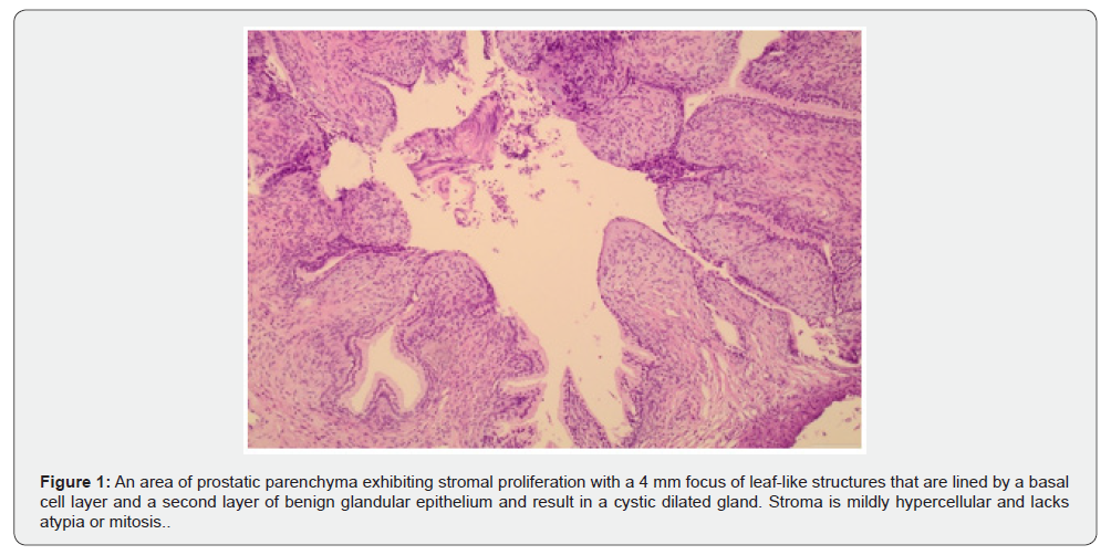
Stromal BPH with a phyllodes pattern, as in our case, exhibit only a small focus of leaf- like structures admixed in an overall background of nodular configuration. The nodules are characterized by mild stromal cellularity, bland spindle cells and prominent round hyalinized vessels. Mitosis or necrosis are absent [3,5]. STUMPs comprise rare neoplastic stromal and glandular prostatic lesions which resemble their counterparts in mammary gland [2,3]. They may exhibit 5 distinct histologic patterns each one with diverse stromal cellularity and atypia. Phyllodes pattern is characterized by a slightly cellular fibromyomatous stroma with leaf-like projections lined by secretory epithelium. However, in contrast with stromal BPH, STUMPs exhibit greater stromal cellularity, more eosinophilic appearance and a diffuse stromal growth without nodularity. Stromal cells may display some atypia. The glandular epithelium is benign and may exhibit proliferative changes [3,5]. Prostatic stromal sarcoma of phyllodes type demonstrates increased stromal cellularity, marked cytologic atypia, severe nuclear pleomorphism and increased mitotic activity. Necrosis may also be encountered. Sarcomatoid carcinoma of the prostate combines a malignant stromal and glandular component and a history of adenocarcinoma of prostate usually precedes [5]. Immunohistochemistry is less helpful for the differential diagnosis since the above-mentioned entities demonstrate stromal expression of vimentin and CD34 and variable expression for desmin and SMA. PR expression is also a common finding [6].
Exoprostatic mesenchymal lesions with prostatic involvement including smooth muscle tumors, inflammatory myofibroblastic tumors, solitary fibrous tumor and Gastrointestinal Stromal Tumors (GISTs) usually lack phyllodes architecture and present identical morphologic and immunohistochemical features.
In conclusion, in our case, the morphologic findings are compatible with stromal BPH with a focus of phyllodes pattern.
Discussion
Diagnosis favouring stromal BPH with a phyllodes focus is clinically significant since STUMP even with low grade histologic features is associated with a remarkable risk of local reccurences (15%) irrespective of the specific histologic pattern. Moroever, in rare cases STUMP coexists with stromal sarcoma implying a probable sarcomatous dedifferentiation [1-5]. In particular, phyllodes pattern, is considered by many studies as a distinct entity previously termed as phyllodes tumor with a potentially agressive behaviour due to local infiltrative growth to colon or urinary bladder, More critically, prostatic stromal sarcoma with a phyllodes pattern (ex. malignant “phyllodes tymor”) is associated with distant metastasis to lung and bones. Consequently, close follow up and an individualized treatment is needed for these entities with total tumor resection or even radical prostatectomy. On the other hand, stromal BPH is totally benign and resection usually comprises the definite therapy.
Acknowledgement
We are grateful to the company “Leriva” for supporting financially the publishing of this article.”
Conflict of interest
“All authors declare no conflict of interest”.
References
- Gaudin PB, Rosai J, Epstein JI, (1998) “Sarcomas and related proliferative lesions of specialized prostatic stroma: a clinicopathologic study of 22 cases Am J Surg Pathol 22(2): 148-162.
- Donna E Hansel, Mehsati Herawi, Elizabeth Montgomery and Jonathan Epstein (2007) “Spindle cell lesions of the adult prostate. Mod Pathol 20: 148-158.
- Jesse K McKennney (2018) “Mesenchymal tumors of prostate” Modern Pathology 31(S1): S133-S142.
- Andreas Bannowsky, Andreas Probst,1 Helmut Dunker, Tillmann Loch, et al. (2009) “Rare and Challenging Tumor Entity: Phyllodes Tumor of the Prostate. J Oncol 241270.
- WHO Classification of tumors, IARC, Urinary and Male genital tract tumors, 5th edition
- Bostwick DG, Hossain D, Qian J, Neumann RM, Yang P, et al. (2004), “Phyllodes tumor of the prostate: long-term follow up study of 23 cases,” J Urol 172(3): 894-899,

























