




 JOJUN Home



Abstract
Bilateral renal pelvic duplication with a ureteral malformation is a highly unusual clinical condition that can be asymptomatic or present with various clinical symptoms. Because of the rarity of this illness, clinicians and patients face a difficult hurdle. We provide the first case report of recurrent urinary tract infections caused by bilateral duplication of the renal pelvis and ureteral deformity. In our example, a 23-year-old woman with leucocytes in her urine was discovered to have bilateral duplication of the renal pelvis and ureteral malformation paired with a renal pelvic cyst. This example demonstrated that congenital genitourinary tract anomalies should be explored in cases of recurrent urinary tract infections.
Keywords:Bilateral duplication of renal pelvis; Ureteral malformation; Renal pelvic cyst; Urinary tract infections
Introduction
Complete ureteral duplication constitutes the most scarce ureteral aberration that can either present bilaterally or unilaterally and represents the 33% of incomplete duplications [1]. The patients who with ureteral duplication may has not presented any further episodes of urinary tract infection or they may show with a variety of clinical manifestations including urinary stones, ureterocele, vesicoureteral reflux, urinary tract infection and obstructive uropathy [2]. It is difficult to evaluate and manage the patients who present with recurrent urinary tract infections (UTI). Other diseases such as voiding dysfunction, overactive bladder, as well as ectopic ureter are some disorders that can manifest with these symptoms [3].
Case Report
A 23-year-old woman was brought to our hospital for an inquiry after recurrent urine tests for leukocytes were positive for three months. The initial physical check revealed no abnormalities. At the presentation, the patient’s blood pressure, pulse rate, and body temperature were 125/80 mmHg, 72 beats per minute, and 37.1°C, respectively.
Prior to 3 months, the patient’s pee routine screening revealed that leukocytes 27(2 +), occult blood, and urinary protein were all negative. He took medicine at this time and did not improve.The pee routine examination revealed that the leukocytes in the urine were (2 + 3+) as expected. A microscopic urine examination revealed a WBC count of 150/high power field and a red blood cell count of 11/high power field, although albumin and occult blood were negative. The laboratory tests confirmed that the urine culture was positive for Escherichia coli and that the renal function was intact. Blood biochemistry, routine, urinalysis, immunological function, antinuclear antibody, and vasculitis tests revealed no abnormalities.
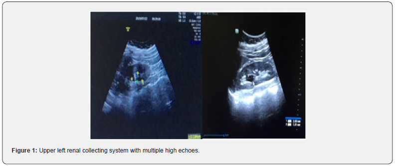
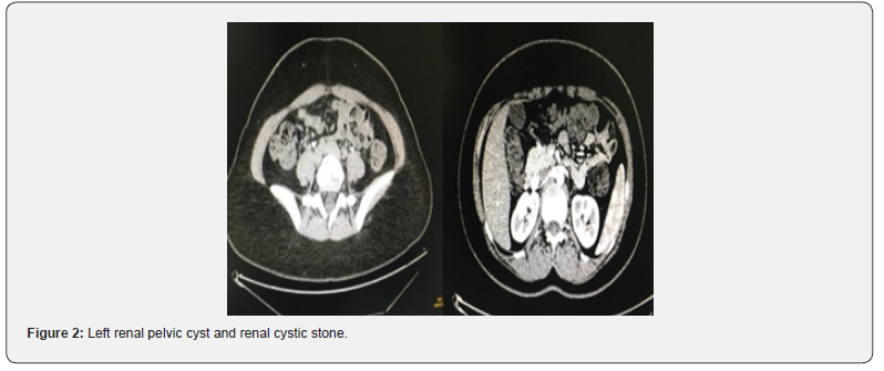
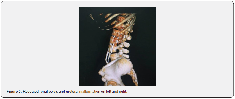
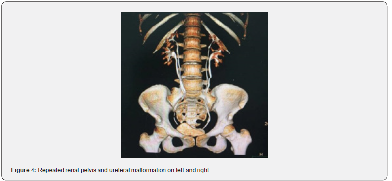
Color Doppler ultrasonography imaging revealed that the upper left renal collecting system had a local extension with several high echoes (kidney stones complicated with infection). Figure 1: A CT scan revealed a left renal pelvic cyst and a renal cystic stone. Figures 2 and 3 The 3D pictures provided crucial information concerning 1) a recurrent renal pelvis and ureteral deformity on both sides and 2) a left renal cyst (Figure 3 and 4).
In examining recurrent urinary tract infections in people of this age, a differential diagnosis of UTI, chronic kidney disease, diabetes, pyelonephritis, and urinary malformation should be addressed. Due to the recovery of awareness impairment and laboratory abnormalities, the patient was discharged from the hospital after several days and is presently getting anti-infection medical treatment. Due to the concomitance with other abnormalities, the patient is currently in outpatient follow-up.
Discussion
This is an uncommon congenital defect characterized by bilateral duplication of the renal pelvis and ureteral deformity, resulting in recurrent urinary tract infections. A urinary tract infection is defined as any microbial invasion that causes an inflammatory response in the urinary system epithelium, which includes infection of the urethra, bladder, and kidneys [4]. Infection can occur at any age in both genders but is more common in women due to anatomy and reproductive function. Causative creatures include Bacteria, fungi, and viruses, with bacteria playing a significant role [4]. Although the risk with UTI has not been clearly demonstrated in clinical trials, several behaviors are thought to increase the risk of an UTI including reduced fluid intake, habitually delaying urination, delaying postcoital urination, wiping from back to front after defecation, douching, and wearing occlusive underwear. In older women, risk factors also include urinary incontinence, history of UTI before menopause, blood group antigen nonsecretor status, and having a cystocele and increased postvoid residual urine [5]. Regularly treating urinary tract infections involves the selective use of sensitive antibiotics against urinary tract bacteria. However, for patients with recurrent urinary tract infections, long-term or excessive antibiotic usage can easily lead to the formation of drugresistant bacteria, resulting in limited antibiotic selection during follow-up therapy. Furthermore, long-term antibiotic treatment increases the risk of flora imbalance, liver and kidney damage, and other side effects. In this regard, several forms of nano-antibiotics or nano medication have been created recently to provide a high therapeutic index, compounded drug release, a longer halflife of drug circulation, and superior pharmacokinetics [6]. It is commonly accepted that individuals with recurrent urinary tract infections and aberrant urinary tract anatomy should be treated as much as feasible. This patient’s recurring urinary tract infection may be due to anatomical anomalies of the renal pelvis and ureter. If patients do not receive surgical therapy, the attacks continue to be prevalent during the duration of recurrent antibiotic use. Furthermore, specific medications can be used to treat urinary tract infections, minimize the frequency of recurrences, and enhance the quality of life. Early detection and diagnosis of urinary tract infection due to congenital urinary system malformation or urinary tract dysfunction, as well as appropriate treatment, are critical. The severity of symptoms, renal function, patient age, and life quality will influence how a urinary tract infection is managed. Conservative therapy is an appropriate approach in adults, but it may not eliminate the problem of recurring urinary tract infections and pose long-term hazards. Surgery is the best treatment for symptomatic individuals since it attempts to address incontinence, avoid future issues, preserve renal function, and remove UTIs. [7]. Biles et al. demonstrated that duplex systems may be treated using either an upper tract strategy, which includes pyelopyelostomy, ureteropyelostomy, and upper moiety heminephrectomy, or lower tract reconstructive procedures, which have ureteral reimplantation and ureteroureterostomy [7].
The primary pathogen that causes recurrent UTI in women and accounts for 80% of all episodes of infection is E. coli. Staphylococcus saprophyticus, Klebsiella pneumonia, and Proteus mirabilis are three more significant pathogens that account for about 4% of all cases of acute cystitis [8]. The fimbria on uropathogenic E. coli promotes binding to the urethral and vaginal epithelium, which increases the bacteria’s capacity to cause cystitis [5].
According to this study, the patient had a double renal pelvis, ureteral malformation, renal pelvic cysts, and intracapsular calculi, with typical clinical symptoms. Due to the limitations of B-ultrasound and CT, it is difficult to discover individuals with urinary anomalies if CTU testing is not available. Finally, CTUs are required for an appropriate diagnosis. CTU procedures are non-invasive, whereas using CT contrast agents necessitates the intravenous injection of the agent to complete the scan [8]. Given the diagnostic significance and immediate appropriate care for patients with urinary tract problems, CT and CTU are becoming more effective instruments for assessing urinary tract disorders, and CTU has emerged as a viable technique for identifying urinary tract abnormalities. Each imaging approach has advantages and limitations, even though both give imaging excellent clarity for normal and diseased states in clinical settings [9].
The clinical performance of ureteral duplication is highly diverse and depends on the patient’s age. They are expected to function asymptomatically in adults or to manifest with hematuria, stomach, or flank discomfort, and to predispose to blockage, vesicoureteral reflux, and recurrent urinary infections. Furthermore, those specific congenital abnormalities will enhance the likelihood of ureteral damage during the operation [10].
Our study has some limitations, including its retrospective methodology, limited patient sample size, absence of a reference group, and single institution. Furthermore, large sample sizes and long-term follow-up studies are required. Finally, surgery should be considered if a blockage is discovered or a conservative strategy fails.
Ethics Approval and Consent to Participate
This research was approved by the ethics committee of The Affiliated Hospital of Weifang Medical University and the patient gave written informed consent prior to obtain the data. All authors have no competing interests or conflicts of interest declare.
References
- Scantling D, Ross C, Altman H (2013) A 52-year-old male with bilaterally duplicated collecting systems with obstructing ureteral stones: a case report. Curr Urol 7(2): 104-106.
- Karakose A, Aydogdu O, Atesci YZ (2013) Unilateral complete ureteral duplication with distal ureteral stone: A rare entity. Can Urol Assoc J 7(7-8): E511-512.
- Demir M, Ciftci H, Kılıçarslan N, Gümüş K, Oğur M, et al. (2015) A case of an ectopic ureter with vaginal insertion diagnosed in adulthood. Turk J Urol 41(1): 53-55.
- Devi N, Rani K, Kharb P, Prasad M (2021) Herbal Medicine for Urinary Tract Infections with the Blazing Nanotechnology. J Nanosci Nanotechnol 21(6): 3495-3512.
- Vahlensieck W, Perepanovab T, Bjerklund TE, Johansenc TEB, Tenke P, et al. (2016) Management of Uncomplicated Recurrent Urinary Tract Infections. European Urology Supplements 15(4): 95-101.
- Van Giau, V, Soo A An S, Hulme J (2019) Recent advances in the treatment of pathogenic infections using antibiotics and nano-drug delivery vehicles. Drug Des Devel Ther 13: 327-343.
- Biles MJ, Finkelstein JB, Silva MV, Lambert SM, Casale P, et al. (2016) Innovation in Robotics and Pediatric Urology: Robotic Ureteroureterostomy for Duplex Systems with Ureteral Ectopia. J Endourol 30(10): 1041-1048.
- Bafaraj SM (2018) Value of Magnetic Resonance Urography Versus Computerized Tomography Urography (CTU) in Evaluation of Obstructive Uropathy: An Observational Study. Curr Med Imaging Rev 14(1): 129-134.
- Hilton S, Jones LP (2014) Recent advances in imaging cancer of the kidney and urinary tract. Surg Oncol Clin N Am 23(4): 863-910.
- Varlatzidou A, Zarokosta M, Nikou E, Theodoropoulos P, Kakaviatos D, et al. (2018) Complete unilateral ureteral duplication encountered during intersphincteric resection for low rectal cancer. J Surg Case Rep 2018(10): p. rjy266.

























