A calcified offender
John Banerji*
Department of Urology, UPMC Horizon, USA
Submission: October 16, 2017; Published: November 21, 2017
*Corresponding author: John Banerji, Department of Urology, UPMC Horizon, USA, Tel: 2065828834; Email: johnsbanerji2002@gmail.com
How to cite this article: Dejan S. Evaluation of Nephrotoxicity using Lysoenzymuria in Patients with Rheumathoid Arthritis Treated with Most used Slow acting Antirheumatic Drugs-Saards and Establishment to the Diagnostic Value as Sifted Test. JOJ uro & nephron. 2017; 4(2): 555636. DOI:10.19080/JOJUN.2017.04.555636
Clinical Image
A 55 year old lady, presented to the urology department with symptoms of urinary frequency and urgency, of 3 months duration. She had an episode of calculuria, 3 years ago, but denied any haematuria. She was postmenopausal, and previously had normal menstrual cycles.
General physical examination was unremarkable. Abdominal examination revealed a firm pelvic mass, in the midline, which was dull on percussion. This could not be differentiated from the bladder. Her urine routine examination revealed numerous red blood cells, with 2-4 leucocytes. Her serum creatinine, electrolytes and CBC were within normal limits.
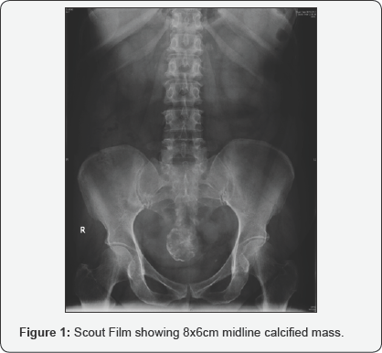
An Intravenous urogram was performed, as her previous history of calculuria and present microscopic haematuria were suggestive of calculous disease again. The scout film showed a 8x6cm, calcific shadow in the midline (Figure1). The IVU films revealed this mass, indenting the dome of the bladder (Figure2), explaining her symptoms of recent onset frequency and urgency. She had no calculi. Large fibroids can sometimes present with urinary complaints [1].


Calcification in fibroids occur in about 4% of fibroids [2]. This calcification is generally amorphous and dense. However, peripheral calcification can sometimes occur, when it is assumed to be due to degeneration with thrombosed veins.
References
- Wallach EE, Vlahos NF (2004) Uterine myomas: an overview of development, clinical features and management. Obstet Gynecol 104(2): 393-406.
- Ueda H, Togashi K, Konishi I, Kataoka ML, Koyama T, et al. (1999) Unusual appearnces of uterine leiomyomas: MR imaging findings and their histopathologic backgrounds. Radiographics 19: 131-135.


