Value of Hounsfield Unit in Prediction of Stone Free Rate in Management of Upper Urinary Tract Stones Using Ct and Eswl
Omer Mohammed Ahmed Eltayeb, Abdelazim Hussein Khalafalla*, Faisal Yousif Amir and Tarig Hassan Hag Ali
Department of Urology, University of Pittsburgh School of Medicine, USA
Submission: March 04, 2017; Published:May 04, 2017
*Corresponding author: Abdelazim Hussein Khalafalla, Department of Urology, University of Pittsburgh School of Medicine, USA, Email: aazeem33@gmail.com
How to cite this article: Omer M A E, Abdelazim H K, Faisal Y A, Tarig H H A. Value of Hounsfield Unit in Prediction of Stone Free Rate in Management of Upper Urinary Tract Stones Using Ct and Eswl. JOJ uro & nephron. 2017; 2(5): 555600. DOI:10.19080/JOJUN.2017.2.555600.
Abstract
Purpose: To evaluate the attenuation value of upper urinary calculi on unenhanced axial computerized tomography (CT) images as a predictor of stone free rate in upper urinary tract after extra corporeal shock wave lithotripsy (ESWL).
Materials and Methods: We included 50 patients with renal calculi up to 20mm in this prospective study. Calculus attenuation value was measured in Hounsfield units on unenhanced CT sections through the calculi. Patients were subsequently treated with ESWL and followed up for 3 months to see the results.
Results: Patients were grouped according to calculus attenuation value as groups:
- less than 500,
- 500 to 1,000 and
- greater than 1,000 Hounsfield units.
Of the 50 patients 36 underwent successful treatment. The rate of stone clearance was 100% (23 of 23 cases) in group 1, 70.5% (12 of 17) in group 2 and 10% (1 of 10) in group 3. The success rate for stones with an attenuation value of greater than 1,000. Hounsfield units was significantly lower than that for stones with a value of less than 1,000 Hounsfield units (1 of 10) 10% versus 35 of 40, 87.5% cases
Conclusion: The CT attenuation value of renal calculi can help to differentiate stones that are likely to fragment easily on ESWL from those that may fail to fragment on ESWL.
Keywords: Extracorporeal shockwave lithotripsy; Lower pole; CT; ESWL; Hounsfield units
Introduction
Since its introduction in the early eighties of the last century by Chaussy et al. [1]. ESWL has been accepted as first election procedure to treat renal stones smaller than 2cm. Due to the effectiveness of ESWL and its minimal side effects,the FDA quickly approved ESWL. Analyzing different series itssuccess rate varies from 60 to 99%. ESWL failure increasessanitary costs. Stone shape, fragility, location and composition, obstructive uropathy, urinary infection, shock wave's physical properties and the distance from skin to stone are described as related to therapeutic result.
The aim of this paper is to evaluate if the stone density measured in Hounsfield Units with a non-contrast-enhancedCT scan (NCCT) is able to predict its fragmentation after extra corporeal shock wave lithotripsy.
Objectives
To evaluate the significance of stone density measured by Hounsfield Units by CT, in prediction of stone-free rate in upper urinary calculi after ESWL.
Patients and Methods
Hospital based comparative, prospective study, between October 2011 and October 2012, conducted in Al-Amal national hospital in Khartoum north, Sudan. The targeted population was all adults with renal and upper ureteric stones. Hospital based comparative, prospective study, between October 2011 and October 2012, conducted in Al-Amal national hospital in Khartoum north, Sudan. The targeted population was all adults with renal and upper ureteric stones.
Data were collected using pre-designed questionnaire. The questionnaire covered all the personal data, stone burden (size and number), presence or absence of hydronephrosis, pre and post ESWL renal functions, presence or absence of (UTI), number of shock waves used, method of localization of stone, outcome, complications and confirmatory tests.
Data were fed to Statistical Package for Social Sciences (SPSS). The results obtained are presented in tables and figures.Data were fed to Statistical Package for Social Sciences (SPSS). The results obtained are presented in tables and figures.
All patients included in this study were suffering from a solitary stone in the upper urinary tract ranging from 5mm to 20mm in size. Patients for whom ESWL was contraindicated because of pregnancy, coagulation disorder, distal mechanical obstruction, multiple stones, Stag horn stones, were excluded from the study.
The patients were examined by hematology, biochemical and urine tests. Before ESWL, patients had Non contrast helical computerized tomography. The longitudinal calculus dimension was calculated using collimation thickness, the reconstruction interval and the number of images in which the calculus could be visualized. The image showing the calculus in the largest width was selected. The maximum diameter and the mean density of the stone were calculated by drawing a region of interest over the calculus. The maximum dimension of the calculus included in the study was either the longitudinal or the transverse diameter, whichever was the largest. The patients were followed-up for 3 months after ESWL by KUB and US.
A total of 50 patients undergoing therapy for upper urinary tract calculi were included in the study, after defining the indications of treatment, the patients were made aware of ESWL and its probable complications. The need for anesthesia, its possible complications, and consent forms were filled to the all patient.
ESWL was performed using the Dornier lithotripter (Dornier Compact Delta II). All patients were positioned supine and the calculi were localized with fluoroscopic guidance.
All patients were given sedatives and analgesics. The fragmentation of the calculus during the therapy was monitored by fluoroscopy. A maximum of 4.0kV was given to each patient, starting at 0.1kV and increasing gradually stepwise after every 500 shock waves till satisfactory stone fragmentation.
The 50 patients received 131 sessions, mean 2.62±1.958.
During each ESWL session no more than 3000 shock waves were given, and an interval of 2 to 4 weeks maintained between ESWL sessions.
An ultrasound was taken after each ESWL session to document fragmentation before the next session to ascertain position and clearance. Clearance was documented by an ultrasound 12 weeks after the last ESWL session, and defined as complete disappearance of the renal calculus; fragments of ≤5mm were defined as clinically insignificant residual fragments (CIRF) and patients with CIRF were subsequently managed conservatively.
Patients were followed-up to assess the success rates and complications of the procedure.
The CT values were analyzed with the outcome of ESWL (the number of sessions required and clearance of the calculi). Result was analyzed using one-way an ova.
Treatment failure was based on the need for further surgical intervention during follow-up or failure to become stone-free within 3 months. At initial follow up, patients were given a questionnaire.
Results
50 patients were treated by ESWL (male/female: 37/13), 30 had a calculus on the right and 20 on the left. Patient's age varied between 20 and 70 years. 14 patients had stone of size 10mm or less while 36 had stones of sizes more than 10mm. The mean stone size was 13.06±4.42mm (range 5 to20).
The calculus was in the pelvis in 21 patients (42%), in the inferior calyx in 14 (28%), in the superior calyx in five (10%), in the middle calyx in 1 patient (2%), and in the upper ureter in 6 (12%).The calculus was in the pelvis in 21 patients (42%), in the inferior calyx in 14 (28%), in the superior calyx in five (10%), in the middle calyx in 1 patient (2%), and in the upper ureter in 6 (12%).
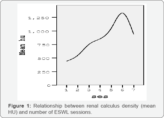
The 50 patients required 131 sessions of lithotripsy. Those who needed less than 3 sessions to clear the stone were 34 patients, while those who underwent more than 3 sessions were 16 patients. The average number of shock waves was 2500 at 1020 kV. The mean calculus density and number of ESWL sessions needed had a linear correlation which was maintained at all levels (Figure 1).

Patients were grouped according to calculus attenuation value as groups
- Less than 500,
- 500 to 1,000 and
- greater than 1,000 Hounsfield units
Of the 50 patients 36 underwent successful treatment. The rate of stone clearance was 100% (23 of 23 cases) in group 1, 70.5% (12 of 17) in group 2 and 10% (1 of 10) in group 3. The success rate for stones with an attenuation value of greater than 1,000 Hounsfield units was significantly lower than that for stones with a value of less than 1,000 Hounsfield units (1 of 10) 10% versus (35 of 40) 87.5% .The CT value of the stone- free group was 511.36±208.405 HU, significantly lower than that of the residual stone group (1057.7±372.839 HU, t = 6.842, P < 0.01).
Stone-free status at 1 month and 3 month were 42% (n=21) and 72% (n=36), respectively. There was no major complication.
Considerable differences with regard to patient satisfaction were noted. Of the patients 40(80%) were satisfied and will recommend the procedure to the others while 10(20%) who required re-treatment would not for recurrence.
Discussion
Plain x-ray has been used to predict the outcome of ESWL treatment by comparing stone density with bone density. However, this method has some disadvantages since the stone diameter and appearance might not be measured accurately, especially in the presence of bowel gas interference or neighboring bony structures and the density measurement is subjective. We used plain CT scan which is a non invasive technique and provides greater density discrimination than plain x-ray. CT can distinguish density differences as low as 0.5% compared to only 5% discrimination using plain x-ray [1].
Joseph et al. [2] suggested that stones with CT attenuation value of greater than 950 Hounsfield units and 7500 shockwaves failed to achieve fragmentation. In our study, the success of ESWL treatment is almost always guaranteed when the CT attenuation value is less than 500 Hounsfield units, while, at the same time, treatment failure is almost certain when the CT attenuation value exceeds 1000. Stone densities in the range of 500 to 1000 HU may, or may not, respond successfully to ESWL treatment. This study revealed that stone diameters of up to 20mm may still (depending on stone density) respond successfully to ESWL treatment. Similar to Joseph et al, the results of this study clearly reveals that stones with densities exceeding 950 Hounsfield units are difficult to fragment. Our study revealed that stone density is the determinant factor of treatment success for stone sizes of 20mm or smaller.
HU measurement of urinary calculi on pretreatment noncontrast computerized tomography may predict the stone- free rate. This information may be beneficial for selecting the preferred treatment option for patients with urinary calculi [3].
In a study of 30 patients, Joseph et al. [2] found that patients with calculi of less than 500 Hounsfield units had complete clearance and required a median of 2500 shockwaves, patients with calculi of 500-1000 Hounsfield units had a clearance rate of 86% and required a median of 3390 shockwaves, and patients with calculi of more than 1000 Hounfield units had a clearance rate of 55% only and required a median of 7300 shockwaves.
Pareek et al. [3] correlated calculus density with stone clearance in their study of 100 patients. They concluded that patients with residual calculi had a mean calculus density of more than 900 Hounsfield units. However, Pareek et al. [3] did not correlate the calculus density with fragmentation. The results of this study concurs with Pareek et al. [3] results in that stone clearance is unlikely when stone density exceeds 1000 Hounsfield units.
The HU value on pretreatment NCHCT of the upper urinary tract stones can be used to predict the stone-free rate after ESWL [4].
The CT attenuation value of renal calculi can help to differentiate stones that are likely to fragment easily on ESWL from those that would require a greater number of shock waves for fragmentation or may fail to fragment on ESWL [5,6].
An escalating voltage treatment strategy produced better stone comminution than a fixed voltage treatment strategy. The study suggests that there may be a protective effect of an escalating treatment strategy. There was significantly increased renal damage in the fixed group, as shown by increased urine micro albumin and macroglobulin [7-10].
Conclusion
Selection of patients and types of the stones are the cornerstone in ESWL success, and when performed by an expert gives the best results. The HU value on pretreatment NCHCT of the upper urinary tract stones can be used to predict the stone- free rate after ESW, treatment failure is almost certain when the HU value exceeds 1000.
References
- Chaussy C, Brendel W, Schmiedt E (1980) Extracorporeally induced destruction of Kidney stones by shock waves. Lancet 2(8207): 12651268.
- Tarawneh E, Awad Z, Hani A, Haroun AA, Hadidy A, et al. Department of Diagnostic, Radiology and Urological Surgery Jordan University Hospita, Amman, Jordan.
- Saudi Center for Organ Transplantation. Saudi J Kidney Dis Transpl 21(4): 660-665.
- Joseph P, Mandal AK, Sharma SK (2002) CT attenuation value of renal calculus: can it predict successful fragmentation of the calculus by extracorporeal shockwave lithotripsy? A preliminary study. J Urol 167(5): 1968-1971.
- Lingeman JE, Newman D, Mertz JH, Mosbaugh PG, Steele RE, et al. (1986) Extracorporeal shockwave lithotripsy: the Methodist Indiana experience. J Urol 135(6): 1134-1137.
- Pareek G, Armenakas NA, Fracchia JA (2003) Hounsfield units on computerized tomography predict stone-free rates after extracorporeal shock wave lithotripsy. J Urol 169(5): 1679-1681.?
- Zhonghua Yi, Xue ZaZhi (2006) Can Hounsfield units on CT Predict Stone Free Rate of Upper Urinary Calculi after ESWL and Stone Composition? 86(4): 276-278.
- Cheng G, Xie L, Department of urology, The First Affiliated Hospital, College of Medicine, Zhejiang University. Hangzhou, China.
- Joseph P, Mandal AK, Singh SK, Mandal P, Sankhwar SN, et al. (2002) Computerized tomography attenuation value of renal calculus: can it predict successful fragmentation of the calculus by extracorporeal shock wave lithotripsy? A preliminary study. J Urol 167(5): 1968-1971.
- Lambert EH, Walsh R, Melissa W, Moreno (2009) Effect of Escalating Versus Fixed Voltage Treatment on Stone Comminution and Renal Injury During Extracorporeal Shock.Wave Lithotripsy: A Prospective Randomized Trial. J Urol 183(2): 580-584.


