




 GJAA Home



Shape and Sexual Dimorphism of Canines in Old World Monkeys and their Significance
Hiroyuki Yamada*
Department of Oral Anatomy, School of Dentistry, Aichi Gakuin University, Japan
Submission:June 23, 2026; Published:July 03, 2026
*Corresponding author: Hiroyuki Yamada, Department of Oral Anatomy, School of Dentistry, Aichi Gakuin University, 1-100, Kusumoto-Cho, Chikusa-Ku, Nagoya 464-8650, Japan
How to cite this article: Hiroyuki Y. Shape and Sexual Dimorphism of Canines in Old World Monkeys and their Significance. Glob J Arch & Anthropol. 2026; 14(5): 555900. DOI: 10.19080/GJAA.2026.14.555900
Abstract
It has been reported that the crown outline of New World monkey canines changes in response to their diet. However, this has not yet been clarified for Old World monkeys. This study examined whether similar changes occur or not in the canines of Old World monkeys. It also examined the factors influencing the shape morphology of Old World monkey canines. The results revealed that the canine teeth of males in both Cercopithecidae subfamilies were fang-like, with sharply pointed inner surfaces. It was considered that the canines of Cercopithecidae monkeys evolved as weapons rather than as adaptations to leaf-eating due to strong selective pressure for high aggression arising from the need for defense against predators and intense competition among males. This characteristic was considered a primitive trait (symplesiomorphy) shared by all Cercopithecidae members. The female canines were distorted into a rhomboid shape. This morphology is due to the fact that the female canine teeth are not evolutionarily important as weapons. The cervical ridge was more developed in females than in males. The deciduous canines resembled equilateral triangles. The lower canines were also discussed.
Keywords: Canine Outline; Fanged Shape; Weapons; Diet; Old World Monkeys
Introduction
The morphology of mammalian teeth reflects their evolutionary history, ecological niche, and diet, exhibiting remarkable diversity. Canines, in particular, serve as weapons for intimidating enemies or inflicting fatal, deep wounds on predators during combat, while also playing a role in intraspecific sexual selection. In evolutionary studies of paleoprimatology and paleoanthropology, research on canines is invaluable for understanding phylogeny, diet, social structure, and sex determination. In particular, the reduction in canine size is strongly linked to the emergence of humans, and numerous studies have been reported to date focusing on this aspect [1-9]. However, research based on canine tooth shape is not particularly abundant. The shape of canine teeth is just as important as their size. This is because the shape of an animal’s tooth is highly heritable and often reflects its lineage and diet.
Canine teeth are originally simple, conical shapes with little variation in the external contour of the crown, and there are also few noticeable phenotypic characteristics on the tooth surface. Consequently, with the exception of a few researchers [2,3, 10-12], the external morphology of canine teeth has received little attention to date. A detailed examination of canine morphology reveals subtle differences—both between phylogenetic groups and between sexes—that cannot be captured by quantitative analyses [13-20]. This is because differences in tooth shape strongly contribute to sexual dimorphism. Even when there are no statistically significant differences and almost no variation in size, differences in shape are highly likely to be effective for sex determination.
The present study was conducted based on the hypothesis that the crown morphology of Old World monkey canines, like that of New World monkeys, is likely influenced by factors such as diet, social structure, sexual dimorphism, and phylogeny. It involved a detailed observation of Old World monkey canine morphology to test this hypothesis. Furthermore, the study examined the factors that determine canine morphology.
Materials and Methods
The objective of the study was the examination of two subfamilies of Cercopithecidae. The study included six Cercopithecinae genera (Cercopithecus, Chlorocebus, Erythrocebus, Papio, Cercocebus and Macaca) and seven Colobinae genera (Colobus, Piliocolobus, Procolobus, Trachypithecus, Presbytis, Nasalis and Simias). The materials used were skulls held by the Center for the Evolutionary Origins of Human Behavior at Kyoto University (EHUB) and the Japan Monkey Centre (JMC). Impressions were taken using a silicone-based dental impression material (Provil, Heraeus Kulzer GmbH), after which plaster models were made. For the photographs, the camera was positioned so that its optical axis was perpendicular to the coronal axis of the canines and images were captured from the lingual side [20].
The overall size of the canine crown was assessed based on its area. The area was calculated by multiplying the mesiodistal diameter (MD) and the labio-lingual diameter (LL) of the crown, and the results were classified into one of three categories. Values less than 10.0 mm² were classified as “small,” values between 10.0 and 20.0 mm² as “medium,” and values of 20.0 mm² or greater as “large,” following the method adopted for New World monkeys [20]. The degree of canine size dimorphism was calculated by dividing the average area value for males by that for females; values of 1.00–1.09 were classified as “none,” 1.10–1.19 as “mild,” 1.20–1.29 as “moderate,” and 1.30 or higher as strong Data on the average size of canines in Old World monkeys were cited from the following references [21-24]. The dimorphism of canine shape was classified into four categories—“none,” “mild,” “moderate,” and “strong”—based on the degree of difference in canine shape between males and females.
The crown of a canine tooth typically consists of five features: the mesial and distal crests, the mesial and distal marginal ridges, and the cervical ridge. During observation, attention focused on the outline of the crown defined by these characteristics. In many previous studies on crown morphology, the MD/LL ratio (“crown index”), which describes tooth shape as viewed from the occlusal aspect, has been used [6, 7, 16, 17, 22, 25]. However, the crown index alone is insufficient to explain detailed changes in tooth morphology. Although three-dimensional analysis is a suitable method for detailed analysis of tooth morphology, this approach is labor-intensive and time-consuming because it requires processing a vast amount of data for each specimen [14-16]. In contrast, classical methods involving detailed examination and observation of teeth have the advantage of allowing for close inspection of individual specimens, but they have the disadvantage of being prone to subjectivity in the results.
In this study, specific examples were provided to ensure greater objectivity. The shape of the canine was described using specific terms, such as dagger-, fang-, hook-, sickle-, blade-, and diamond-shaped. The phylogenetic classification of primates followed JMC and Fleagle [26], and sex determination was based on records from the research institution.
Results
Cercopithecinae
Upper canines
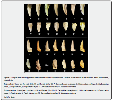
When viewed from the lingual side, the external contour of the male M. nemestrina (genus Macaca) upper canine resembled a fang, and its inner surface was as sharp as a sickle. The mesial crest was arched and the distal crest was gently concave. On the mesial surface of the crown, a deep and wide mesial groove ran from the apex toward the cervical region; it was not interrupted by a mesial marginal ridge and extended all the way to the root. Distal to the mesial groove, a well-developed, rounded mesial lingual ridge extended in a flared pattern to the root. A central groove ran along the lingual ridge, forming a shallow, wide depression that extended to the root. The lingual fossa showed significant wear from grinding against the lower third premolar. The mesial shoulder was underdeveloped, making the mesial tubercle difficult to visualize. The distal shoulder was slightly developed, and a mild bulge was present. The cervical ridge was extremely thin, making the cervical line difficult to discern. The enamel-cementum junction transitioned smoothly.
The females were much smaller than the males and were shaped differently. Their crown outline was rhomboidal, with a short basal portion. A moderately deep mesial groove ran from the crown apex toward the cervical region along the mesial crest. It was interrupted by the mesial marginal ridge in some cases and extended beyond it in others. The mesial lingual ridge was well-developed and rounded. It extended to the root. The shallow, broad central groove either terminated in a V-shaped notch at the cervical ridge or extended beyond it to the root. The cervical ridge was more developed than in males and ran in a U- or V-shape. The enamel-cementum junction was clearly visible. Similar morphology was also exhibited by other genera (Cercopithecus, Chlorocebus, Erythrocebus, Papio, Theropithecus, and Cercocebus).
Lower canines
The crown of the male M. nemestrina appeared pentagonal or hexagonal lingually. A high, triangular region was observed mesially and a heel-like structure was observed distally. The mesial crest was gently curved, while the distal crest was straight. The mesial marginal ridge ran obliquely downward, and the cervical ridge ran nearly horizontally. The mesial groove was deep and either interrupted or crossed by the marginal ridge in the cervical region. The lingual ridge was thick and rounded. It ran straight down along the mesial groove and merged with the cervical ridge at its base. The distal lingual groove was shallow and faint, extending from the apex to the cervical region. The poorly developed distal shoulder formed a broad depression. Severe wear was observed in this area. The cervical ridge was well developed, with a visible bulge in the distal region. Due to the bulge of the cervical ridge, the enamel-cementum junction on the lingual surface was flexed.
The females were considerably smaller than the males. Their crown outline was pentagonal to hexagonal, similar to the males', and their heel-like structure was only slightly developed. The mesial crest was straight, and the distal crest was slightly concave. The mesial groove was shallow and interrupted by the marginal ridge. In contrast, the distal groove was relatively deep and extended from the crown apex to the distal lingual fossa and sometimes extended as far as the cervical ridge. The lingual ridge was well-developed and merged with the cervical ridge. The mesial tubercle curvature was notably more pronounced. The mesial shoulder was located approximately two-thirds of the way up the crown from the cervical line. Similar morphological features were observed in other genera and species (Figure 1).
Deciduous canines
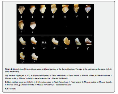
Lingually, the upper deciduous canine in M. fasicularis resembled an equilateral triangle in both males and females but showed asymmetry between the directions of the left and right. The lingual ridge was well-developed and descended from the crown apex to merge with the cervical ridge. The mesial shoulder was located approximately one-third of the height of the crown from the cervical line, and the distal shoulder was located approximately one-quarter of the height of the crown. The cervical ridge was strongly prominent and ran obliquely downward from mesial to distal. The enamel-cementum junction was markedly constricted. Other genera and species generally exhibited similar morphology.
The lower deciduous canine had a hexagonal outline similar to the lower canine of adult females. The distal region was well developed and heel-shaped. The lingual ridge extended obliquely downward from the apex and converged with the cervical ridge at its base. The mesial tubercle was more curved than in adult females. The cervical ridge ran obliquely downward and encircled the entire crown before reversing direction at its lowest point. A pronounced constriction was observed at the enamel-cementum junction. The crown tended to be longer in the anteroposterior direction. Similar morphology was observed in other genera (Figure 2).
Colobinae
Upper canines
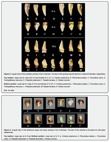
The upper canines of male Colobus polykomos closely resembled those of the Cercopithecinae, because they had a fanged crown morphology. The female canines also closely resembled those of females in the Cercopithecinae.
Lower canines
The crown morphology of the lower canines in male C. polykomos closely resembled that of the Cercopithecinae. The crown outline was pentagonal or hexagonal. However, these morphological features were significantly less pronounced (Figure 3).
Deciduous canines
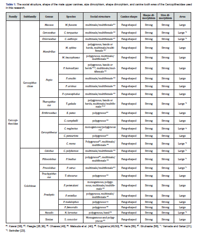
The deciduous canines showed a strong resemblance to those of Cercopithecinae in both the upper and lower jaws. The differences lay in the upper jaw, where the morphological phenotype was less pronounced and the symmetry was stronger than in the Cercopithecinae. The Cercopithecinae exhibited longer anteroposterior crowns, whereas the Colobinae had longer vertical crowns (Figure 4).
Discussion
Old World monkeys and apes are thought to have diverged from New World monkeys during the late Eocene to early Oligocene, approximately 35 to 40 million years ago [27,28]. Cercopithecidae evolved to have bilophodont molars in the lower jaw, whereas apes evolved to retain the archaic Y5 molar structure. The Cercopithecidae family includes the Cercopithecinae and Colobinae subfamilies. These subfamilies are more closely related to apes than to New World monkeys, as evidenced by the presence of two premolars on each side of the jaw [29]. The Cercopithecinae and Colobinae are thought to have diverged between 16.2 million and 14.5 million years ago [30].
Many extant primates exhibit pronounced sexual dimorphism, with males typically having larger bodies and canines than females. Their diet is primarily herbivorous and consists of leaves, fruits, and tree sap [31]. The Old World monkeys included in both Cercopithecinae and Colobinae are essentially plant-based omnivores. However, Cercopithecinae have a broader dietary range and consume a diverse array of foods, including fruits, leaves, seeds, young shoots, mushrooms, insects, spiders, and small vertebrates [31]. The Japanese macaque (Macaca fuscata), for example, primarily eats plants, such as fruits, leaves, young shoots, and seeds, as well as insects, frogs, small bird eggs, and fish during the winter [32]. The hamadryas baboon (P. hamadryas) eats grass, fruit, leaves, young shoots, insects, and small vertebrates. In contrast, the mandrill primarily eats fruit, as well as seeds and leaves [33-35]. Additionally, C. neglectus feeds on fruit, leaves, flowers, mushrooms, and insects, including beetles and termites [36,37].
The colobines, on the other hand, are primarily folivorous, feeding mainly on tree leaves. They digest these leaves using a stomach structure similar to that of ruminants. Leaf monkeys are the most strictly folivorous primates, consuming mostly leaves and a small amount of insects. The king colobus (C. polykomos) eats not only leaves but also fruits and flowers. S. concolor is primarily folivorous but also eats fruit, seeds, and flowers occasionally [38]. The proboscis monkey (N. larvatus) is folivorous and eats leaves and fruit. It shows a preference for young leaves [39, 40].
Although both the Cercopithecinae and the Colobinae subsist on plant-based diets, their dietary compositions differ significantly. Examining their dental structures reveals that fruit-eating species in the Cercopithecinae have low cusps, shorter shearing crests, and thicker enamel. In contrast, leaf-eating Colobinae species have the opposite structure, with taller cusps, longer shearing crests, and thinner enamel, characteristics of more folivorous species [29]. However, the canines of both male subfamilies are similar in that they are fang-like. This suggests that this trait is not related to dietary intake.
Canine teeth size, social structure, and shape dimorphism
The size dimorphism of canines in primates is closely associated with intragroup and intergroup competition among males, as well as with sexual selection [1,2,41-44]. Kay et al. [13] and Izawa [45] reported a positive correlation between body size, canine tooth size, and sexual dimorphism in South American monkeys. Sexual dimorphism in canines is weakest in monogamous and polyandrous species and strongest in species with hierarchical, male-dominant social structures. Species with fission-fusion social structures are said to fall between these two extremes.
Many species in the Cercopithecinae are polygynous and form groups consisting of multiple males and females. The hamadryas baboon (P. hamadryas), for instance, has a hierarchical social structure in which small groups (clans) unite to create larger groups (bands). The Japanese macaque (M. fuscata) forms multimale/multifemale groups. These groups exhibit a hierarchical structure among males [46-48]. In contrast, C. neglectus exhibits monogamous or polygynous social structures [26]). Despite this diversity in social structure, all Cercopithecinae species have uniformly fang-shaped upper canines that exhibit significant sexual dimorphism in size and shape.
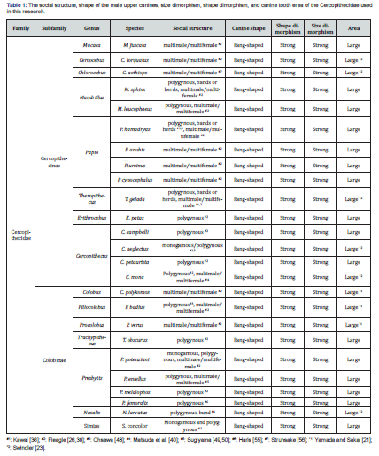
The Colobus monkeys exhibit monogamous or polygynous mating systems. The leaf monkey (P. entellus) forms harems of one male and multiple females, or groups of multiple males and females [49-51]. In contrast, the proboscis monkey (N. larvatus) forms groups of one male and multiple females [26, 40, 52]. The leaf monkey P. potenziani, however, forms monogamous, polygynous, and harem-like groups [26,53]. S. concolor exhibits both monogamous and polygynous behavior, forming social groups known as "bands" [26]. Even among the diverse social structures of the Colobinae, all species have fang-shaped upper canines. The dimorphism in the crown morphology of these canines is striking in terms of both size and shape. The canines are large in all species. New World monkeys that live in monogamous societies, such as the Callitrichidae and Aotidae, do not exhibit sexual dimorphism in canine size or shape [20, 46-48]. However, monkeys in monogamous Cercopithecidae societies, including C. neglectus, P. potenziani, and S. concolor, exhibit significant sexual dimorphism in canine size and shape; their teeth are also larger [43] (Table 1).
The fifteen species across eight genera in the Cercopithecinae and the ten species across seven genera in the Colobinae of the Cercopithecidae examined in this study all had uniformly fang-shaped canines that showed significant sexual dimorphism in size and shape. The external shape of the canines was uniformly fang-like even among species from different lineages, and their inner surfaces were sharply pointed, resembling sickles. Based on these findings, the original hypothesis was rejected. Currently, it is believed that the shape of the canine teeth in Old World monkeys is not strongly related to their dietary habits.
Relationship with the lineage
The ability to eat leaves first emerged late in the evolution of anthropoids, evolving independently in catarrhines and platyrrhines [31]. New World monkeys exhibit a wide range of dietary habits. For instance, titi monkeys prefer the seeds of hard, unripe fruits; spider monkeys primarily eat ripe fruits; marmosets use their teeth to scrape sap from trees; and howler and woolly monkeys are primarily folivorous, feeding mainly on young leaves, flowers, and fruits. Their canines come in various shapes adapted to their dietary habits [20]. On the other hand, Cercopithecinae monkeys eat a variety of foods, while Colobinae monkeys are leaf-eaters whose diet consists mainly of tree leaves. Both groups have fang-shaped canines that are good for tearing and harvesting leaves. This allows them to easily obtain fibrous foods. The shape of these canines is nearly identical in both subfamilies [24]. Howler monkeys and muriqui are folivorous New World monkeys, but their teeth are slightly different from those of monkeys in the Cercopithecidae. However, they both share characteristics common to the canine fangs of folivorous monkeys.
Of the 24 species in 16 genera of New World monkeys, only three have hook-shaped canines adapted for eating leaves [20]. In contrast, fanged and sharply curved canines are found in both the Colobinae and the Cercopithecinae, which are both part of the Cercopithecidae. This morphology was observed in all 25 species across the 15 genera included in the study.
The relationship between the shape of canines in anthropoid primates and their diet has been studied. It has been suggested that the crown morphology of canines either changes in response to dietary adaptations [11, 16, 20, 49, 50] or is unrelated to die [24]. However, examining the canines of New World and Old World monkeys separately is not enough to determine which theory is correct. Both arguments have merit. New World monkeys evolved canines as an adaptation to their diet. In contrast, the Cercopithecidae developed fang-like canines for attack and defense that are almost entirely independent of their diet.
Old World monkeys are diurnal and have more opportunities to defend their groups from predators. They also tend to form larger groups. They have highly developed defensive mechanisms, and their large, fang-like canines were used not only for feeding but rather as weapons. These canines resemble fangs like sickles and are a distinctive feature common to all monkeys in the Cercopithecidae. This shape is a primitive trait (symplesiomorph) inherited from a common ancestor and shared by all monkeys in the Cercopithecidae.
Female canine teeth are generally smaller than males. This is believed to be because their canines have lower evolutionary value as weapons [11]. In the Cercopithecidae, the upper canines of females are distorted into a rhombus or diamond shape. This characteristic is unrelated to diet, social structure, or phylogenetic relationships.
A distinguishing feature of the lower canine is its "heel"-like structure, which is associated with well-developed honing wear. This region serves as the cutting surface when the upper canine bites into the sectorial surface of the lower third premolar (P3). Consequently, marked wear is evident on the lingual surface of the upper canine. This structure is present in all taxonomic groups, albeit to varying degrees [54-56]. The lower canines of male Cercopithecidae are hexagonal in shape and are similar in both subfamilies. The "heel"-like structure is more developed than in New World monkeys. The female lower canines are similar, but the "heel"-like structure is less developed. The mesial tubercle is positioned closer to the crown apex and has a more acute angle of curvature than in males.
Deciduous teeth are said to be more primitive than permanent teeth. Traits of a primitive nature tend to persist in deciduous teeth even when they are absent in permanent teeth [41]. In both subfamilies of Cercopithecidae, the upper deciduous canines are roughly equilateral triangular, while the lower deciduous canines are hexagonal. The mesial tubercle of the lower deciduous canines is more curved than that of adult female lower canines. The size and shape of deciduous canines in Cercopithecidae resemble canines in adult females.
Conclusion
The tooth crown shapes of the upper and lower canines were compared across many extant Old World monkeys. The upper canines of male Old World monkeys were fang-like with sharply pointed, sickle-shaped inner edges. These canines evolved to serve as weapons regardless of lineage or social structure. The upper canines of females were distorted and took on a rhomboid or diamond shape. These canines were not used as weapons. This characteristic was unrelated to lineage, diet, or social structure. In addition, the cervical ridge was more developed in females than in males. Their lower canines were hexagonal, and the "heel"-like structure was more pronounced than that of New World monkeys. The lower canines of female were similar in shape to those of males, but the mesial tubercle was located closer to the crown apex and had a more acute angle of flexion. The deciduous canines were roughly equilateral triangles in the upper jaw and hexagons in the lower jaw. Unlike New World monkeys, sexual dimorphism in the size and shape of canines was evident among monogamous Cercopithecidae monkeys.
References
- Leutenegger W, Shell B (1987) Variability and sexual dimorphism in canine size of Australopithecus and extant hominoids. Journal of Human Evolution 16: 359-367.
- Plavcan JM, van Schaik CP (1992) Intrasexual competition and canine dimorphism in anthropoid primates. American Journal of Physical Anthropology 87(4): 461-477.
- Plavcan JM, van Schaik CP (1994) Canine dimorphism. Evolutionary Anthropology 2(6): 208-214.
- Semaw S, Simpson SW, Quade J, Renne PR, Butler RF, et al. (2005) Early Pliocene hominids from Gona, Ethiopia. Nature 433: 301-305.
- Ward CV, Leaky MJ & Walker AW (2001) Morphology of Australopithecus anamensis from Kanapoi and Allia Bay, Kenya. Journal of Human Evolution 41(4): 255-368.
- Kimbel W, Rak Y & Johanson DC (2004) The Skull of Australopithecus afarensis. Oxford University Press. Oxford, pp. 254.
- Moggi-Cecchi J, Grine FE & Tobias PV (2006) Early hominid dental remains from Members 4 and 5 of the Sterkfontein formation (1966-1996 excavations): Catalogue, individual associations, morphological descriptions and initial metrical analysis. Journal of Human Evolution 50(3): 239-328.
- Suwa G, Kono RT, Simpson SW, Asfaw B, Lovejoy CO, et al. (2009) Palaeobiological implication of the Ardipithecus ramidus dentition. Science 326: 94-99.
- Suwa G, Sasaki T, Semaw S, Rogers MJ, Simpson SW, et al. (2021) Canine sexual dimorphism in Ardipithecus ramidus was nearly human-like. Proceedings of the National Academy of Sciences of the United States of America 118(49): 1-11.
- Sakuma M, Irish JD & Morris DH (1991) The Bushman maxillary canine of the Chewa Tribe in east-central Africa. Journal of Anthropological Society of Nippon 99(4): 411-417.
- Greenfield LO (1992) Origin of the human canine: A solution to an old enigma. Yearbook of Physical Anthropology 35(S15): 153-185.
- Hershkovitz I, Weber GW, Quam R, Duval M, Grün R, et al. (2018) The earliest modern humans outside Africa Israel. Science 359(6374): 456-459.
- Kay RF, Plavcan JM, Glander KE & Wright PC (1988) Sexual selection and canine dimorphism in New World monkeys. American Journal of Physical Anthropology 77(3): 385-397.
- Calandra I, Schulz E, Pinnow M, Krohn S & Kaiser TM (2012) Teasing apart the contributions of hard dietary items on 3D dental microtextures in primates. Journal of Human Evolution 63(1): 85-98.
- Buti L, Cabec IL, Panetta D, Tripodi M, Salvadori PA, et al. (2017) 3D enamel thickness in Neandertal and modern human permanent canines. Journal of Human Evolution 113: 162-172.
- Berthaume MA, Vincent Lazzari V & Guy F (2020) The landscape of tooth shape: Over 20 years of dental topography in primates. Evolutionary Anthropology 29(5): 245-262.
- Yamada H, Nakatsukasa M, Kunimatsu Y & Ishida H (2022) Evolution of humans in view of maxillary canine morphology. Anthropological Science (Japanese Series) 130(1): 21-54.
- Yamada H (2024a) Morphology of deciduous canines in African apes. Global Journal of Archaeology & Anthropology 13(4): 1-7.
- Yamada H (2024b) Morphology of deciduous canines in Asian apes. Global Journal of Archaeology & Anthropology 14(1): 1-8.
- Yamada H (2025) Shape and sexual dimorphism of canines in New World monkeys. Global Journal of Archaeology & Anthropology 14(3): 1-13.
- Yamada H & Sakai T (1983) Tooth size and sexual dimorphism in Colobus monkeys. The Journal of the Anthropological Society of Nippon 91(1): 79-98.
- Plavcan JM (2001) Sexual dimorphism in primate evolution. Yearbook of Physical Anthropology 116 (S33): 25-53.
- Swindler DR (2002) Primate Dentition. Cambridge University Press. Cambridge, pp. 296.
- Plavcan JM & Ruff CB (2008) Canine size, shape, and bending strength in Primates and Carnivores. American Journal of Physical Anthropology 136(1): 65-84.
- Kinzey WG (1972) Canine teeth of the Monkey, Callicebus moloch: Lack of sexual dimorphism. Primates 13(4): 365-369.
- Fleagle JG (2013) Hominidae. In: Fleagle JG (ed.), Primate Adaptation and Evolution. (3rd edition). Academic Press, Amsterdam.
- Perelman P, Johnson WE, Roos C, Seuánez HN, Horvath JE, et al. (2011) A Molecular Phylogeny of Living Primates. PLOS Genetics 7(3): e1001342.
- Schrago CG, Russo CAM (2003) Timing the Origin of New World Monkeys. Molecular Biology and Evolution 20(10): 1620-1625.
- Rasmussen DT, Frisciab AR, Gutierrezc M, Kappelmand J, Millerf ER, et al. (2019) Primitive Old World monkey from the earliest Miocene of Kenya and the evolution of cercopithecoid bilophodonty. Proceedings of the National Academy of Sciences of the United States of America 116(13): 6051-6056.
- Nakatsukasa M & Kunimatsu Y (2012) Evolution of Miocene Old World monkeys in Africa: Implications for the evolution of modern hominoids. Anthropological Science (Japanese Series) 120(2): 99-119.
- Kay RF, Ross C & Williams BA (1997) Anthropoid Origins. Science 275: 797-804.
- Tsuji Y (2012) Intraspecific variation in food habits of Japanese macaques. Primate Research 28(2): 109-126.
- Jolly CJ (1970) The seed-eaters: A new model of hominid differentiation based on a baboon analogy. Man (Lond.) 5: 5-26.
- Jolly CJ (2001) A proper study for mankind: analogies from the papionine monkeys and their implications for human evolution. Yearbook of Physical Anthropology 44: 177-204.
- Hongo S, Nakashima Y, Akomo-Okoue EF & Mindonga-Nguelet FL (2022) Seasonality in daily movement patterns of mandrills revealed by combining direct tracking and camera traps. Journal of Mammalogy 103(1): 159-168.
- Kawai M, Dunbar R, Ohsawa H & Mori U (1983) Social organization of gelada baboon: Social units and definitions. Primates 24: 13-24.
- Fairbanks LA & McGuire MT (1984) Dominance hierarchies, female kin chains, and reproductive success in captive vervet monkeys 7(1): 27-38.
- Fleagle J (1977) Locomotor behavior and muscular anatomy of sympatric Malaysian leaf-monkeys (Presbytis obscura and Presbytis melalophos). American Journal of Physical Anthropology 46(2): 297-307.
- Boonratana R (1993) The ecology and behaviour of the proboscis monkey (Nasalis larvatus) in the lower Kinabatangan, Sabah (PhD). A thesis submitted ㏌ partial fulfillment of the requirements for the degree of doctor of philosophy (biology) ㏌ the faculty of graduate studies of Mahidol University 1993.
- Matsuda K (2012) Proboscis Monkeys-Living with the River-. Tokai University Press, pp. 146.
- Kinzey WG (1971) Evolution of the human canine tooth. American Anthropology 73(3): 680-694.
- Lovejoy CO (1981) The Origin of Man. Science, New Series 211(4480): 341-350.
- Lucas PW, Corlett RT, & Luke DA (1986) Sexual Dimorphism of Tooth Size in Anthropoids. Human Evolution 1: 23-39.
- Plavcan JM, Ward CV, & Paulus FL (2009) Estimating canine tooth height in early Australopithecus. Journal of Human Evolution 57(1): 2-10.
- Izawa K (1991) Another path of evolution. In: Nishida T, Izawa K, and Kanou T (eds.), A Cultural History of Monkeys. Heibonsha Co. Ltd.
- Idani G (1961) The Origin of the Human Family: A Primatological Approach. From the Perspective of Primatology. Ethnological Research 25(3): 119-138.
- Idani G (2014) Origin of the Human Family. Genes and Environment 36(3): 89-94.
- Ohsawa H (1991) A society of polygamy. In: Nishida T, Izawa K, & Kanou T (), A Cultural History of Monkeys, Heibon Publishing, Tokyo, Japan.
- Sugiyama Y (1965) Behavioral development and social structure in two troops of Hanuman langurs (Presbytis entellus). Primates 6(2): 213-247.
- Sugiyama Y (1966) Sociological studies of Presbytis entellus. Kyoto Univ. Research Information Repository 82-85.
- Newton PN (1994) Colobine monkey society. In: Davies AG and Aates JF (eds.), Colobine Monkeys. Their Ecology, Behavior and Evolution. Cambridge University Press, Cambridge, pp. 311-346.
- Yeager CP (1991) Possible antipredator behavior associated with river crossings by Proboscis monkeys (Nasalis larvatus). American Journal of Primatology 24(1): 61-66.
- Watanabe K (1981) Variations in group composition and population density of the two sympatric Mentawaian leaf-monkeys. Primates 22(2): 145-160.
- Haile-Selassie Y, Suwa G, & White T (2009) Hominidae. In: Haile-Selassie Y and WoldeGabriel G (eds.), Ardipithecus Kadabba. Late Miocene Evidence from the Middle Awash, Ethiopia. University of California, pp. 159-236.
- Haris H, Othman N, Kaviarasu M, Najmuddin MF, Abdullah-Fauzi NAF, et al. (2024) Ethnoprimatology reveals new extended distribution of critically endangered banded langur Presbytis femoralis (Martin, 1838) in Pahang, Malaysia: Insights from indigenous traditional knowledge and molecular analysis 86(11): e23631.
- Struhsake TT (1967) Social structure among vervet monkeys (Cercopithecus aethiops). Behaviour 29(2-4): 83-121.

























