- Mini Review
- Abstract
- Introduction
- Examples of Multicultural and Workforce Diversity Frameworks that Could be Utilized to Advance Contemporary Leadership Practices
- The Benefits of Incorporating Multicultural and Workforce Diversity Leadership Attributes and Diversity Efforts in Contempoary Leadership Practices
- Conclusion
- References
Exploring Radio Frequency Techniques for Bone Fracture Detection: A Comprehensive Review of Low Frequency and Microwave Approaches
Ahmad Aldelemy, Ebenezer Adjei1, Prince Siaw, John Buckley, Maryann Hardy, Rami Qahwaji, Raed A Abd-Alhameed*, Joaquim Bastos, Cláudia Barbosa, Issa Elfergani, Dimitrios Lymberopoulos, Spyros Denazis, Georgios Mandellos, Jorge Martins, Luis Campos, Francisco Loureiro and Valdemar Monteiro
1 University of Bradford, UK
2 Instituto de Telecomunicacoes, Portugal
3 University OF Patras, Greece
4 PDM E FC LDA, Portugal
5 Evotel informatica SL, Spain
Submission: September 04, 2023; Published: September 13, 2023
*Corresponding author: Raed A Abd-Alhameed, University of Bradford, UK
How to cite this article: Ahmad A, Ebenezer A, Prince S, John B, Maryann H, et al. Exploring Radio Frequency Techniques for Bone Fracture Detection: A Comprehensive Review of Low Frequency and Microwave Approaches. Ann Rev Resear. 2023; 10(1): 555778. DOI: 10.19080/ARR.2023.10.555778
- Mini Review
- Abstract
- Introduction
- Examples of Multicultural and Workforce Diversity Frameworks that Could be Utilized to Advance Contemporary Leadership Practices
- The Benefits of Incorporating Multicultural and Workforce Diversity Leadership Attributes and Diversity Efforts in Contempoary Leadership Practices
- Conclusion
- References
Abstract
This comprehensive review paper examines bone fracture detection techniques based on time-domain low-frequency and microwave radiofrequency (RF). Early and accurate diagnosis of bone fractures remains critical in healthcare, as it can significantly improve patient outcomes. This review focuses on the potential of low-frequency and microwave RF methods, particularly their combination and application of time-domain analysis for enhanced fracture detection. We begin by providing an overview of the fundamental concepts of RF techniques and then by examining biological tissues’ dielectric properties. We then compare the advantages and limitations of various bone fracture detection techniques, such as low-frequency RF methods, microwave RF methods, ultrasonography, X-ray, and CT scans. The discussion then shifts to hybrid approaches that combine low-frequency and microwave techniques, emphasizing the advantages of such combinations in fracture detection. Machine learning techniques, their applications in bone fracture detection, and the role of time-domain analysis in hybrid approaches are also investigated.
Finally, we examine the accuracy and reliability of simulated models for bone fracture detection. We discuss recent advancements and future directions, such as novel sensor technologies, improved signal processing techniques, integration with medical imaging modalities, and personalized fracture detection approaches. This review aims to comprehensively understand the landscape and future potential of time-domain analysis in low-frequency and microwave RF techniques for bone fracture detection.
Keywords: Radiologists; Ultrasonography; High radiation dose; Computed tomography (CT); Bone texture analysis
Abbreviations: Computed Tomography (CT); Microwave Imaging (MWI); Microwave Radio Frequency (RF); Positron Emission Tomography (PET); Radio Frequency (RF); Ultra-Wideband (UWB); Radio Frequency Identification (RFID); Internet-of-Things (IoT); Electrical Impedance Spectroscopy (EIS); specific absorption Rate (SAR); Magnetic Resonance Imaging (MRI); Diagnose Motor Neuron Disease (MND); Diffusion Tensor Imaging (DTI); Magnetic Resonance Spectroscopy (MRS); Microwave Tomography (MWT); General Data Protection Regulation (GDPR); Health Insurance Portability and Accountability Act (HIPAA); Machine Learning (ML); Texture Analysis (TA); Scale-Invariant Feature Transform (SIFT); Support Vector Machines (SVM); Ultra-High Frequency (UHF); Speed Of Sound (SOS); Convolutional Neural Networks (CNN); Area Under the Curve (AUC); Negative predictive value (NPV); Positive predictive value (PPV); Deep Neural Networks (DNNs); Artificial Intelligence (AI); Machine Learning (ML); Texture Analysis (TA)
- Mini Review
- Abstract
- Introduction
- Examples of Multicultural and Workforce Diversity Frameworks that Could be Utilized to Advance Contemporary Leadership Practices
- The Benefits of Incorporating Multicultural and Workforce Diversity Leadership Attributes and Diversity Efforts in Contempoary Leadership Practices
- Conclusion
- References
Introduction
Bone fracture is a common and significant medical condition that requires accurate and timely diagnosis for effective treatment and management. Detecting fractures in a timely manner is crucial, as missed or delayed diagnosis can lead to prolonged pain, impaired function, and long-term disabilities. Missed fractures are one of the most common diagnostic errors, of which up to 80% end in emergency departments, contributing to a substantial burden on healthcare systems and compromised patient outcomes [1]. Conventional techniques such as X-rays, ultrasound, and computed tomography (CT) have been widely used for fracture detection. While these methods have been valuable in clinical practice, they are accompanied by certain limitations. X-rays, for instance, are the primary imaging tool for identifying fractures. Still, they may only sometimes provide clear and definitive results, especially in complex cases or when fractures are small or occult. Additionally, the interpretation of X-ray images requires expertise, and the process can take time, leading to potential delays in diagnosis and treatment initiation [2]. The limitations of existing techniques highlight the need for advancements in bone fracture detection. Recent years have witnessed significant progress in radio frequency (RF) techniques, machine learning, and microwave imaging, offering promising avenues for more accurate, efficient, and non-invasive fracture diagnosis [3].
RF techniques, such as electrical impedance spectroscopy, have shown potential in measuring the dielectric properties of biological tissues, including bones. These properties, such as relative permittivity and conductivity, can vary in the presence of fractures, potentially providing a basis for detecting and characterizing fractures. By utilizing RF signals and analyzing their interactions with tissues, it becomes possible to develop techniques that can enhance the accuracy and reliability of fracture detection, enabling early interventions and improved patient outcomes. Machine learning, a subset of artificial intelligence, has emerged as a powerful tool in various medical applications, including bone fracture detection. By leveraging large datasets and complex algorithms, machine learning techniques can analyze medical images, such as X-rays or CT scans, to recognise patterns and signs of fractures. Deep learning, a subset of machine learning, has shown promising results, exhibiting high diagnostic accuracy comparable to general physicians [4,5]. These advancements in machine learning offer the potential for automated and efficient fracture detection, reducing the burden on radiologists and enabling faster diagnoses.
In parallel, microwave imaging (MWI) has gained attention as a non-ionizing and cost-effective method for bone fracture detection. MWI techniques leverage the unique properties of microwave signals and their interaction with bones to identify fractures. This approach provides an alternative to traditional X-ray technologies and can be particularly useful in scenarios where X-ray use is not viable or recommended [6]
Real-world examples and statistics underscore the significance of accurate and timely fracture detection. For instance, studies have shown that missed fractures can lead to delayed treatment and long-term disability. Emergency departments’ most common diagnostic error involves missed fractures, highlighting the urgent need for improved detection methods [1,7]. Additionally, fractures in areas such as the wrist or spine can be challenging to diagnose accurately, further emphasizing the importance of advancements in fracture detection techniques.
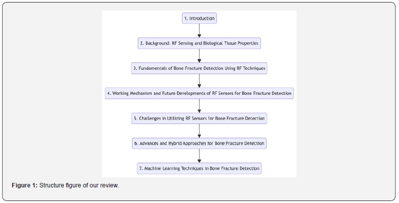
Considering the limitations of existing techniques and the potential benefits offered by RF techniques, machine learning, and microwave imaging, this review aims to explore and analyze recent advancements in bone fracture detection. We will evaluate these techniques’ effectiveness, limitations, and practical applications by examining the existing literature, providing valuable insights for researchers, clinicians, and healthcare professionals. The article also delves into the significance of time-domain analysis in hybrid approaches and evaluates simulated models used for fracture detection regarding their accuracy and reliability. Overall, this paper aims to summarize the latest developments and identify potential avenues for future research, including innovative sensor technologies, improved signal processing methodologies, integration with medical imaging modalities, and customized strategies for fracture identification (Figure 1).
- Mini Review
- Abstract
- Introduction
- Examples of Multicultural and Workforce Diversity Frameworks that Could be Utilized to Advance Contemporary Leadership Practices
- The Benefits of Incorporating Multicultural and Workforce Diversity Leadership Attributes and Diversity Efforts in Contempoary Leadership Practices
- Conclusion
- References
The Objectives and Scope of the Review
This study:
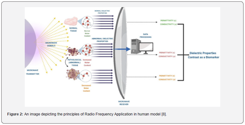
i. Examines various methods for detecting bone fractures using low-frequency and microwave radio frequency (RF) techniques. It covers the fundamental principles underlying RF techniques and an overview of the dielectric properties of biological tissues. Additionally, it compares the advantages and disadvantages of these methods with other procedures like ultrasonography, X-ray, and CT scan for detecting bone fractures, as shown in Table 1 & Figure 2.
ii. Investigates hybrid techniques integrating lowfrequency and microwave radio frequency (RF) methods. The focus is on the potential of time-domain analysis to improve fracture detection.
iii. This study reviews the literature highlighting the application of machine learning techniques in detecting bone fractures. They emphasize their incorporation within the RF methods and time-domain analysis framework.
iv. This research provides an overview of the precision and reliability of simulated models, as reported in the literature, for detecting bone fractures using the mentioned methodologies.”.
v. Discusses the latest developments and potential future directions in the field. These include advancements in sensor technology, signal processing techniques, integration with medical imaging modalities, and personalised fracture detection methods.
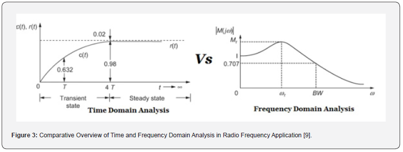
vi. It focuses exclusively on research and methodologies utilising time-domain low-frequency and microwave RF techniques to detect bone fractures. Consequently, the primary focus of this review will be on something other than studies utilising frequency-domain analysis or other imaging modalities. However, they may be referenced for comparative purposes as illustrated in Figure 3.

Background and Motivation
Bone fractures, resulting from trauma, overuse, or underlying medical conditions that weaken the bones, are commonplace injuries that require timely and accurate detection for appropriate treatment and effective healing. Traditionally, the detection of bone fractures primarily relies on imaging techniques such as X-rays, CT scans, and MRI scans. X-rays are often the first line of diagnosis due to their wide availability, speed, and cost-effectiveness. However, they have notable limitations. For instance, X-rays may struggle to detect certain types of fractures, such as hairline or stress fractures, as these may not be discernible in the X-ray images [10]. CT scans, on the other hand, provide more detailed images of the bone structure and can detect fractures that might be invisible on an X-ray. Despite these advantages, CT scans have their own set of drawbacks. They expose the patient to a high radiation dose and are typically more expensive than X-rays, making them feasible for only some patients [11].
MRI scans are a viable alternative as they do not involve ionizing radiation and can provide intricate images of soft tissue and bone. This makes them particularly useful for detecting bone bruises or other injuries accompanying a fracture. However, MRI scans are generally more expensive than X-rays and CT scans, limiting their availability to only certain medical facilities [12,13]. Given these challenges, there is a clear impetus for developing more accessible, cost-effective, and less invasive methods for bone fracture detection. Integrating technologies such as RF sensing into clinical practice offers a promising solution, potentially enhancing the accuracy of diagnosis, improving patient experience, and contributing to more effective treatment strategies.
Bone Fracture Detection Techniques
In recent years, technological advancements have introduced non-invasive approaches for monitoring the process of bone fracture healing as the novel methodology to replace the conventional techniques in use. Traditional methods such as radiography, ultrasound, computed tomography (CT), magnetic resonance imaging (MRI), and positron emission tomography (PET) have been widely used for the diagnosis of bone fractures [14]. However, the merits and demerits associated with each technique can be identified through the comparative evaluation of the techniques, providing insight into the choice of method for specified jobs
Limitations of Traditional Monitoring Methods (X-rays, CT scans, and MRI)
X-ray and computed tomography (CT) scans are conventional modalities for surveillant bone fractures. Although these techniques are extensively employed and can provide valuable insights, they have certain constraints. For example, according to [15], a constraint of X-ray imaging is its potentially reduced efficacy in areas where multiple structures overlap. In addition, it has been noted that certain anomalies affecting the left and right collar bone, heart, and lung may appear less visible on an X-ray of these areas compared to an X-ray of the forearm [16]. Computed tomography (CT) scans are a valuable diagnostic tool for detecting bone fractures; however, they possess certain limitations. One of these limitations is that no prescribed threshold exists for the quantity of CT scans a patient can undergo [17]. Furthermore, Factors such as the dimensions of the X-ray detector and the sample size can influence the resolution of 3D scans in computed tomography [17].
The examples above are just a few of the limitations inherent in the constraints of conventional techniques employed to monitor bone fractures. The existence of additional limitations is contingent upon the circumstances of each case. Other constraints of traditional monitoring techniques, such as X-rays and CT scans, encompass the potential for radiation exposure and financial expenses [13]. Research has found that exposure to ionizing radiation from X-rays and CT scans can increase the risk of cancer development in patients [18]. Although the potential harm from a solitary X-ray or CT scan is minimal, the likelihood of adverse effects may escalate with repeated exposure [19]. An additional constraint associated with these methodologies pertains to their financial implications. Computed tomography (CT) scans can incur significant costs and may not be universally covered by insurance policies [20]. The accessibility of these tests may pose a challenge for certain patients requiring them.
The Need for a Non-Invasive, Cost-Effective, and Accessible Monitoring Method
There is a pressing need for imaging methods that are non-invasive, cost-effective, and widely accessible within the healthcare industry. Current mainstream medical imaging techniques, such as X-rays, CT scans, and MRI scans, have several drawbacks that present challenges to both healthcare providers and patients. These costly procedures may expose patients to ionizing radiation, which poses health risks. Furthermore, they may not be readily available in all healthcare settings, particularly remote or underserved areas, exacerbating healthcare disparities [13].
The advent of new technologies that can surmount these challenges holds immense potential for improving patient care. RF sensing technology, for instance, presents a non-invasive approach to fracture detection, causing no discomfort to the patient. It is potentially lower cost than traditional imaging modalities and could make it more accessible, alleviating the financial burden on patients and healthcare systems. Furthermore, with developments towards more portable or wearable RF sensors, this technology could be used in a wide range of healthcare settings - from large hospitals to remote clinics and even in home-based care. This widespread accessibility could democratize health monitoring, providing valuable diagnostic information irrespective of location or socioeconomic status. The integration of such noninvasive, cost-effective, and universally accessible technology into healthcare workflows has the potential to significantly enhance the quality of patient care, facilitate early and accurate diagnosis, and reduce strain on healthcare providers and systems.
RF Sensing as an Alternative Method
A novel method for detecting fractures involves using Radio Frequency (RF) sensors. These sensors can accurately detect and measure changes in electromagnetic fields, making them suitable for various applications, including monitoring bone fracture healing. RF sensors can effectively identify bone fractures by employing an RF antenna, showing great potential for future use. Research indicates that fracture recognition can be achieved by analyzing using reactive impedance surfaces [21]. This suggests that RF sensors could circumvent the constraints of traditional imaging techniques, offering a non-invasive and potentially more precise mode for fracture detection. However, applying RF sensing to fracture identification and monitoring is an emerging field with several challenges. These include sensor design and placement issues, signal processing and analysis, and ensuring desirable sensitivity and specificity levels within the complex human anatomy context
RF sensors work by discerning alterations in the dielectric properties of biological tissues caused by radio frequency waves. Factors such as bone fractures can induce these changes. When a fracture occurs, dielectric property alterations in the surrounding tissues mainly result from fluid accumulation, like blood. These changes affect the material’s permittivity and conductivity, subsequently altering the behaviour of the radio frequency waves traversing the tissue [22]. RF sensors are engineered to sense these propagation changes, yielding valuable data about the physical alterations within the tissue.
This data can provide detailed insights into a bone fracture’s existence, size, and severity. Notably, the specific operating mechanisms of RF sensors can vary based on their design and application. Some may focus on changes in the transmitted signal, whereas others might rely on reflected or scattered signals for detection. By employing these mechanisms, RF sensors can offer a non-invasive, potentially real-time tool for detecting and monitoring bone fractures. This opens significant possibilities for improving patient care in orthopaedic and trauma scenarios
How RF Sensors Overcome Traditional Limitations
RF sensors employ ultra-wideband (UWB) technology and Radio Frequency Identification (RFID) methodologies to overcome the limitations of traditional monitoring methods. UWB technology, operating over short distances, enables precise indoor positioning and real-time device movement and motion tracking. It outperforms conventional methods with fewer than 50 centimeters of accuracy under optimal conditions [23]. Similarly, RFID sensor tags are crucial for the future of Internet-of-Things (IoT) applications. They are contactless, wireless, lightweight, capable of non-line-of-needs to be clarified mission, and flexible [24]. From a design perspective, RF sensors are relatively diminutive in size. They do not require supplementary components like magnetic circuits, coils, or magnets and can fit onto a small silicon wafer [25]. They show their strongest response at specific optical frequencies, but broadband sensors can measure a wide spectrum of frequencies [24].
Applying RF sensors in medical settings, particularly in detecting bone fractures, has emerged as a viable and promising approach. For example, Utilizing Electrical Impedance Spectroscopy (EIS) in the radio frequency range in medical settings, especially for detecting and monitoring bone fractures, has proven to be a promising and effective method. While traditional imaging techniques like X-rays, CT scans, DEXA scans, and ultrasounds are available, EIS offers a non-intrusive, portable, and potentially more accurate alternative for identifying bone fractures. Moreover, EIS has demonstrated its capability to monitor the healing process of fractures, providing more precise and sensitive data compared to conventional methods [26]. These conventional methods have limitations. For instance, X-rays are only effective at later stages of repair and correlate poorly with bone strength. Despite their improved diagnostic capabilities, CT and DEXA scans have limited clinical use due to their cost and high radiation dose [27]. Meanwhile, physical examinations by physicians, though commonly relied upon, can result in imprecise assessments
In contrast, MRI provides superior soft tissue contrast without radiation and is frequently used for imaging various organ systems, including the musculoskeletal system [27]. While MRI can deliver valuable information for diagnostics, 3D modelling, and treatment planning across multiple anatomical regions [27], RF sensors can objectively measure fracture healing. This can help guide clinical decision-making for patients, although their use is more specific and less versatile than MRI [28]. RF sensors offer significant advantages in bone fracture detection compared to alternative imaging techniques, including X-rays, MRI, and CT scans, as summarized in Table 1. This underscores the potential of RF sensing technology in reshaping healthcare diagnostics and patient treatment approaches (Table 2).

- Mini Review
- Abstract
- Introduction
- Examples of Multicultural and Workforce Diversity Frameworks that Could be Utilized to Advance Contemporary Leadership Practices
- The Benefits of Incorporating Multicultural and Workforce Diversity Leadership Attributes and Diversity Efforts in Contempoary Leadership Practices
- Conclusion
- References
Background: RF Sensing and Biological Tissue Properties
Fundamentals and Medical Applications of RF Sensing
Radio Frequency (RF) sensing leverages the radio frequency spectrum to detect and analyze environmental changes. This innovative technology intersects various domains, including medicine and healthcare, where it offers unparalleled advantages. Here, we provide a comprehensive guide to the rudiments of RF sensing and illuminate its pivotal role in the medical field.
RF Sensing Basics: At its core, RF sensing relies on the principle that radio frequency signals interact dynamically with their surroundings. By meticulously examining reflected or modulated RF signals, environmental changes are detected. Consequently, these variations offer invaluable insights into the subjects’ or objects’ characteristics and conditions proximal to the RF sensor. A strict frequency range doesn’t bind RF sensing. It can be deployed over a spectrum ranging from low frequencies (kHz) to extremely high frequencies (GHz). The choice of operating frequency is influenced by the specific application and the target’s characteristics being sensed. A distinguishing feature of RF sensing is its non-contact nature, allowing for noninvasive measurements. This attribute is notably attractive in the medical field, where contact-based sensing may need to be more practical and comfortable for patients [29]. RF signals can permeate various materials, including clothing and body tissues, obtaining information through obstacles. The signals reflected offer a wealth of information about the sensed object’s structure and composition. Complex signal processing algorithms and data analysis techniques underpin RF sensing. These approaches distil meaningful information from the captured RF signals. Integrating machine learning and artificial intelligence techniques significantly enhances the sensitivity and accuracy of the sensing system.
Medical Implications of RF Sensing: One of the promising applications of RF sensing in the medical field is non-invasive wound monitoring. RF sensing can analyze RF signals reflected from the wound area, detecting changes in the healing processes, such as inflammation and oedema. This aids in prompt intervention and assessment [30]. Vital sign monitoring has also been a benefactor in RF sensing. Noncontact RF sensors can monitor heart, respiration, and blood pressure. Patients can be continuously monitored without physical contact by measuring minute changes in the reflected signals due to bodily movements [30]. In sleep medicine, RF sensing has shown potential in sleep pattern monitoring and sleep apnea detection. Technology can identify changes in breathing patterns and movements by analyzing RF signals reflected from a sleeping individual, thus assisting in diagnosing and treating sleep-related disorders [31].
RF sensing is harnessed for surgeon’s gesture recognition in the operating theatre. Detecting hand movements and gestures, RF sensors can manage medical equipment like robotic surgical instruments, obviating the need for direct touch or physical contact [32]. Temperature monitoring, particularly skin temperature, can be conducted non-invasively using RF sensing. Any changes in skin temperature are reflected in the variations in the RF signals, providing real-time updates on the patient’s health [33]. Additionally, RF sensing has shown promise in detecting and monitoring bone fractures. By analyzing the characteristics of the RF signals reflected from the bone, fractures can be identified, and the healing process can be monitored over time. This provides clinicians with a contactless and non-invasive method to track recovery, leading to personalized treatment plans and improved patient outcomes [34].
Dielectric Properties and RF Signal Behaviour in Biological Tissues
The dielectric properties of a material can either reflect or penetrate RF signals and significantly affect the electromagnetic characteristics of a substance. This phenomenon is known as dielectric dispersion, a well-studied occurrence that significantly alters the dielectric properties within a specific frequency range [35]. These properties significantly impact the behaviour of RF signals within biological systems, necessitating knowledge of the frequency-dependent dielectric properties of biological tissues [36]. Understanding these properties is crucial as human tissues have varying dielectric characteristics at different frequencies. Understanding this interaction between electromagnetic fields and biological tissues is very important, particularly in medical imaging and telecommunications. Several variables, such as frequency, temperature, density, water content, salt content, and the physical state of a substance, can influence the dielectric characteristics of a substance [36].
Dielectric dispersion in biological tissues can impact RF signals’ attenuation and phase velocity as they travel through the medium. The dielectric characteristics of biological tissues, such as their relative permittivity (ε ′) and conductivity (σ), play a crucial role in determining their ability to polarize in response to an external electric field and their capacity to conduct electrical current [37]. The dielectric characteristics of biological tissues, namely relative permittivity (ε′) and conductivity (σ), play a crucial role in influencing the behaviour of radio frequency (RF) signals during their interaction with these tissues [38]. They are essential in determining how easily a material can become polarised by an applied electric field and how well it can conduct an electric current.
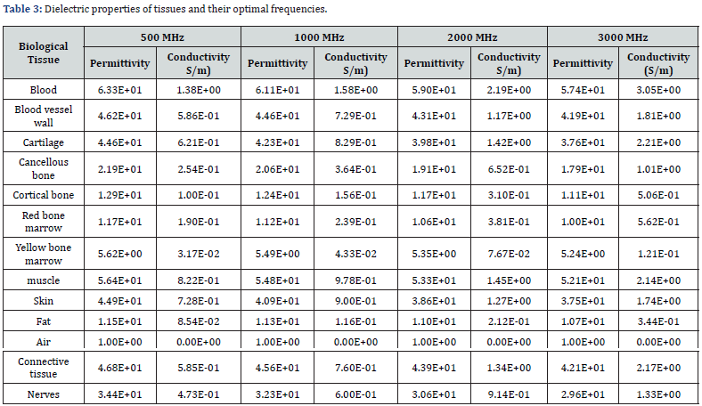
Moreover, these properties significantly impact medical imaging modalities, such as Magnetic Resonance Imaging (MRI), utilizing RF signals to interact with biological tissues. An accurate understanding of tissue dielectric properties is crucial for improving specific absorption rate (SAR) estimates and reducing undesired tissue heating [39]. It is also critical to the design of new electromagnetic-based imaging and therapeutic technologies. Detecting changes in bone tissue composition is significantly assisted by determining these essential dielectric properties: relative permittivity (ε′) and conductivity (σ). Techniques like electrical impedance spectroscopy can measure these properties related to bone tissue, such as conductivity and structure. The properties can vary substantially across various biological tissues due to dielectric dispersion, and this variance can be quite substantial over a frequency range [40]. A significant database maintained by the IT’IS Foundation provides dielectric properties of biological tissues, including values for permittivity and electrical conductivity for over 100 human tissues at frequencies of 10 Hz to 100 GHz [41]. These properties are invaluable in biological and medical applications, notably in investigations involving tissue imaging, therapeutic interventions, and electromagnetic field interactions. The distinctive features of their respective permittivity (ε) and conductivity (σ) determine the response of biological tissues when exposed to an electric field. However, these tissue properties vary according to the operating frequency [42]. Table 2 illustrates the dielectric properties of the possible tissues encountered when studying a fracture in the human thigh based on the Gabriel dispersion relationship (Table 3).
- Mini Review
- Abstract
- Introduction
- Examples of Multicultural and Workforce Diversity Frameworks that Could be Utilized to Advance Contemporary Leadership Practices
- The Benefits of Incorporating Multicultural and Workforce Diversity Leadership Attributes and Diversity Efforts in Contempoary Leadership Practices
- Conclusion
- References
Fundamentals of Bone Fracture Detection Using RF Sensor Techniques
Low-Frequency RF Techniques
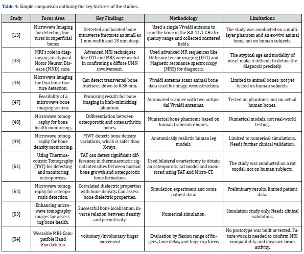
Within RF technology, frequencies below 300 MHz are categorized as low-frequency spectrum. This spectrum is utilized in various applications, including medical equipment and communication systems. Study [43] employed modern magnetic resonance imaging (MRI) techniques such as Diffusion Tensor Imaging (DTI) and Magnetic Resonance Spectroscopy (MRS) to diagnose motor neuron disease (MND). These MRI techniques are analogous to RF technology, utilizing distinct frequencies and signal generators for precise medical diagnosis. RF signals from communication systems are processed to extract meaningful data, utilizing techniques such as modulation and demodulation. For instance, received RF signals in communication systems undergo IQ-demodulation to extract the actual information, which is then processed using MATLAB Signal Processing Toolbox functions [44].
In most cases, antenna designs like loop antennas, dipole antennas, and monopole antennas are generally preferred for converting electrical signals to magnetic waves in a lowfrequency range. These antennas can receive low-frequency RF signals and process them through filtering, demodulation, and decoding techniques based on application requirements. It’s worth noting that the safety of these low-frequency techniques has been thoroughly investigated, as highlighted by a Study [45] which examines human exposure to electromagnetic fields across various frequencies.
Microwave RF Techniques
Microwave RF techniques, operating between 300 MHz to 300 GHz, are increasingly being applied in healthcare, particularly in detecting bone fractures. Studies [46-48] have shown that Microwave Imaging (MWI) offers distinct advantages such as non-ionization, portability, and cost-effectiveness, making it an attractive alternative to traditional X-ray technologies. Study [49] uses Microwave Tomography (MWT) to monitor bone density, highlighting its potential for diagnosing osteoporosis and tracking the disease’s progression. Another noteworthy contribution to the field comes from a preliminary numerical analysis, as detailed in the study [50]. This research explores the potential of Microwave Tomography in monitoring bone density, thereby opening new avenues for diagnosing conditions like osteoporosis. The study employs numerical simulations to validate the efficacy of this technology, although it is preliminary and further clinical validation is needed.
The research paper mentioned in Study [51] highlights significant progress in Microwave RF Techniques, which focuses on the ‘Detection and Monitoring of Osteoporosis in a Rat Model by Thermoacoustic Tomography.’ This study introduces a novel approach to osteoporosis detection, which could be a significant leap in early diagnosis and disease monitoring. The methodology involves thermoacoustic tomography, a technique that could offer a more nuanced understanding of bone health. However, it’s worth noting that the study’s limitations include its focus on rat models, which may not directly translate to human applications. Despite these advancements, there are limitations to consider. For instance, study [52] points out that the dielectric properties of surrounding tissues like muscles, fats, and skin have not been adequately considered, making the clinical acceptance of these findings challenging. Study [53] introduces the concept of using prior information to enhance microwave tomography images, which could potentially address some of these limitations. Of interest in recent times is the use of Microwave Imaging (MWI) as a revolutionary method for detecting bone fractures. MWI offers distinct advantages, including nonionization, portability, and cost-effectiveness, which makes it an attractive alternative to traditional X-ray technologies. Study [13] further supports the feasibility of using MWI for bone fracture detection, thereby adding to the growing body of evidence favoring this technology.
In wearable technology, the development of MRI-compatible hand exoskeleton robots marks a significant advancement [54]. These wearable systems are designed to be compatible with MRI environments, allowing for real-time imaging during movement tasks. This innovation could revolutionize how clinicians approach diagnostics and treatment planning, particularly in orthopaedic and neurological conditions. Lastly, the advancements in MWI, as evidenced by studies [53, 54], signify a shift towards more noninvasive and cost-effective health diagnostics. However, further research and clinical trials are necessary to fully ascertain its efficacy and accuracy, especially in orthopaedic diagnostics (Table 4).
- Mini Review
- Abstract
- Introduction
- Examples of Multicultural and Workforce Diversity Frameworks that Could be Utilized to Advance Contemporary Leadership Practices
- The Benefits of Incorporating Multicultural and Workforce Diversity Leadership Attributes and Diversity Efforts in Contempoary Leadership Practices
- Conclusion
- References
Challenges in Utilizing RF Sensors for Bone Fracture Detection
Technical Challenges in RF Sensing for Bone Fracture Detection
Signal Interference and Attenuation: The effectiveness of Radio Frequency (RF) sensing in detecting bone fractures hinges on the accurate interpretation of signals. This process can be notably affected by environmental elements such as noise, interference, and signal attenuation. Furthermore, the dielectric properties of tissues surrounding the bone can exhibit variations that may influence the RF signal. Consequently, these variations could impact the precision of fracture detection measurements [28]. Therefore, it is paramount to understand and mitigate the effects of environmental interference and signal attenuation. For instance, addressing these challenges through developing robust signal processing and error correction algorithms is crucial for ensuring the successful application of RF sensing in detecting bone fractures. Only by achieving high signal fidelity and accuracy can we ensure that this technology reliably aids in the diagnostic process and ultimately enhances patient care.
Therefore, it is paramount to understand and mitigate the effects of environmental interference and signal attenuation. Addressing these challenges through the development of robust signal processing and error correction algorithms, for instance, is crucial for ensuring the successful application of RF sensing in detecting bone fractures. Only by achieving high signal fidelity and accuracy can we ensure that this technology reliably aids in the diagnostic process and ultimately enhances patient care
Sensor Calibration and Placement The precision of RF sensors and their appropriate placement are critical factors affecting the effective detection of bone fractures. Sensor calibration is a meticulous procedure involving the transfer of a calibration factor or efficiency from a standard to the sensor, ensuring accurate measurements of RF and microwave power. This process is vital to maintain the consistency and reliability of the data captured by the sensors. Likewise, the positioning of the RF sensor is of paramount importance. The sensor’s location with the bone fracture can significantly influence the amplitude and fidelity of the received signal. If appropriately positioned, the resulting data may lead to accuracy in fracture detection, impacting the effectiveness of diagnosis and subsequent treatment plans [55].
The Complexity of Interpretation: Interpreting the data derived from RF sensors for detecting bone fractures represents a multifaceted challenge. This task requires an indepth understanding of RF signal processing and the interaction of RF signals with various biological tissues. Additionally, the increasingly complex and crowded electromagnetic environment can further compound these challenges, adding layers of difficulty to data interpretation [56]. These complexities are particularly prominent when the data comes from intricate devices such as network analyzers, which may contain substantial noise and interference, making extracting high-quality, usable data a demanding task [57].
Advanced computational methodologies, such as deep learning, can be utilized to address these complexities. These techniques leverage the computational power of modern hardware and sophisticated algorithms to parse complex data, identify patterns, and extract actionable information. Deep learning methodologies have shown considerable promise in signal detection and classification tasks. They can enhance the precision of fracture detection by reducing false positives and negatives, even in the presence of complex and noisy data. Moreover, deep learning methodologies can help to transform raw, noisy data into valuable, actionable insights. These insights can significantly enhance the efficacy of RF sensing technology in detecting bone fractures, leading to more accurate diagnostics and more effective patient care [56]. Furthermore, as these methodologies evolve, they will offer even more refined tools for interpreting the data from RF sensors, promising future advancements in bone fracture detection and monitoring. This underlines the importance of ongoing research and development in RF sensing technology and the advanced computational techniques used to interpret these sensors’ data.
Data Quality: Ensuring data quality is one of the significant challenges when utilizing RF signals for bone fracture detection. The data obtained from RF sensors, mainly sourced from complex devices such as network analyzers, can be significantly impacted by various factors. These include environmental interference, signal noise, signal attenuation, and variability in signals associated with different types of bone fractures. These issues may complicate the data interpretation and extraction process of highquality, actionable information, ultimately affecting the reliability of the machine learning models used for fracture detection [58]. Addressing these issues necessitates the incorporation of advanced strategies for signal processing. Noise filtering techniques can be employed to reduce the impact of environmental interference and signal noise. Signal amplification can enhance the strength of relevant signals, improving the overall signal-to-noise ratio [58]. Data cleaning techniques can further ensure that only relevant, high-quality data is used for training machine learning models, thereby improving the models’ performance and reliability [59].
Furthermore, the large volumes of data generated by these RF sensors can pose challenges regarding storage, retrieval, and processing. This is particularly significant given that healthcare institutions are already grappling with large volumes of data. As such, it is necessary to consider practical and efficient data management strategies, which may involve using cloud computing solutions or advanced data compression techniques [59]. Moreover, it is also crucial to develop robust quality assurance mechanisms to monitor and validate the data’s integrity continuously. This can involve employing outlier detection techniques to identify and address anomalies in the data and implementing rigorous testing protocols to validate the accuracy and consistency of the data. In combination, these strategies can significantly improve the quality of data derived from RF sensors, leading to more reliable and accurate machine-learning models for bone fracture detection. This underlines the importance of investing in advanced signal processing and data management techniques when integrating RF sensing technology into clinical workflows for fracture detection and monitoring [60].
Variability in Biological Response: Individual diversity in biological reactions is an additional barrier for RF sensors. There is variability in the reaction of human bones to RF radiation. Various factors, including age, sex, health state, and genetic composition, might substantially affect these reactions, posing challenges in achieving standardization of sensor readings and interpretations. It is imperative for researchers to devise advanced machinelearning models capable of accommodating these variations while maintaining accurate predictions of bone fractures. To ensure the comprehensive training of these models, gathering a diversified array of data and a wide variety of responses is imperative [60].
Technology Limitations: While RF sensors offer groundbreaking advancements in bone fracture detection, their limitations can affect clinical utility. For instance, these sensors may encounter difficulties in accurately diagnosing specific types of fractures, such as hairline fractures, especially those situated in anatomically complex regions. These challenges are not unique to RF sensors but are also faced by other diagnostic methods like DXA, quantitative computed tomography, and quantitative ultrasound. Even MRI, which is cited as the most sensitive and specific imaging test for diagnosing stress fractures, has its limitations, as mentioned in [61,62]. This highlights the ongoing need for improved diagnostic tools. Furthermore, the performance of RF sensors is not uniform across all patients and can be impacted by many patient-specific factors. These include age, which can affect bone density and tissue properties; bone density itself, which can influence how RF signals interact with the bone; and underlying medical conditions, which can alter the physical properties of bones and surrounding tissues, thereby affecting the RF signal characteristics. The patient’s body composition and the presence of implants or other medical devices could also affect the RF signals, adding another layer of complexity to the data interpretation and potentially affecting the accuracy of fracture detection [63].
These limitations necessitate careful consideration when planning and implementing the clinical application of RF sensing technology. It’s essential to have a comprehensive understanding of these limitations to inform clinicians, guide patient selection, and manage expectations regarding the performance of this technology. Ongoing research and development efforts are critical to deepen our understanding of these limitations and devise strategies to overcome them. Enhancements in signal processing techniques, data interpretation algorithms, and machine learning models could mitigate some of these limitations [8]. Optimizing the performance of RF sensors will require a multifaceted approach, considering both technological advancements and the unique characteristics of individual patients. Through continued research and innovation, we can strive to enhance the accuracy, reliability, and inclusivity of RF-based fracture detection, ultimately contributing to improved patient outcomes and advancing the field of bone health diagnostics
Broader Implementation Challenges
Integration with Clinical Workflow: Incorporating RF sensing technology into the existing clinical workflow presents numerous challenges that demand thorough planning and management. One of the primary considerations is ensuring that healthcare providers are proficiently trained in using the technology and interpreting its results. This education and training aspect is critical to maximize the potential benefits of the technology and reduce the possibility of user-related errors.
In addition, modifications to current clinical procedures may be necessary to ensure a smooth integration process. These changes might involve altering certain routines, processes, or systems to accommodate the new technology. Optimal utilization of RF sensors necessitates a workflow design that aligns with the technology’s functional requirements while maintaining or enhancing the efficiency of clinical operations.
Interdisciplinary collaboration plays a crucial role in addressing these challenges. The combined efforts of healthcare providers, technologists, and administrators can guide the successful implementation process, addressing any technological, operational, or organizational barriers that may arise [64]. The goal is to integrate RF sensing technology seamlessly into the clinical workflow, providing value in enhanced bone fracture detection and monitoring while causing minimal disruption to existing practices. This balance is critical to ensuring that technology becomes a valuable and effective tool in delivering healthcare services.
Validation and Regulatory Approval: One of the significant challenges in the broader integration of RF sensing technology into clinical settings is the uncompromising approach required towards testing, validation, and regulatory approval. Obtaining approval can be long and arduous, involving extensive testing and rigorous evaluation to demonstrate the device’s safety, efficacy, and reliability, mainly when used for detecting bone fractures. This stringent review occurs under controlled and varied conditions, ensuring the technology functions accurately and consistently as intended [45].
Moreover, this evaluation extends to the potential risks associated with the technology’s usage, and measures are taken to identify and mitigate them [64]. As a medical device, an RF sensor must comply with specific safety and performance standards before gaining approval for clinical use. This laborious process, while ensuring the technology’s integrity and quality, can pose a significant barrier to the rapid adoption of this technology in the healthcare system. [64] Once the regulatory approval is achieved, it signifies that the technology has met the specific predefined standards and guidelines set by recognised health authorities. This approval is not just a testament to the integrity and quality of the technology but also ensures that the RF sensing technology is safe for patient use, performs effectively, and adheres to ethical standards such as privacy, confidentiality, and equal access. Despite the challenges, adherence to these comprehensive procedures is paramount. They are essential to promote trust among healthcare professionals and patients and ensure that the technology contributes positively to patient outcomes and the healthcare system. Therefore, while the path to regulatory approval can indeed be challenging and time-consuming, it is a necessary hurdle to ensure the responsible deployment of RF sensing technology in clinical practice.
Privacy and Data Security: The widespread implementation of RF sensing technology in clinical settings necessitates rigorous testing, validation, and regulatory approval. An extensive evaluation process is imperative to affirm its safety, effectiveness, and reliability, specifically in detecting bone fractures. Ensuring the technology’s compliance with regulatory standards and guidelines is a fundamental requirement that serves to maintain the technology’s integrity and quality
In addition to these crucial aspects, addressing privacy and security concerns is paramount. Applying RF sensing technology in clinical settings must be performed with stringent data protection measures to safeguard patients’ personal and medical information. Secure data transmission and storage protocols should be in place to prevent unauthorized access and potential breaches [65]. Moreover, privacy regulations, such as the Health Insurance Portability and Accountability Act (HIPAA) in the United States or the General Data Protection Regulation (GDPR) in the European Union, should guide the implementation and use of this technology. Adherence to these regulations will help ensure patient data is handled responsibly and confidentially. These comprehensive measures, from technical performance to data security, foster trust in this technology among healthcare professionals and patients. This confidence is vital for RF sensing technology’s safe, efficient, and ethical application in a clinical environment.
Ethical Considerations: Integrating RF sensing technology in healthcare necessitates a comprehensive evaluation of ethical implications, including equal access and potential unintended consequences. It is paramount to ensure the equitable distribution of this technology’s benefits, addressing potential disparities in access across diverse populations. Such an approach is vital for maintaining social justice in healthcare, where the advantages of innovative technologies should not be limited to specific socioeconomic or geographic segments. Beyond ensuring access, it’s critical to identify and mitigate potential unintended consequences of this technology’s deployment. For instance, overreliance on technology could inadvertently lead to diminished human interaction, altering the patient-care provider dynamic. Similarly, potential biases inherent in the decision-making algorithms of these systems could lead to disparities in diagnosis or treatment outcomes [66].
Establishing ethical frameworks and guidelines to govern the application of RF sensing technology in fracture detection is of utmost importance. These guidelines should encompass informed consent, privacy, data security, accountability, and transparency. By upholding these standards, the responsible and ethical use of RF sensing technology can be ensured, fostering trust among healthcare professionals and patients and facilitating its successful integration into clinical practice.
Financial Considerations: Another significant challenge involves the financial aspects related to implementing RF sensing technology in clinical settings. The cost of acquiring and maintaining these systems may be a barrier for many healthcare providers, particularly in low and middle-income regions. Also, there are costs associated with training healthcare providers on using this technology and interpreting its results. Therefore, strategies for cost reduction and efficient resource allocation need to be considered, such as developing more cost-effective RF sensors and providing online training programs for healthcare providers [67].
Future Prospects and Strategies for Overcoming Challenges
Despite the mentioned challenges, the prospects of RF sensing technology for bone fracture detection are immense. With continuous technological advancements and a growing understanding of the human bone anatomy, RF sensors are expected to significantly revolutionize fracture detection [68]. Addressing technical challenges will largely depend on technological advancements and research breakthroughs. For instance, developing more sophisticated signal processing algorithms can help to minimize the impact of signal interference and attenuation, improving the accuracy and reliability of RF sensors. Also, integrating machine learning and artificial intelligence techniques can enhance the data interpretation process, enabling more precise fracture detection, even amidst complex and noisy data [69]. The implementation challenges can be addressed through careful planning and management. One key aspect will be educating healthcare providers about the technology and providing them with the necessary training. Ensuring compatibility with existing healthcare systems and practices can also ease the integration of RF sensors into the clinical workflow. Moreover, developing cost-effective RF sensors can make the technology more accessible and alleviate financial constraints [70].
In terms of regulatory approval, manufacturers of RF sensors will need to work closely with regulatory bodies to ensure that the technology meets the required standards. This process should begin at the early stages of product development to streamline the approval process and ensure regulatory compliance. Applying robust data protection measures can address privacy and data security challenges. These may include secure data transmission protocols, encrypted data storage, and training of healthcare providers on data protection policies and practices. Furthermore, ensuring compliance with privacy regulations will protect patient data and maintain trust [70,71].
Addressing these challenges will be challenging and will require a concerted effort from all stakeholders, including researchers, healthcare providers, technology developers, and regulatory bodies. However, the potential benefits of RF sensing technology in detecting bone fractures make it a worthwhile endeavor. By overcoming these challenges, we can pave the way for this technology to become an integral part of clinical practice, significantly improving patient outcomes and advancing the field of bone health diagnostics [71].
- Mini Review
- Abstract
- Introduction
- Examples of Multicultural and Workforce Diversity Frameworks that Could be Utilized to Advance Contemporary Leadership Practices
- The Benefits of Incorporating Multicultural and Workforce Diversity Leadership Attributes and Diversity Efforts in Contempoary Leadership Practices
- Conclusion
- References
Advancement in Innovative Approaches for Bone Fracture Detection
Hybrid Techniques
Complementary information may be obtained when combining different imaging modalities to detect bone fractures. The combination of low-frequency and microwave methods is one such hybrid strategy. Electrical impedance tomography (EIT) and electrical conductivity measurements are low-frequency methods that offer information about the electrical characteristics of tissues. Because of bone structure and mineralization changes, fractured bones have different electrical characteristics than healthy bones. It is feasible to diagnose fractures and analyze the healing process by measuring the electrical conductivity of the bone. Microwave methods, on the other hand, such as microwave radiometry or microwave tomography, use electromagnetic waves in the microwave frequency range. These methods may offer information regarding tissue dielectric characteristics, such as changes in water content, which can be symptomatic of bone fractures [72].
A comprehensive assessment of bone fractures can be achieved by integrating low-frequency and microwave techniques. While low-frequency measurements can shed light on structural alterations in the bone, microwave measurements offer insights into water content and other dielectric properties. This is similar to how the contrast in dielectric properties detected by microwave imaging in breast cancer is largely due to variations in water content between normal and malignant tissues [73]. Combining these strategies can potentially increase the accuracy and dependability of fracture diagnosis and monitoring [74]. A hybrid technique proposed by [75] utilized image processing and machine learningbased methods in classifying fracture detection using images from x-rays. The research demonstrated a commendable integration of machine learning algorithms and image processing techniques for facilitating fracture detection based on the severity and type of the fracture. However, this technique has the potential for broader clinical applicability due to the reliance on existing X-ray data. Diagnosing medical images in real-life clinical scenarios remains challenging due to the subjective nature of interpreting these images. The lack of a comprehensive approach that integrates image processing and machine learning techniques further complicates the process, particularly in identifying fractures [75].
Another research was conducted by [76] on “A study on the sensitivity of microwave imaging for detecting small-width bone fractures”. The study’s value rests in its narrow emphasis on smallwidth fractures and quantitative evaluation of microwave imaging sensitivity. The study provides insights into the technique’s potential by methodically altering fracture widths and examining the generated photographs. However, the complexity of phantom models raises concerns about their ability to replicate genuine clinical circumstances. The lack of real-world validation using actual bone specimens is a restriction that may impact the study’s usefulness
Signal Processing and Feature Extraction
Signal processing and feature extraction techniques are vital in analyzing data received from various imaging modalities in hybrid methods for identifying bone fractures. These strategies assist in extracting useful information and features from the recorded signals, making fracture diagnosis and evaluation easier
In hybrid methods, signal processing techniques are used to pre-process the obtained data to eliminate noise, increase the signal quality, and improve the overall picture quality [75]. Following this process, the data used for the upcoming analysis will be clean and reliable.
Feature extraction methods derive helpful information after pre-processing the signals or pictures. These characteristics may be quantitative data or unique patterns that are indicative of bone fractures or the healing process [77]. In fracture identification, some of the characteristics often employed include fracture line direction, bone mineral density, callus development, and local alterations in the properties of the tissue.
In recent times, the use of machine learning techniques to analyze the retrieved characteristics and construct models for fracture diagnosis has gained the attention of researchers. It is believed that these models gain the ability to categorize incoming data as either healthy or broken based on the patterns and correlations that they learn from the training data. It is possible to accomplish precise and automated fracture diagnosis via signal processing and feature extraction methods in hybrid systems [78,79]. A study demonstrated that combining machine learning techniques with signal processing and feature extraction methods enabled the creation of accurate and automated fracture diagnosis models. These models could categorize incoming data as healthy or fractured based on learned patterns and correlations, facilitating precise fracture identification
Time-Domain Analysis in Hybrid Approaches
Analyzing signals in the time domain is called time-domain analysis. This analysis investigates changes in the signal’s amplitude and phase over time. In hybrid methods for detecting bone fractures, time-domain analysis is often used to extract useful information linked to the healing process compared to the frequency-domain technique. This is because, unlike frequency domain analysis, analysis in the time domain makes it feasible to trace the development of fracture healing by observing the temporal changes in the observed signals or pictures. For instance, in techniques based on ultrasound, the time-domain analysis may indicate the development of the fracture callus, which includes its genesis, growth, and remodeling. It is possible to evaluate the effectiveness of bone healing and its rate by examining the temporal patterns. The study demonstrated that time-domain analysis in hybrid imaging modalities allowed for the tracking and evaluation the temporal changes in fracture healing, providing valuable insights into the development of the fracture callus, its growth, and remodeling. This approach proved more effective than frequency domain analysis in observing dynamic changes during the healing process [80]. Time-domain analysis may also be used in other imaging modalities, such as magnetic resonance imaging (MRI) or computed tomography (CT), to analyze dynamic changes in tissue characteristics, vascularization, or inflammation surrounding the fracture site. These temporal differences provide helpful insights into the wound-healing process and have the potential to contribute to the diagnosis and ongoing monitoring of bone fractures [81].
In conclusion, hybrid methods for detecting bone fractures often require the utilization of various imaging modalities, signal processing, feature extraction strategies, and time-domain analysis. These techniques use the beneficial aspects of each method to thoroughly analyze fractures, improve diagnostic accuracy, and more effectively monitor the healing process. When combined with frequency-domain methods through Fourier transformation, time-domain analysis offers a more comprehensive evaluation of wave propagation and scattering phenomena. This hybrid approach allows for high accuracy and efficiency in solving timedependent problems, potentially offering insights into various applications, including medical imaging techniques for assessing bone fractures and healing rates [82].
UHF Antenna-Based Detection Techniques
A recent development in the field employs ultra-high frequency (UHF) antennas and s-parameters for the non-invasive detection of bone fractures [83]. The concept involves using the UHF antenna to scan a bone phantom modelled after a human femur with a fracture. The difference in reflected and transmitted signals is then used to identify the presence and location of the fracture. This innovative approach offers low-power reflection and high-power transmission coefficients, signaling the presence of a fracture. In simulations, this technique has demonstrated the ability to detect fractures at varying locations on the bone phantom, with the s-parameters displaying differences according to the fracture’s position. This could indicate the technique’s potential for detecting fractures in different parts of the bone, thus enhancing the method’s versatility. However, using this technique in real-world scenarios requires further investigation, as the research has only encompassed simulations.
Dual-Polarized Microwave Sensor for Fracture Detection
A recent human bone fracture detection development involves a novel, compact sensor system that employs a patch antenna, reactive impedance surfaces, and a dielectric plano-concave lens [21]. This innovative system, about 30% smaller than traditional designs, is tailored to the human body to enhance impedance matching and optimize sensor performance. The methodology employed for this system includes designing and optimizing sensor components using CST Microwave Studio software, fabrication via PCB technology and 3D printing, and performance validation on a semi-solid phantom. The sensor operates at a centre frequency of 2.45 GHz with a bandwidth of 12.5%, and this sensor system demonstrates impressive sensitivity and accuracy in detecting and imaging narrow cracks in bone tissue and determining their location and orientation. The system’s effectiveness surpasses traditional methods such as X-ray or ultrasound.
However, despite its innovative design and impressive capabilities, the system’s implementation in real-world scenarios is yet to be explored. Future work can focus on enhancing the accuracy and robustness of the sensor system, testing on more realistic phantoms or animal models, and exploring applications in other biomedical fields, such as tumour detection or osteoporosis screening.
- Mini Review
- Abstract
- Introduction
- Examples of Multicultural and Workforce Diversity Frameworks that Could be Utilized to Advance Contemporary Leadership Practices
- The Benefits of Incorporating Multicultural and Workforce Diversity Leadership Attributes and Diversity Efforts in Contempoary Leadership Practices
- Conclusion
- References
Application of Artificial Intelligence in Bone Fracture Detection
The challenge of accurately diagnosing fractures in emergency departments is significant, with missed fractures being the most common diagnostic error. This failure can result in delayed treatment and long-term disability [84]. This task becomes further complicated due to the variability in fracture appearances based on factors such as the affected bone, regional anatomy, and radiographic projection. However, the evolution of machine learning and deep learning techniques, particularly CNN, has heralded a new era in fracture detection, significantly improving speed, efficiency, and error rates in diagnostics [85].
Machine Learning in Bone Fracture Detection
Machine learning is making considerable progress in healthcare, particularly in the early detection of bone fractures. Bone fractures can lead to significant morbidity if not diagnosed and treated promptly. Sophisticated machine learning algorithms, such as Support Vector Machines (SVM) and Neural Networks, can analyze imaging data from CT scans or X-rays to recognise patterns and signs indicative of fractures [85, 86]. Machine learning also synergizes with advances in radio frequency (RF) technology, yielding non-invasive methods for fracture detection. These methods leverage the unique changes in RF signals that occur due to alterations in bone structure, opening a new avenue for early fracture detection
A study has demonstrated that deep learning is a dependable method for diagnosing fractures, with a high diagnostic accuracy comparable to that of general physicians [5]. The study aimed to investigate the accuracy and reliability of deep learning in detecting orthopaedic fractures. The pooled sensitivity and specificity for the whole group (17 trials, 5,434 images) were 0.87 and 0.91, respectively. The AUC was 0.95. Eight trials (1,574 images) were included in the long-bone group, which contained seven studies. The pooled sensitivity was 0.96, and the specificity was 0.94. The AUC was 0.99. Another study [87] proposed a novel machine learning-based technique to detect bone fractures by analyzing bone contours. Several machine-learning algorithms, including Naïve Bayes, Decision Tree, Nearest Neighbors, Random Forest, and SVM, were employed on a dataset of 270 x-ray images. The accuracy measures varied, with SVM exhibiting the highest accuracy. Accuracy measures for the various algorithms used in the study range from 0.64 to 0.92, with values obtained for Naïve Bayes, Decision Tree, Nearest Neighbors, Random Forest, and SVM. Statistically, the accuracy for SVM was the highest in this research
Another study finds that bone contours can detect fractures when removing discontinuity processed by segmentation [88]. This method offers a nuanced approach that addresses the limitations of a previous study [89], which solely relied on texture analysis. Specifically, the study [88] employs various algorithms, including backpropagation and Scale-Invariant Feature Transform (SIFT), achieving a high accuracy rate of 90%. This suggests that the method not only overcomes the shortcomings of texture-only analysis but also opens avenues for further research in machine learning and digital geometry to optimize diagnostic methods. The earlier study [89] focused on the effectiveness of bone texture analysis (TA) and machine learning (ML) in identifying patients at risk for vertebrae fractures from CT scans. While this method was significantly more accurate than traditional methods in identifying at-risk patients, it struggled to pinpoint individual at-risk vertebrae accurately. Human assessment could have been more accurate overall. The study concludes that bone texture analysis combined with machine learning allows for high accuracy in identifying patients at risk for vertebral body insufficiency fractures on standard CT scans, thereby improving fracture risk prediction compared to mere Hounsfield unit measurements on CT scans. This analysis can identify vertebrae at risk for insufficiency fracture and may increase the diagnostic value of standard CT scans.
Deep Learning and Neural Networks
While earlier deep-learning systems for fracture detection were limited, focusing on single bones or specific anatomical regions, recent advancements have considerably broadened this scope, making them powerful tools for addressing fracture detection in medical imaging. Artificial Intelligence (AI) comprises a range of technologies, including Deep Learning, which uses artificial neuron layers to improve machines’ ability to understand and interpret complex data.
In recent advancements in orthopedics and traumatology, Deep Neural Networks (DNNs) have emerged as a transformative technology for automating the diagnosis of bone fractures. Specifically, a DNN model has been developed to analyze X-ray images accurately [90]. Traditional manual diagnosis methods, although indispensable, are time-consuming and susceptible to human error. To address these limitations, the proposed DNN model is an automated diagnostic tool capable of distinguishing between healthy and fractured bones. One of the challenges encountered during the model’s development was the issue of overfitting due to the small size of the available dataset. Data augmentation techniques were employed to mitigate this, effectively enlarging the dataset for more robust training. The model’s performance was rigorously evaluated using 5-fold cross-validation, achieving an impressive accuracy rate of 92.44%. Moreover, when subjected to further testing on subsets comprising 10% and 20% of the test data, the model demonstrated even higher accuracy rates of over 95% and 93%, respectively. These results validate the model’s efficacy and signify a substantial improvement over existing methods. However, it is worth noting that additional research is required to adapt these innovative techniques for detecting and classifying fractures in computed tomography (CT) scans.
Another study [84] evaluated whether a Food and Drug Administration-cleared deep learning system that identifies fractures in adult musculoskeletal radiographs would improve diagnostic accuracy for fracture detection across different types of clinicians. The study found that clinicians were more accurate at diagnosing fractures when aided by the deep learning system, particularly those clinicians with limited training in musculoskeletal image interpretation. Reducing the number of missed fractures may allow for improved patient care and increased patient mobility. The study shows that a deep-learning system can accurately identify fractures throughout the adult musculoskeletal system. The overall AUC of the deep-learning system was 0.974 (95% CI: 0.971–0.977), sensitivity was 95.2% (95% CI: 94.2–96.0%), specificity was 81.3% (95% CI: 80.7– 81.9%), positive predictive value (PPV) was 47.4% (95% CI: 46.0– 48.9%), and negative predictive value (NPV) of 99.0% (95% CI: 98.8–99.1%).
The integration of deep learning techniques has significantly propelled recent advancements in the field of medical imaging. One groundbreaking application lies in the realm of orthopedics, particularly in the automated detection of thighbone fractures. A novel study employs a two-stage, region-based convolutional neural network to accurately identify thighbone fractures in X-ray images. Utilizing a dataset comprising 3842 thighbone X-ray radiographs, the study achieved an Average Precision of 88.9%, thereby outperforming other state-of-the-art methods. This research validates the efficacy of deep learning in medical diagnostics and takes us one step closer to its practical application in healthcare settings [91]. This approach enhances diagnostic accuracy and addresses the time-consuming nature of traditional manual methods [91]. Moreover, the use of deep learning extends beyond orthopedics into other healthcare domains, such as neuroscience for Alzheimer’s disease classification [92] and bioinformatics for pharmacogenomics [93].
However, the implementation of these automated systems is not without challenges. One significant hurdle is the issue of observer bias in creating labelled datasets. To mitigate this, some studies propose using a ‘gold standard’ dataset incorporating input from multiple expert radiologists [94]. Another challenge is the limitation of small datasets, which can lead to model overfitting. To address this, data augmentation techniques and transfer learning have been suggested as viable solutions [91]. Furthermore, the rapid advancements in deep learning are not solely confined to algorithmic improvements. Hardware innovations, such as GPUs, have also played a crucial role in enabling effective CNN training [95]. As we move forward, cloud-based platforms are anticipated to become increasingly integral to developing computational Deep Learning applications [95]. In conclusion, integrating Deep Learning, particularly CNNs, into healthcare diagnostics holds immense promise. However, it requires a multi-faceted approach that addresses algorithmic challenges, dataset limitations, and hardware requirements. Future research should focus on creating more comprehensive labelled datasets and developing more efficient models that can be applied across various anatomical regions [95,91,93].
In another study, Deepa Joshi, Thipendra P. Singh, and Anil Kumar Joshi developed a deep neural network to detect, localize and divide the wrist region into segments to identify fractures around the wrist joint in radiographs [96]. Their study aims to assist investigators in developing models that automatically detect fractures in human bones. The authors discuss the data preparation stage and present various image-processing techniques for fracture detection. Then, they analyze conventional and deep learning-based techniques for diagnosing bone fractures. They make a comparative analysis of existing techniques. The proposed model achieved an average precision of 92.278 on 50° and 79.003 on a strict scale of 75° for fracture detection. Similarly, the average accuracy of 77.445 on 50° and 52.156 on a strict scale of 75° was reported for fracture segmentation.
Several examples of using deep learning techniques, such as Convolutional Neural Networks (CNN), for automated fracture detection and localization on radiographs. For instance, a study [97] utilized a modest dataset size of 7,356 radiographic studies but achieved remarkable results. Specifically, the study reported a high sensitivity of 95.7% for the frontal view of the wrist radiographs. Additionally, the frontal view’s specificity and Area Under the Curve (AUC) were 82.5% and 0.918, respectively. For the lateral view, the sensitivity was even higher at 96.7%, with a specificity of 86.4% and an AUC of 0.933. These results underscore the potential of deep learning in enhancing the accuracy of fracture detection in medical imaging (Table 5, Table 6 & Table 7).
- Mini Review
- Abstract
- Introduction
- Examples of Multicultural and Workforce Diversity Frameworks that Could be Utilized to Advance Contemporary Leadership Practices
- The Benefits of Incorporating Multicultural and Workforce Diversity Leadership Attributes and Diversity Efforts in Contempoary Leadership Practices
- Conclusion
- References
Advancements in RF Sensing Technology for Fracture Detection: A Future Perspective
Emerging computational techniques like artificial intelligence (AI), machine learning, and deep learning could revolutionize RF sensors’ usage in bone fracture detection. By analyzing radio frequency signals emitted from bones and identifying patterns or anomalies, these techniques could significantly improve fracture detection precision and sensitivity [98]. This study utilized a multi-channel residual network (MResNet) based on ultrasonic radiofrequency (RF) signals to discriminate fragile fractures in postmenopausal women. The MResNet demonstrated discriminant ability for all fragile, vertebral, and non-vertebral fractures.
The results showed that the MResNet model significantly improved the QUS device’s ability to recognise previous fragile fractures compared to other measurements like the speed of sound (SOS). This suggests that analyzing radio frequency signals through the MResNet model is an effective fracture detection and risk assessment approach. Real-time monitoring could further enhance this area, enabling prompt tracking of the healing process and timely detection of complications. Such capabilities would improve clinical decision-making and patient management, resulting in better outcomes. Predictive algorithms could facilitate anticipatory analysis, providing insights into future fracture risks and enabling preventive measures, especially beneficial for conditions like osteoporosis, where fracture risk is considerably elevated. This allows timely interventions and tailors preventive strategies, reducing fractures and enhancing patient outcomes. Technology advancements continually improve the accuracy and effectiveness of these predictive algorithms [99].


Moreover, integrating RF sensor data with comprehensive patient information like medical history, demographics, and data from other imaging modalities can enrich the overall understanding of a patient’s health status. This integrated view can aid in crafting more personalized treatment plans, thereby increasing the effectiveness of interventions and hastening recovery times. In line with technological advancements, future RF sensors could be more compact, portable, and cost-effective, enhancing accessibility in diverse healthcare settings. These developments could dramatically refine bone fracture diagnosis, monitoring, and management, signaling a new era in orthopaedic care. Inventive sensor technologies could significantly enhance fracture diagnosis. Flexible and wearable sensors, for instance, which adapt to the body’s contours and allow continuous healing process monitoring, can be integrated into clothing or bandages, enabling real-time data collection without disturbing everyday activities. Miniaturized sensors embedded in implantable devices can facilitate direct healing process monitoring [100].
Advanced signal processing techniques, such as wavelet transforms and Fourier analysis, play a pivotal role in elevating the accuracy of bone fracture diagnosis. These techniques can significantly enhance diagnostic precision by harnessing the potential of machine learning and AI for pattern detection and categorization of fracture-related signals. Machine learning algorithms, meticulously trained on extensive and diverse datasets, exhibit an impressive ability to distinguish between normal bone patterns and those indicative of fractures. This discrimination capability substantially amplifies the accuracy of fracture detection.
In signal processing, exploring complex feature extraction methodologies holds excellent promise. Analyzing signal frequency content, energy distribution, statistical properties, and other advanced features allows for a deep understanding of fracture characteristics. These insights can be instrumental in refining diagnostic algorithms and ultimately improving the effectiveness of fracture diagnosis. With continued advancements and refinements in these signal-processing approaches, the future of bone fracture diagnosis appears promising, paving the way for enhanced healthcare outcomes and patient care. Combining sensor data with conventional imaging methods, such as X-rays, MRI, CT scan, or ultrasound, can comprehensively analyze bone fractures. Integrating sensor data and MRI scans, for example, provides a more accurate depiction of the fracture location, granting a greater understanding of the fracture’s severity, alignment, and healing progress [101], [102].
Individualized approaches for fracture detection can adjust to unique patient characteristics such as age, bone density, and medical history, leading to more accurate identification and tailored treatments. Patient-specific biomechanical models considering the patient’s anatomy, bone characteristics, and loading conditions can provide more precise fracture predictions and diagnoses [98]. Detecting bone fractures could also be improved by investigating multi-modal systems combining several sensing techniques, such as RF, ultrasound, or optical imaging. Non-linear analytic approaches like chaos theory or fractal analysis could identify minor changes in fracture-related signals that standard linear methods may not capture. Modern data fusion and fusion algorithms can integrate information from different sensors or imaging modalities, facilitating an all-encompassing analysis of bone fractures [90].
Significant strides in bone fracture detection research require collaborative efforts between researchers, clinicians, and industry partners. Establishing partnerships could translate research results into practical clinical solutions and foster the development of commercial devices convenient for regular clinical practice. Standardized methods and benchmarks should be implemented to assess and compare various fracture detection systems, ensuring research and clinical application consistency and reproducibility. Further research to advance RF-based bone fracture detection could explore enhanced RF techniques. Frequency-domain analysis can offer detailed insights into fracture features and healing process progression. Techniques such as impedance spectroscopy or time-reversal imaging could improve RF-based fracture detection’s precision and sensitivity. Extensive clinical research could evaluate the efficacy and reliability of RF-based approaches for monitoring fracture healing and therapy outcomes
Collaboration with radiologists and orthopaedic surgeons is crucial to ensure RF-based fracture diagnostic systems’ clinical relevance and practical usability. Partnerships with industry stakeholders could assist in the development of cost-effective, portable devices convenient for regular clinical use. Comparisons between RF-based fracture detection approaches, other imaging modalities, and clinical standards are necessary, providing a solid base to evaluate their precision and efficacy. By focusing on these future directions and suggestions, researchers could facilitate breakthroughs in bone fracture detection, improving patient outcomes by providing accurate, timely diagnoses and monitoring.
- Mini Review
- Abstract
- Introduction
- Examples of Multicultural and Workforce Diversity Frameworks that Could be Utilized to Advance Contemporary Leadership Practices
- The Benefits of Incorporating Multicultural and Workforce Diversity Leadership Attributes and Diversity Efforts in Contempoary Leadership Practices
- Conclusion
- References
Conclusion
In conclusion, this comprehensive review has examined the latest bone fracture detection methods advancements, specifically focusing on time-domain low-frequency and microwave radio frequency (RF) techniques. These innovative technologies have shown promise in improving diagnostic accuracy, efficiency, and safety, potentially enhancing patient outcomes. Our detailed analysis of RF techniques highlights their ability to precisely measure the dielectric properties of biological tissues, which could provide critical insights for detecting fractures. This represents a significant leap over traditional detection methods. Additionally, the integration of machine learning and artificial intelligence has proven crucial in identifying complex patterns and anomalies, enabling faster and more accurate fracture diagnosis, thus reducing the pressure on healthcare systems.
Microwave imaging techniques offer a more affordable and safer alternative to traditional methods, as they do not use ionizing radiation, thereby benefiting healthcare facilities where X-ray usage could be more practical and encouraged. We also spotlighted the advantages of innovative hybrid techniques that merge low-frequency and microwave approaches, with the incorporation of time-domain analysis presenting an exciting opportunity to magnify diagnostic accuracy further. While we have navigated the challenges of utilizing RF sensors for bone fracture detection, further research is encouraged to overcome these challenges and optimize the performance of these techniques. This review underscores the immense potential of new sensor technologies, improved signal processing techniques, integration with current medical imaging methods, and personalized fracture detection approaches. As we refine these techniques and broaden our understanding of their practical applications, we will likely see enhanced patient outcomes, eased burdens on healthcare systems, and improved overall healthcare delivery.
- Mini Review
- Abstract
- Introduction
- Examples of Multicultural and Workforce Diversity Frameworks that Could be Utilized to Advance Contemporary Leadership Practices
- The Benefits of Incorporating Multicultural and Workforce Diversity Leadership Attributes and Diversity Efforts in Contempoary Leadership Practices
- Conclusion
- References
Acknowledgements
This work is partially supported by the innovation programme under grant agreements H2020-MSCA-RISE-2022-2027 (ID: 101086492) and H2020-MSCA-RISE-2019-2024 (ID 872878), Marie Skłodowska-Curie, Research and Innovation Staff Exchange (RISE), and the financial support from the UK Engineering and Physical Sciences Research Council (EPSRC) under grant EP/ X039366/1.
- Mini Review
- Abstract
- Introduction
- Examples of Multicultural and Workforce Diversity Frameworks that Could be Utilized to Advance Contemporary Leadership Practices
- The Benefits of Incorporating Multicultural and Workforce Diversity Leadership Attributes and Diversity Efforts in Contempoary Leadership Practices
- Conclusion
- References
References
- Deakin A, Schultz TJ, Hansen K, Crock C (2015) Diagnostic error: missed fractures in emergency medicine. Emerg Med Australas 27(2): 177-178.
- Wahyuni F, Sakti SP, Santjojo D J D H, Juswono UP and Santoso DR (2020) The effect of delay time processing on exposure index in x-ray examination. AIP Conference Proceedings 2296(1): 020117.
- X Hu K, Naya P, Li T Miyazaki and K Wang (2017) Non-invasive sleep monitoring based on RFID," 2017 IEEE 19th International Conference on e-Health Networking, Applications and Services (Healthcom) p: 1-3.
- Liu X, Faes L, Kale A U, Wagner SK, Fu D J, et al. (2019) A comparison of deep learning performance against health-care professionals in detecting diseases from medical imaging: A systematic review and meta-analysis. The Lancet Digital Health 1(6): e271-e297.
- Yang S, Yin B, Cao W, Feng C, Fan G, et al. (2020) Diagnostic accuracy of deep learning in orthopaedic fractures: a systematic review and meta-analysis. Clin Radiol 75(9): 713.
- Santos KC, Fernandes CA, Costa JR (2022) Feasibility of Bone Fracture Detection Using Microwave Imaging; IEEE open journal of antennas and propagation 3(1): 836-847.
- Mounts J, Clingenpeel J, McGuire E, Byers E, Kireeva Y (2011) Most frequently missed fractures in the emergency department. Clinical pediatrics 50(3): 183-186.
- Gopalakrishnan K, Adhikari A, Pallipamu N, Singh M, Nusrat T, et al. (2023) Applications of Microwaves in Medicine Leveraging Artificial Intelligence: Future Perspectives. Electronics 12(5): 1101.
- Electronics Club (2020) Difference between Time Domain Vs Frequency Domain Analysis [online] Electronics Club.
- Ardran GM (1979) The Application and Limitation of the Use of X-Rays in Medical Diagnosis. Philosophical Transactions of the Royal Society of London. Series A, Mathematical and Physical Sciences 292(1390): 147-156.
- Fred HL (2004) Drawbacks and limitations of computed tomography: views from a medical educator. Texas Heart Institute Journal 31(4): 345-348.
- Nascimento D (2014) The role of magnetic resonance imaging in the evaluation of bone tumours and tumour-like lesions. Insights into Imaging 5(4): 419-440.
- Santos KC, Fernandes CA, Costa JR (2022) Feasibility of bone fracture detection using microwave imaging. IEEE Open Journal of Antennas and Propagation 3: 836-847.
- Meena T, Roy S (2022) Bone Fracture Detection Using Deep Supervised Learning from Radiological Images: A Paradigm Shift, Diagnostics 12(10): 2420.
- Nicholson JA (2020) Monitoring of Fracture healing. Update on Current and Future Imaging Modalities to Predict Union. Injury p. 52.
- Kajla V, Gupta A, Khatak A (2018) Analysis of X-Ray Images with Image Processing Techniques: A Review," International Conference on Computing Communication and Automation (ICCCA), Greater Noida India p. 1-4.
- Elkhoury JE, Shankar R, Ramakrishnan TS (2019) Resolution and Limitations of X-Ray Micro-CT with Applications to Sandstones and Limestones. Transport in Porous Media 129: 413-425.
- Rodgers CC (2020) Low-dose X-ray imaging may increase the risk of neurodegenerative diseases. Medical Hypotheses 142: 109726.
- Power SP, Moloney F, Twomey M, James K, O'Connor OJ, et al. (2016) Computed tomography and patient risk: Facts, perceptions and uncertainties. World journal of radiology 8(12): 902-915.
- YL Low, E Finkelstein (2021) Cost-Effective Analysis of Dual-Energy Computed Tomography for the Diagnosis of Occult Hip Fractures Among Older Adults. Value in Health 24(1).
- Nouri Moqadam A, Kazemi R (2023) Design of a novel dual-polarized microwave sensor for human bone fracture detection using reactive impedance surfaces. Sci Rep 13: 10776.
- Mohammed Essaid Achour (2016) Dielectric Materials and Applications. Materials Research Forum LLC.
- Dabove P, Di Pietra V, Piras M, Jabbar AA, Kazim SA (2018) Indoor positioning using Ultra-wide band (UWB) technologies: Positioning accuracies and sensors' performances. In IEEE/ION Position, Location and Navigation Symposium (PLANS) Monterey, CA, USA: IEEE pp. 175-184.
- Costa F (2021) A Review of RFID Sensors, the New Frontier of Internet of Things. Sensors 21(9): 3138.
- Butterfly B (2019) Radio Frequency Sensors for Long Distance Detection. FindLight Blog.
- Lin MC, Hu D, Marmor M (2019) Smart bone plates can monitor fracture healing. Scientific Reports 9: 2122.
- MC Florkow, K Willemsen, VV Mascarenhas, EHG Oei, M Stralen, et al. (2022) Magnetic Resonance Imaging Versus Computed Tomography for Three‐Dimensional Bone Imaging of Musculoskeletal Pathologies: A Review. Journal of Magnetic Resonance Imaging 56(1).
- Safecom (2020) Radio Frequency Interference Best Practices Guidebook.
- Mustafa A, Farman Ullah, Mobeen Ur Rehman, Muhammad Bilal Khan, Shujaat Ali Khan Tanoli, et al. (2023) Non-intrusive RF sensing for early diagnosis of spinal curvature syndrome disorders,” Non-intrusive RF sensing for early diagnosis of spinal curvature syndrome disorders pp. 106614-106614.
- Lubna L, Hameed H, Ansari S, Zahid A, Sharif A, et al. (2022) Radio frequency sensing and its innovative applications in diverse sectors: A comprehensive study. Frontiers in Communications and Networks 3: 1010228.
- Yang, Yang (2017) Radio frequency-based wireless monitoring of sleep apnoea patient. Monash University. Thesis.
- Ahmed S (2021) Hand Gestures Recognition Using Radar Sensors for Human-Computer-Interaction: A Review. Remote Sens 13: 527.
- Chen J, Li T (2019) Design of Radio Frequency Temperature Measurement System for Precursor Ceramic Sensor Based on FPGA, IEEE 2nd International Conference on Electronics and Communication Engineering (ICECE), Xi'an, China
- L Cui, Z Zhang, N Gao, Z Meng, Z Li (2019) Radio Frequency Identification and Sensing Techniques and Their Applications—A Review of the State-of-the-Art. Sensors 19(1): 4012.
- Schwan HP (1983) Dielectric Properties of Biological Tissue and Cells at RF- and MW-Frequencies. In: Grandolfo M, Michaelson SM, Rindi A (eds) Biological Effects and Dosimetry of Nonionizing Radiation. NATO Advanced Study Institutes Series, vol 49. Springer, Boston, MA.
- Editors of Encyclopaedia Britannica. (2023) Dielectric. Encyclopedia Britannica.
- Gabriel C, S Gabriel,E Corthout (1996) The dielectric properties of biological tissues: I. Literature survey.
- Bulumulla SB, Lee SK, Yeo DTB (2012) Conductivity and permittivity imaging at 3.0T,” Concepts in magnetic resonance. Part B, Magnetic resonance engineering 41B(1): 13-21.
- Herman P Schwan (2020) Dielectric Properties of Biological Tissue and Cells at RF- and MW-Frequencies.
- Dean DA, Ramanathan T, Machado D, Sundararajan R (2018) Electrical impedance spectroscopy study of biological tissues, Journal of Electrostatics 66(3-4): 165-177.
- (2010) ITIS Foundation, Tissue Frequency Chart» IT’IS Foundation. itis.swiss.
- Gabriel C, Peyman A (2018) Chapter 69 - Dielectric Properties of Biological Tissues; Variation with Age.
- Ugga L, Coppola C, Cocozza S, Saracino D, Caranci F, et al. (2017) Diagnostic contribution of magnetic resonance imaging in an atypical presentation of motor neuron disease.
- Introduction to IQ-demodulation of RF-data.Staebler P (2017) Human exposure to electromagnetic fields: from extremely low frequency (ELF) to radiofrequency. London: Iste Ltd; Hoboken, NJ.
- Staebler, P (2017) Human exposure to electromagnetic fields: from extremely low frequency (ELF) to radiofrequency. Iste Ltd. Print ISBN:9781786301215
- Santos KC, Fernandes CA, Costa JR (2022) Experimental Evaluation of Thin Bone Fracture Detection Using Microwave Imaging, 2022 16th European Conference on Antennas and Propagation (EuCAP), Madrid, Spain p: 1-3.
- Ruvio G, Cuccaro A, Solimene R, Brancaccio A, Basile B (2016) Microwave bone imaging: a preliminary scanning system for proof-of-concept. Health Technol Lett 3(3): 218-221.
- Amin B, Shahzad A, O’Halloran M, Elahi MA (2020) Microwave Bone Imaging: A Preliminary Investigation on Numerical Bone Phantoms for Bone Health Monitoring. Sensors 20(21): 6320.
- Alkhodari M, Mohanad, Amer Z, Nasser Q (2021) Monitoring Bone Density Using Microwave Tomography of Human Legs: A Numerical Feasibility Study. Sensors (Basel) 21(21): 7078.
- Alkhodari M, Zakaria A, Qaddoumi N (2018) Preliminary Numerical Analysis of Monitoring Bone Density Using Microwave Tomography," 2018 Asia-Pacific Microwave Conference (APMC), Kyoto, Japan pp: 563-565.
- Staebler Staebler Chi Z, Liang X, Wang X, Huang L, Jiang H, et al. (2020) Detection and Monitoring of Osteoporosis in a Rat Model by Thermoacoustic Tomography. IEEE Journal of Electromagnetics, RF and Microwaves in Medicine and Biology 4(4): 234-239.
- (2011) Microwave tomography for bone imaging, Proceedings / IEEE International Symposium on Biomedical Imaging: from nano to macro. IEEE.
- Alkhodari M, Zakaria A, Qaddoumi N (2022) Using prior information to enhance microwave tomography images in bone health assessment. Biomed Eng Online 21(1): 8.
- Liu, K. et al. (2017) Design of a Wearable MRI-Compatible Hand Exoskeleton Robot. In Intelligent Robotics and Applications. ICIRA 2017. Lecture Notes in Computer Science 10462.
- Jeyaraman M, Jayakumar T, Jeyaraman N (2023) Sensor Technology in Fracture Healing. Indian J Orthop 57(8): 1196-1202.
- Youssef K, Bouchard L, Haigh K, Silovsky J, Thapa B (2018) Machine Learning Approach to RF Transmitter Identification, IEEE Journal of Radio Frequency Identification 2(4): 197-205.
- Chandra R, Zhou H, Balasingham I, Narayanan RM (2015) On the Opportunities and Challenges in Microwave Medical Sensing and Imaging, IEEE transactions on bio-medical engineering 62(7): 1667-1682.
- Chen J, Benesty J, Huang Y (2007) On the optimal linear filtering techniques for noise reduction, Speech Communication, 49(4): 305-316.
- Naghib A, Jafari N, Hosseinzadeh N (2023) A comprehensive and systematic literature review on the big data management techniques on the internet of things. Wireless Netw 29: 1085-1144.
- Alanazi O, Abdullah AH, Qureshi KN (2017) A Critical Review for Developing Accurate and Dynamic Predictive Models Using Machine Learning Methods in Medicine and Health Care, J Med Syst 41(4): 69.
- Diez-Perez A, Brandi ML, Al-Daghri N, Branco JC, Bruyère O, et al. (2019) Radiofrequency echo graphic multi-spectrometry for the in-vivo assessment of bone strength: state of the art-outcomes of an expert consensus meeting organized by the European Society for Clinical and Economic Aspects of Osteoporosis, Osteoarthritis and Musculoskeletal Diseases (ESCEO). Aging clin Exp Res 31(10): 1375-1389.
- Wright AA, Hegedus EJ, Lenchik L, Kuhn KJ, Santiago L (2016) Diagnostic Accuracy of Various Imaging Modalities for Suspected Lower Extremity Stress Fractures: A Systematic Review With Evidence-Based Recommendations for Clinical Practice. The Am J Sports Med 44(1): 255-263.
- Golestanirad L, Rahsepar AA, Kirsch JE, Suwa K, Collins JC, et al. (2019) Changes in the specific absorption rate (SAR) of radiofrequency energy in patients with retained cardiac leads during MRI at 1.5T and 3T. Magnetic resonance in medicine 81(1): 653-669.
- Goldsack JC, Coravos A, Bakker JP, Bent B, Dowling AV, et al. (2020) Verification, analytical validation, and clinical validation (V3): the foundation of determining fit-for-purpose for Biometric Monitoring Technologies (BioMeTs). NPJ digital medicine 3: 55.
- Liu J, Xiao C, Cui K, Han J, Xu X (2023) Behavior Privacy Preserving in RF Sensing, in IEEE Transactions on Dependable and Secure Computing 20(1): 784-796.
- Cochran PL, Tatikonda MV, Magid JM (2007) Radio Frequency Identification and the Ethics of Privacy, Organizational Dynamics 36(2): 217-229.
- Figueiredo P (2013) A financial viability analysis of a RFID system in the microcomputer supply chain. GEPROS. Production, Operations and Systems Management 8: 9-29.
- Cui L, Gao N, Meng Z, Li Z (2019) Radio Frequency Identification and Sensing Techniques and Their Applications - A Review of the State-of-the-Art p: 19.
- Sharma S (2023) Artificial intelligence for fracture diagnosis in orthopedic X-rays: current developments and future potential. SICOT J 9: 21.
- Grimwood T, Snell L (2020) The use of technology in healthcare education: a literature review. MedEd Publish 9: 137.
- Motti VG, Berkovsky S (2022) Healthcare Privacy. In BP Knijnenburg, X Page, P Wisniewski, HR Lipford, N Proferes, & J Romano (Eds.), Modern Socio-Technical Perspectives on Privacy pp: 189-206.
- Meaney P (2012) Microwave Imaging and Emerging Applications. Int J Biomed Imaging 252093.
- Meaney PM, Fanning MW, Li D, Poplack SP, Paulsen KD (2000) A clinical prototype for active microwave imaging of the breast. IEEE Transactions on Microwave Theory and Techniques 48(11): 1841-1853.
- Rydberg A, Guo Y, Velander J, Humblot P, Andersson G, et al. (2018) Theragnostic Instrument based on the Combination of Low and High Frequency EM-bio interaction for Bone Defects Analysis and Healing.
- Yadav DP, Sharma A, Athithan S, Bhola A, Sharma B (2022) Hybrid SFNet Model for Bone Fracture Detection and Classification Using ML/DL. Sensor(Basel) 22(15): 5823.
- Santos KC, Fernandes CA, Costa JR (2021) A study on the sensitivity of microwave imaging for detecting small-width bone fractures. IEEE Xplore
- Muhammet ES (2023) Image processing and machine learning‐based bone fracture detection and classification using X‐ray images 33(3): 853-865
- Boger I, Jay C, Kenneth C, Yuntao W, John R et al. (2020) Induced Acoustic Resonance for Noninvasive Bone Fracture Detection Using Digital Signal Processing and Machine Learning. In IEEE Global Humanitarian Technology Conference (GHTC) pp. 1-4.
- Mallikarjunaswamy MS, Rajesh R (2015) Detection of Bone Fracture using Image Processing Methods. International Journal of Computer Applications (0975 – 8887). National Conference on Power Systems & Industrial Automation (NCPSIA 2015)
- Dai Y, Huang X, Chen Z (2021) Application of Wavelet Denoising and Time- Frequency Domain Feature Extraction on Data Processing of Modulated Signals.
- Johnson B, Hamza A, Molly D, (2021) Fast field echo resembling a CT using restricted echo-spacing (FRACTURE): A novel MRI technique with superior bone contrast. Skeletal Radiol 50(8): 1705–1713.
- Anderson TG (2020) Hybrid Frequency-Time Analysis and Numerical Methods for Time-Dependent Wave Propagation (Doctoral dissertation, California Institute of Technology). Defended August 17,
- Riaz M, Gianluigi T, Humphrey A, Mohammad G, Sandra D, et al. (2020) A Non-Invasive Bone Fracture Monitoring Analysis using a UHF Antenna. In 2020, the 12th International Symposium on Communication Systems, Networks, and Digital Signal Processing (CSNDSP), Porto, Portugal
- Jones RM (2020) Assessment of a deep-learning system for fracture detection in musculoskeletal radiographs, npj Digital Medicine 3(1): 1-6.
- Meena T, Roy S (2022) Bone Fracture Detection Using Deep Supervised Learning from Radiological Images: A Paradigm Shift, Diagnostics 12(10): 2420.
- Rana M, Bhushan M (2023) Machine learning and deep learning approach for medical image analysis: diagnosis to detection. Multimedia Tools and Applications 26731-26769.
- Dlshad AK, Hawezi R (2023) Detection of bone fracture based on machine learning techniques. Measurement Sensors27: 100723.
- Sharma A, Mishra A, Bansal A, Bansal A (2021) Bone Fractured Detection Using Machine Learning and Digital Geometry. In: Mobile Radio Communications and 5G Networks. Lecture Notes in Networks and Systems.
- Muehlematter UJ, Mannil M, Becker AS (2019) Vertebral body insufficiency fractures: detection of vertebrae at risk on standard CT images using texture analysis and machine learning. Eur Radiol 29(5): 2207-2217.
- Yadav DP, Rathor S (2020) Bone Fracture Detection and Classification using Deep Learning Approach, 2020 International Conference on Power Electronics & IoT Applications in Renewable Energy and its Control (PARC), Mathura, India pp: 282-285.
- Guan B, Jinkun Y, Shaoquan W, Guoshan Z, Yueming Z, et al. (2022) Automatic detection and localization of thighbone fractures in X-ray based on improved deep learning method. Computer Vision and Image Understanding 216.
- Logan R, Williams BG, Indani A, Schcolnicov N, Ganguly A, et al. (2021) Deep Convolutional Neural Networks With Ensemble Learning and Generative Adversarial Networks for Alzheimer’s Disease Image Data Classification. Frontiers in Aging Neuroscience 13: 720226.
- Vaz JM, Balaji S (2021) Convolutional neural networks (CNNs): concepts and applications in pharmacogenomics. Mol Divers 25(3): 1569–1584
- Budd S, Day T, Simpson J, Lloyd K, Matthew J, et al. (2021) Can Non-specialists Provide High Quality Gold Standard Labels in Challenging Modalities.
- Alzubaidi L, Zhang J, Humaidi A J, Duan Y, Santamaría J, et al. (2021) Review of deep learning: Concepts, CNN architectures, challenges, applications, future directions. 8(1): 53.
- Joshi D, Thipendra P, Anil KJ (2022). Deep learning-based localization and segmentation of wrist fractures on X-ray radiographs. Neural Comput & Applic, 34: 19061–19077.
- Yee LT, Yiting L, Pooja J, David S, Robby T, et al. (2019) Convolutional Neural Networks for Automated Fracture Detection and Localization on Wrist Radiographs 1(1).
- Ding Y, Luo W, Chen Z (2023) Prediction of Fragile Fracture Using Deep Learning Based on Ultrasound Radiofrequency Signal: A Pilot Study. Orthop Procs 105-B(SUPP_7): 106-106.
- Julia H, Carol C, (2012) Derivation and validation of updated Q Fracture algorithm to predict the risk of osteoporotic fracture in primary care in the United Kingdom: prospective open cohort study. BMJ 22: 344: e3427.
- Pu X, An S, Tang Q, Guo H, Hu, C (2021). Wearable triboelectric sensors for biomedical monitoring and human-machine interface. I Science 24(1): p.102027.
- Yilmaz EB, Buerger C, Fricke T, Sagar MMR, Peña J, et al. (2021) Automated Deep Learning-Based Detection of Osteoporotic Fractures in CT Images. In Lecture Notes in Computer Science LNIP 12966.
- Schwarzenberg P, Darwiche S, Yoon R.S (2020) Imaging Modalities to Assess Fracture Healing. Curr Osteoporos Rep Jun;18(3): 169-179.


