Extra Canalicular Osteoma of Temporal Bone: Chisel and Hammer has a Bearing?
Roshan Verma2, Sourabha K Patro1,2*, P Lokesh Kumar2, Debajyoti Chatterjee3 and Ashim Das3
1Department of ENT and Head and Neck Surgery, AIIMS, India
2Department of ENT and Head and Neck Surgery, PGIMER, India
3Department of Histopathology, PGIMER, India
Received: January 22, 2018; Published: February 13, 2018
*Corresponding author: Sourabha K Patro, Assistant Professor, Department of Otolaryngology, Head and Neck surgery, All India Institute of Medical Sciences, Jodhpur, India, Tel: 91-9464552557; Email: sourabhlipi@gmail.com
How to cite this article: Roshan Verma, Sourabha K Patro, P Lokesh Kumar, Debajyoti Chatterjee, Ashim Das. Extra Canalicular Osteoma of Temporal Bone: Chisel and Hammer has a Bearing?. Open Access J Surg. 2018; 8(1): 555729. DOI: 10.19080/OAJS.2018.08.555729.
Abstract
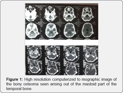
Osteomas are benign tumors of bone seen most commonly in the frontoethmoidal region in head and neck. In temporal bone external auditory canal is the most common site of osteoma. However there are few cases of extra canalicular osteomas of temporal bone that have been reported in the literature. We present a case of a 13 year old child who presented to us with a hard swelling of post auricular region. Radiologic examination revealed features of osteoma arising out of mastoid part of temporal bone. This patient was operated with excision of the lesion for cosmetic reasons. Though rare, this is an important diagnosis for bony hard swellings of temporal bone and needs to be in the differentials of the clinicians, thus we present this case.
Keywords: Retro auricular swelling; Bony swelling of head and neck; Osteoma; Osteomas in head and neck; Mastoid osteoma; Temporal bone osteoma; Extra canalicular osteoma of temporal bone
Background
Osteomas are benign tumors of lamellar bone seen rarely in head and neck region [1]. Common site for osteomas in head and neck region is frontal and ethmoids [2]. In the temporal bones, they are more common in external ear canal (EAC) [3]. There have also been few reports in the literature showing EAC cholesteatoma and even cerebellar abscess in cases of canalicular osteomas [4,5]. In the extra canalicular part they are rare and involve the mastoid part or the squamous part of the temporal bone [6]. They commonly present with bony hard swelling at time causing auricular protrusion [7]. In this report we have presented a case of mastoid bone osteoma in a teenager with its management and postoperative follow up.
Case Report
A 13 year old male child presented to our outpatient department with complaints of a hard swelling behind his left ear for last 4 years which started as a peanut sized swelling and gradually progressed. His was concerned with the unsightly appearance though he had no pain, ear discharge and impairment of hearing. Child denied history of trauma. On examination there was 4x3 cm hard well defined swelling in the post aural region which was pushing pinna anteriorly without causing any narrowing of EAC. Rest of otolaryngologic examination was normal. Audiometry was within normal limits. High resolution computerized tomography (HRCT) of temporal bone revealed a bony lesion confined only to the mastoid part of temporal bone (Figure 1).

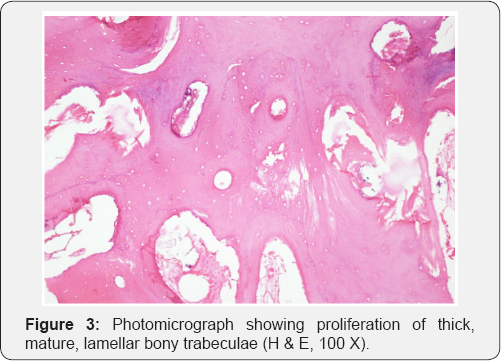
Rest of the EAC, middle ear cleft, ossicles and inner ear were normal. Child was taken up for excision of the lesion under general anesthesia. A modified post aural incision (Figure 2) was made over the prominence of the lump. Soft tissues are separated and lump was adequately exposed. With the help of chisel and hammer whole of the lump was excised with minimal bleeding. Remnant lesion was drilled out with diamond burr. Wound closed in layers. Histopathology showed proliferation of thick, mature, lamellar bony trabeculae (Figure 3). On subsequent follow up for three months child was doing well.
Discussion
Osteomas are benign bone tumors most common in long bones. In head and neck region, frontal and ethmoid regions are relatively common sites [8]. External auditory canal being the most common site in the temporal bone [9], few reports does mention about extra canalicular osteomas in the temporal bone mostly in the mastoid and the squamous part [10]. It has a female predominance [11] with 2nd and 3rd decade [12] being the most common age unlike our case who is in the early adolescent years. As described in our case, osteomas are mostly asymptomatic except for minimal complains of heaviness and cosmetic deformity [13].
Osteomas are very slow growing and remain stable over long duration. Skin overlying is usually free and swelling is bony hard in consistency. In the presented case the patient had also presented with a cosmetic deformity only. Temporal osteomas might lead to pain by stretching of nerve fibers and widening of periosteum [14]. They can also produce canal cholesteatoma [15] with suppuration at times leading to intracranial complications also [16]. The most widely accepted theories on the etiopathogenesis of mastoid osteoma include: embryogenesis, metaplasia, inflammation, hormonal changes and trauma [10,17]. However in most of the cases etiology is unknown. There was no factor in our case which can be attributed as an etiological factor.
Multiple osteomas may occur in Gardner's syndrome in which colonoscopy may be indicated to look for colonic polyposis. Small asymptomatic osteomas require no intervention. High resolution tomography of temporal bone is the imaging modality of choice to assess the extent of involvement. It reveals anatomical relationship with deeper vital structure [18]. In the absence of any complication, surgery is done for cosmetic reasons in mastoid osteomas as in our case. Osteomas can be easily chiseled out with chisel and hammer [19]. The remnant lesion can be drilled with diamond burr till the normal bone appears. Drilling of the attachment till normal mastoid bone prevents further recurrences [20]. Histologically osteomas show dense lamellae with organized haversian canals with the inter trabecular stroma containing osteoblasts, fibroblasts, and giant cells in the absence of hematopoietic marrow.
Osteomas can be of four types based of cellular contents in histology such as compact, cartilaginous, spongiotic, and mixed. Compact osteomas are dense, ivory-like, round and the most common type [21] which is either pedicled or wide based; in contrast the spongiotic types [22] are rare and formed by spongiotic bone and fibrous cellular tissue. In our case, postoperative histology showed features of compact osteoma with proliferation of thick, mature, lamellar bony trabeculae. Postoperative period was uneventful. Child is under follow up and is doing well.
Conclusion
Mastoid osteomas are of rare occurrence. Unsightly appearance is the most common symptom for which surgery is indicated. High resolution computerized tomography of temporal bone confirms the diagnosis and guides during surgical excision. Differentials like osteosarcoma and osteoblastic metastasis should be excluded. Asymptomatic small osteomas without any significant cosmetic deformity do not require any intervention. Chiseling of osteomas with drilling of remnant lesion has given good results.
References
- Abdel Tawab HM, Kumar VR, Tabook SM (2015) Osteoma presenting as a painless solitary mastoid swelling. Case Rep Otolaryngol 2015: 590783.
- Burton DM, Gonzalez C (1991) Mastoid osteomas. Ear, nose, & throat journal 70(3): 161-162.
- Das AK, Kashyap RC (2005) Osteoma of the mastoid bone-A case report. MJAFI 61(1): 86-87.?
- Viswanatha B (2007) A case of osteoma with cholesteatoma of the external auditory canal and cerebellar abscess. International Journal of Pediatric Otorhinolaryngology Extra 2(1): 34-39.
- Orita Y, Nishizaki K, Fukushima K, Akagi H, Ogawa T, et al. (1998) Osteoma with cholesteatoma in the external auditory canal. International journal of pediatric otorhinolaryngology 43(3): 289293.
- Cheng J, Garcia R, Smouha E (2013) Mastoid osteoma: A case report and review of the literature. Ear, nose, & throat journal. 92(3): E7-E9.
- El Fakiri M, El Bakkouri W, Halimi C, Ait Mansour A, Ayache D (2011) Mastoid osteoma: report of two cases. European annals of otorhinolaryngology, head and neck diseases 128(5): 266-268.
- Dominguez Perez AD, Romero RR, Duran ED, Riquelme Montano P, Alcantara Bernal R, et al. (2011) The mastoid osteoma, an incidental feature. Acta otorrinolaringologica Espanola 62(2): 140-143.
- Kim CW, Oh SJ, Kang JM, Ahn HY (2006) Multiple osteomas in the middle ear. Eur Arch Otorhinolaryngol 263(12): 1151-1154.
- D'Ottavi LR, Piccirillo E, De Sanctis S, Cerqua N (1997) Mastoid osteomas: review of literature and presentation of 2 clinical cases. Acta otorhinolaryngologica Italica 17(2): 136-139.
- Ohhashi M, Terayama Y, Mitsui H (1984) Osteoma of the temporal bone-a case report. Nihon Jibiinkoka Gakkai kaiho 87(5): 590-595.
- Gupta OP, Samant HC (1972) Osteoma of the mastoid. The Laryngoscope 82(2): 172-176.
- Pereira CU, de Carvalho R, de Almeida A, Dantas RN (2009) Mastoid Osteoma. Consideration on two cases and literature review. Intl Arch Otorhinolaryngol 13(3): 350-353.
- Sikarwar V, Lavania A, Saxena R (2011) Osteoma Temporal Bone- Rare Case. Nepalese Journal of ENT Head and Neck Surgery 2(1): 25-26.
- Lee DH, Jun BC, Park CS, Cho KJ (2005) A case of osteoma with cholesteatoma in the external auditory canal. Auris, nasus, larynx 32(3): 281-284.
- Van Dellen JR (1977) A mastoid osteoma causing intracranial complications. A case report. S Afr Med J 51(17): 597-598.
- Guerin N, Chauveau E, Julien M, Dumont JM, Merignargues G (1996) Osteoma of the mastoid: apropos of 2 cases. Revue de laryngologie otologie rhinologie 117(2): 127-132.
- Ben Yaakov A, Wohlgelernter J, Gross M (2006) Osteoma of the lateral semicircular canal. Acta oto-laryngologica 126(9): 1005-1007.
- Gulia J, Yadav S (2009) Mastoid osteoma: a case report. The Internet Journal of Head and Neck Surgery 3(1): 22-23.
- Probst LE, Shankar L, Fox R (1991) Osteoma of the mastoid bone. The Journal of otolaryngology 20(3): 228-230.
- Gungor A, Cincik H, Poyrazoglu E, Saglam O, Candan H (2004) Mastoid osteomas: report of two cases. Otology & neurotology: official publication of the American Otological Society, American Neurotology Society and European Academy of Otology and Neurotology 25(2): 95-97.
- Denia A, Perez F, Canalis RR, Graham MD (1979) Extracanalicular osteomas of the temporal bone. Arch Otolaryngol 105(12): 706-709.


