Teaching NeuroImages: Balo’s Concentric Sclerosis
Peter Akpunonu*
Department of Neurology, University of Kentucky, Lexington, KY 40536, US
Submission: May 10, 2024; Published: September 26, 2024
*Corresponding author: Dr Peter Akpunonu, Department of Neurology, University of Kentucky, 800 S Rose St\r\nM-53\r\nLexington, KY 40536, US
How to cite this article: Peter Akpunonu*. Teaching NeuroImages: Balo’s Concentric Sclerosis. Open Access J Neurol Neurosurg 2024; 19(2): 556007. DOI: 10.19080/OAJNN.2024.19.556007.
Abstract
Keywords: NeuroImages; MRI; Corticosteroids; Plasma exchange; Intravenous immunoglobulin; Monoclonal antibodies
Clinical Image
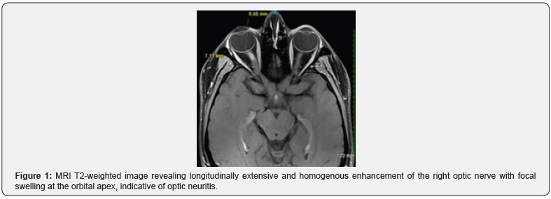
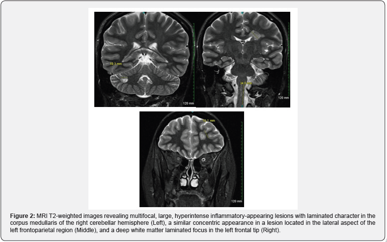
A 24-year-old woman presented for rapidly progressive right-sided monocular vision loss over the course of three days associated with photopsia and eye pain worsened with extraocular movement. Physical examination was significant for hand-motion only visual acuity in right eye, right-sided afferent pupillary defect, and edema of the right optic nerve head evident on fundoscopy. MRI revealed inflammation of the right optic nerve consistent with optic neuritis in addition to multifocal, concentric, hyperintense foci most suggestive of Balo’s concentric sclerosis (BCS). She was treated with high-dose intravenous methylprednisolone for three days with only mild improvement in visual acuity in the right eye and subsequently treated with Ocrelizumab in the outpatient setting.
Considered a rare variant of multiple sclerosis (MS), BCS is identified by concentrically layered lesions on MRI representative of alternating bands of demyelination and myelin preservation. Patients can present with any typical MS symptoms: focal weakness, ataxia, or diplopia; but most commonly report symptoms consistent with any intracerebral mass lesion: headache, cognitive difficulties, and/or hemiparesis. Approximately half of those presenting with a Balo or Balo-like lesion have typical MS lesions elsewhere on their MRI [1]. Treatment strategies consist of corticosteroids, plasma exchange, intravenous immunoglobulin, and monoclonal antibodies [2] (Figures 1 & 2).


