Single Photon Computed Tomography (SPECT) Findings in Cases with Japanese Adult- Attention-Deficit/Hyperactivity Disorder (ADHD): Special Attention to Cerebellar Posterior Lobe Hypoperfusion on Iodoamphetamine (IMP) SPECT using 3 Dimensional Stereotactic Surface Projection (3D-SSP) Analysis
Nobuhiko Miyazawa1* and Toyoaki Shinohara2
1Department of Neurosurgery, Fuefuki central hospital, Japan
2Department of Neurosurgery, Kofu Neurosurgical hospital, Japan
Submission: August 28, 2023; Published: September 08, 2023
*Corresponding author: Nobuhiko Miyazawa MD, PhD, 47-1 Yokkaichiba Fuefuki-city Yamanashi-pref. 406-0032 Japan
How to cite this article: Nobuhiko M, Toyoaki S. Single Photon Computed Tomography (SPECT) Findings in Cases with Japanese Adult-Attention-Deficit/ Hyperactivity Disorder (ADHD): Special Attention to Cerebellar Posterior Lobe Hypoperfusion on Iodoamphetamine (IMP) SPECT using 3 Dimensional Stereotactic Surface Projection (3D-SSP) Analysis.Open Access J Neurol Neurosurg 2023; 18(4): 555991. DOI: 10.19080/OAJNN.2023.18.555991.
Abstract
Although a number of case with adult ADHD has been increasing in Japan , helpful diagnostic method of it has yet not existed. Recently, effective and supportive findings on cerebral SPECT with statistical methods have been fund in cases with Japanese adult ADHD.
During 2 years, 15 cases with Japanese ADHD diagnosed by DSM-5 were examined with IMP-SPECT using statistical methods. ROIs were placed on anterior and posterior lobe of cerebellum on IMP SPEC with vbSEE level 2 software and other regions with that level 5 software.
Significant decrease was observed on cerebellum in 8 cases (bilateral in 6 cases, unilateral in 2 cases), upper posterior lobe, thalamus in 4 cases, middle posterior lobe, lower posterior lobe, pons in 2 cases, cuneus, lingualis gyrus in 1 case. In total 15 cases, there were significant decrease in posterior lobe of cerebellum compared with anterior lobe (left side: p=2.24E-05, right side: p=2.63E-05).
Those results were in accordance with those of studies with SPECT with statistical methods. Hypoperfusion on posterior lobe of cerebellum on SPECT with statistical methods might facilitate the diagnosis of adult ADHD in Japan.
Keywords: Adult ADHD; IMP SPECT; 3D-SSP; Cerebellum
Abbreviations: ADHD: Attention Deficit Hyperactivity Disorder; SPECT: Single Photon Computed Tomography; 3D-SSP: Three Dimensional- Stereotactic Surface Projection; IMP: Iodoamphetamine; HM-PAO: Hexamethylpropyleneamine oxime; ECD: Ethyl Cysteinate Dimer; FDG: Fluolo-Deoxy Glucose; PET: Positron Emission Tomography; MRI: Magnetic Resonance Imaging; rCBF: Regional Cerebral Blood Flow; fMRI: Functional Magnetic Resonance Imaging; MRA: Magnetic Resonance Angiography; DSM: Diagnostic and Statistical Manual of Mental Disorders; ROI: Region of Interest. CBA: Computed Brain Atlas; vbSEE: Voxel-Based Stereotactic Extraction Estimation; SPM: Statistical Parametric Mapping; MMSE: Mini-Mental State Examination; EOAD: Early-Onset Alzheimer’s Disease; MPH: Methylphenidate
Introduction
Until recent days, attention-deficit/hyperactivity (ADHD) has been treated as child and adolescent disorders, however, adult ADHD has been a focus of discussion nowadays [1-4]. As for symptoms of it are unique that hyperactivity are rare, on the other hand, deficit of attention or execution become apparent [1-3]. Sometimes, because differential diagnosis of early-onset Alzheimer’s disease and them are very difficult, patients with adult-ADHD have been examined by doctors of dementia in Japan. Moreover, there has been a diagnostic criteria of DSM-5 [5], exact diagnosis of it is still matter of discussion. Cerebral perfusion SPECT have been utilized for help of diagnosis [6]. HM-PAO or ECD have been used frequently in Europe and United states, however, statistical analysis were rarely done and reports of area in hypoperfusion were not consistent [6]. In Japan, IMP have been utilized in a lot of cases because of significant correlation with cerebral blood flow examination with O2-PET.
In this study, total of 15 cases with adult ADHD in Japan were examined by IMP SPECT with statistical analysis (3D-SSP), helpful findings in terms of cerebellum were emerged and we discuss the hypoperfusion on SPECT in cases adult ADHD comparing with reported cases [7] by special attention to cerebellum.
Patients and Methods
From January 2020 to December 2021, 15 patients with age of 30-59 years old had symptoms of forgetfulness and lack of concentration were diagnosed by DSM-V guides clinical interview, internal DSM-V-guided symptom checklist and Conners continuous performance test [5,8] as adult ADHD. Brain tumor or other brain disorders were excluded by 3.0 tesla MRI. Neurological examination, MMSE, MRI were performed on the same day, cerebral blood flow SPECT were done within 7-30 days after examination. Informed consent was taken after precise explanation from all patients. MMSE-Japanese version was used and total and subscale were evaluated. MRI was performed by Siemens Co. 3.0 tesla with T1-weighted, T2-weighted, DWI, MRA [9]. SPECT was performed by 3D-camera Toshiba Co. After injection of 135Mq IMP/Kg, 30 minutes scan was executed with eye-closed. Usual SPECT images was performed with a triple-head gamma camera (Toshiba GCA93000R, Toshiba, Tokyo, Japan). Images were acquired on a 128x128 matrix. Reconstruction was performed using the filtered back projection technique, and ramp filters were utilized for smoothing and statistical images were taken with 3D-SSP image by NEUROSTAT-version 6. Software on level 5. of vbSEE was used for setup of ROI and calculating z-scores and -1.5 S.D. was judged as significant decrease [10- 12]. Student’s t-test was utilized for differences between z-score of anterior and posterior lobe of cerebellum, Pearson’ s test was used for correlation between z-score of each ROIs of cerebellum and MMSE score (both total and subscales). P<0.05 was judged as significant.
This study was approved by the institutional ethical committee. Written informed consent was obtained from patients. This study was explained to patients and relatives in detail.
Results
Out of 15 cases, Female: 7 and Male: 8. Age distribution was from 30-59 years old, average was 43.5 years old. All patients met the criteria of DSM-V. MMSE score ware 19-30, average of them was 27.2. Subscales of MMSE; orientation ranged from 5-10(average:9.10), attention from 2-3 (2.93), calculation from 1-5 (3.80), recent memory from 2-3(2.80), language from 8-11 (10.5) and visuospatial recognition from 0-1 (0.933). Among them, level of calculation tended to lower. Level of MMSE was considered as same as Minimal cognitive impairment. MRI and MRA study showed that 4 cases had mild white matter lesions, 5 cases had mild atherosclerosis. There was no cerebral infarction nor hematoma, other space taking lesions, nor significant arterial stenosis, occlusions.
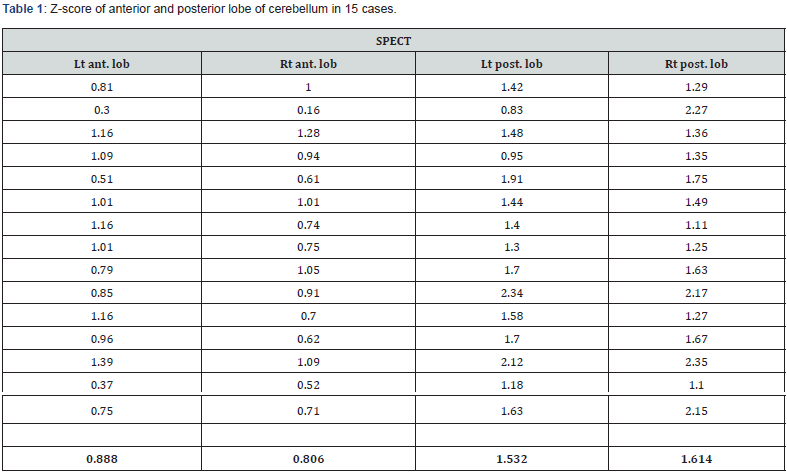
In terms of SPECT findings, significant hypoperfusion were recognized in 8 cases with cerebellum, bilateral hypoperfusion in 6 cases and unilateral in 2cases (Table 1).

Lt: left, ant. lob: anterior lobe, post. lob: posterior lobe, Rt: right Bottom cells showed the average of each ROIs of cerebellum.
Out of 15 cases, z-score of bilateral posterior lobe of cerebellum were significantly lower than that of anterior lobe of cerebellum. (left side; p=2.24E-05, right side; p=2.6267E-05) Interestingly, in 7 cases with non-significant hypoperfusion on cerebellum, there were significant decreases in posterior lobe of cerebellum on both side. (left side; p=0.00708, right side; p=0.0000173) Significant decreases were observed on upper occipital lobe in 4 cases, thalamus in 4cases, middle occipital lobe in 2 cases, lower occipital lobe in 2 cases, pons in 2 cases, cuneus in 1 case and lingual lobe in 1case. Out of 4 cases with thalamus hypoperfusion, 2 cases also showed hypoperfusion in cerebellum.
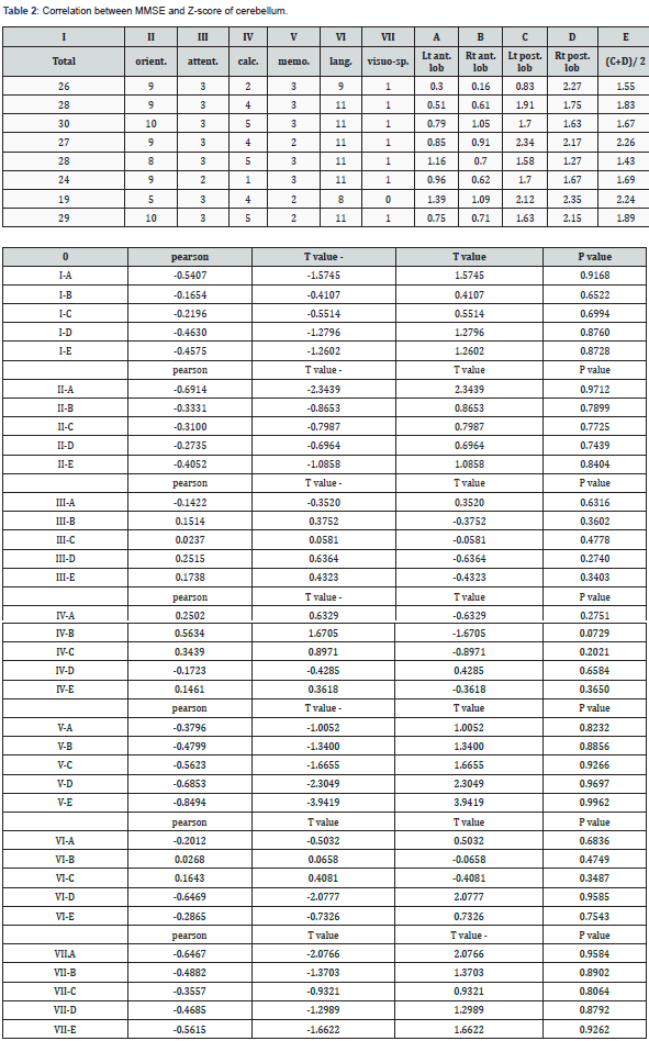
There was no significant correlation between hypoperfusion on cerebellum and MMSE total nor subscales (Table 2).

orient.: orientation, attent.: attention, calc.: calculation, memo.: recent memory, lang.: language, visuo-sp.: visuospatial recognition.
Case Presentations

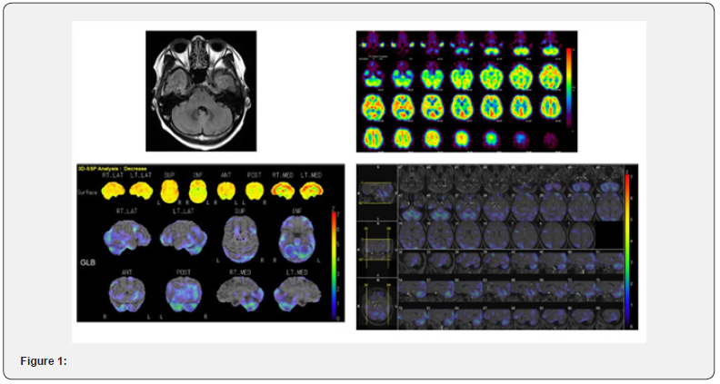
Case 1. 52 years old female had symptoms of forgetfulness and difficulties of daily life. She also had difficulty in usual work. She was examined by MRI, MMSE, DSM-V and SPECT. There was no abnormality on MRI, total score of MMSE was 26 (orientation -1, calculation -3), DSM-V: 5/7 (inattention type) IMP-SPECT showed hypoperfusion in cerebellum on usual image and significant decreases in bilateral cerebellum mainly left side of posterior lobe (z-score; lt-posterior=1.91, rt-posterior=1.75) (Figure 1).
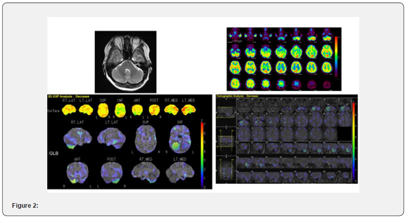
Case 2. 55 years old female suffered from forgetfulness. She had major trouble in official work. She was examined by MMSE, MRI, DSM-V and IMP-SPECT. No abnormality was found on MRI, MMSE was 27 (calculation -3), DSM-V:6/7 (inattention type). IMP-SPECT revealed marked hypoperfusion in right cerebellum on usual image and significant decrease in right posterior lobe of cerebellum. (z-score; lt-posterior=0.83, rt-posterior=2.27) (Figure 2).

Discussion
ADHD is often diagnosed at childhood and adolescence and major problem in these days, however, adult ADHD have not been the main problem nor matter of discussion until recent era [13]. Adult-ADHD has accounted for 2-4% of adult age [14] and 14-72 % in criminal populations [15]. Symptoms and clinical sign are apart from those of childhood, main symptoms are inattention and disorganization rather than hyperactivity and impulsivity [16]. Although the DMS-V has been used as standard of diagnosis, sometimes correct diagnosis is problematic. Moreover, exact core lesions of ADHD have not still identified and several candidates have been reported [17,18]. Fronto-subcortical network and cerebellum have been major and key regions for pathogenesis of ADHD [19-21].
As regard to SPECT study of ADHD, 16 studies reported abnormal findings of ADHD, HMPAO were utilized in 12 studies, 3 studies by ECD and only one study by IMP [6,7,22,23]. Out of 16 studies, only 3 studies dealt with adult-ADHD. Only 3 studies reported the image by statistical methods with SPM and 3D-SSP. There have been hypoperfusion in prefrontal lobe, temporal lobe, parietal lobe, basal ganglia in 16 studies, hypoperfusion in cerebellum was reported in 5 studies and hypoperfusion of cerebellum have observed in all studies with statistical methods including this study [7,22,24,25].
Out of studies with statistical method, Amen et al. reported that significant hypoperfusion were recognized in prefrontal orbit and frontal pole at concentration state (p=0.0007, 0.0009, 0.003, 0.006) using HMPAO in cases with 27 patients over 50 years old and moreover, on both resting and concentration state, significant hypoperfusion were observed in bilateral cerebellum (concentration state p=0.005, 0.005, resting state p=0.004, 0.004), cerebellar hypoperfusion was important and helpful finding of adult-ADHD [22] . Gardner et al. also stated that cerebellar hypoperfusion was crucial point of adult ADHD analyzing 30 cases with depression with ADHD and depression only using HMPAO SPECT with SPM2 and CBA method and concluded that these findings confirm the previous observation of a cerebellar involvement in ADHD [24]. In 2021, Amen et al. revealed that analyzing 1006 cases with adult ADHD and 129 cases with control, although hypoperfusion in medial anterior prefrontal cortex, left anterior temporal lobe, right insular cortex was helpful for differential diagnosis, also hypoperfusion in cerebellar posterior lobe (7b, 8, 9, crus 1,2) was significantly useful (p<0.001) for differential diagnosis with statistical analysis of post-hoc ROI analysis. One of the predictive values in distinguishing adult ADHD could be cerebellum [7]. In this study, also significant hypoperfusion were recognized in posterior lobe of cerebellum by statistical analysis. As for methods of statistical analysis, 3D-SSP method is superior than that of SPM in cases with cerebral ischemia, or same level [26,27]. In terms of psychiatric diseases, findings of SPECT are helpful of judgement of treatment effect of various psychiatric diseases including ADHD [28]. Importantly, areas of hypoperfusion in ADHD have been debatable, frontostriatal- cerebellar circuit has pivotal role in adult-ADHD [13]. In this study, not only significant hypoperfusion in cerebellum but also, that in thalamus were observed. There still have been no consensus which areas are main part or from which part begin with hypoperfusion in cases adult-ADHD.
In terms of relationship between z-score on SPECT and MMSE, there is no report before, our results showed no correlation between them, further investigation will be required. ECD and HM-PAO have been frequently used, IMP was used in only one study without statistical analysis. Results with HM-PAO showed significantly higher cerebral flow was calculated in basal ganglia and cerebellum than that of rCBF-PET [29]. Also results with ECD showed significantly higher cerebral blood flow was reported in occipital lobe [30]. On the other hand, results with IMP has significantly good correlation between rCBF-PET [30]. Correlation between results with IMP and that of FDG-PET were significantly high (r=0.82 p<0.001) in cases with Alzheimer’s disease [31]. Moreover, findings of IMP SPECT is useful for distinguish Parkinson’s disease and MSA [32]. At moment, it is reasonable to use IMP SPECT with statistic analysis for diagnosing adult ADHD.
Functional MRI (fMRI) also have played pivotal role for diagnosis of ADHD. One of the studies with large cases revealed that anterior cingulate and cerebellum had significant strong connectivity with executive control network by comparing 99 cases with adult ADHD and 113 cases with healthy control [33]. Another study by fMRI showed that abnormality of default mode network in cerebellum had correlation with abnormality of frontoparietal and visual network and region of silence, dorsal attention and sensorimotor network regions using 46 cases with adult ADHD and control matched in age, gender, IQ [34]. In terms of comparison with childhood and adult ADHD, abnormality of cortico-cortical and cortico-subcortical connectivity are important in childhood, however, abnormality of cortico-cortical connectivity is critical for adult ADHD [35].
Interesting study reported that level of creatine increased only on cerebellum by administrating MPH in 60 cases with ADHD without no increase on prefrontal cortex, striatum, anterior cingulate cortex, and concluded that role of cerebellum was most important in cases with adult ADHD [36].
Finally, results of this study had accordance with past studies especially hypoperfusion in posterior lobe of cerebellum is one of the most important findings in cases with adult ADHD. This study is the first study using IMP SPECT with 3D-SSP in cases with adult ADHD. Although fMRI has been utilized frequently nowadays, however, analysis of fMRI is time consuming and is not easily done in clinical state. On the other hand, 3D-SSP analysis of IMP-SPECT require only 10 minutes to get results. It is suitable in clinical diagnosis. As for differential diagnosis of adult ADHD and early onset Alzheimer’s disease, this method provide crucial point that no hypoperfusion in cerebellum is seen in case with EOAD opposite for in case with adult ADHD [9,37].
There are several limitations of this study, number of cases are small and patients with ADHD and other psychiatric disease like depression cloud be included in this study. ROI in statistical method in cerebellum are only two (anterior and posterior), more precise ROI should be placed on cerebellum.
Conclusion
IMP-SPECT with 3D-SSP analysis is very helpful to diagnose adult ADHD and special attention should be taken to hypoperfusion in posterior lobe of cerebellum. Future studies with a lot of cases with adult ADHD by statistical SPECT are warranted.
Funding
This research did not receive any specific grant from funding agencies in the public, commercial or not-for-profit sectors.
References
- Volkow ND, Swanson JM (2013) Adult attention deficit-hyperactivity disorder. N Engl J Med 369(20): 1935-1944.
- Schneider H, Thornton JF, Freeman MA, McLean MK, van Lierop MJ, et al. (2014) Conventional SPECT versus 3D thresholded SPECT imaging in the diagnosis of ADHD; a retrospective study. J Neuropsychiatry Clin Neurosci 26(4): 335-343.
- Caye A, Roca TBM, Anselmi L, Murray J, Menezes AMB, et al. (2016) Attention-deficit/hyperactivity disorder trajectories from childhood to young adulthood; evidence from a birth cohort supporting a late-onset syndrome. JAMA Psychiatry 73(7): 705-712.
- Thorell LB, Holst Y, Christiansen H, Kooji JJS, Bijlenga D, et al. (2017) Neuropsychological deficits in adults age 60 and above with attention deficit hyperactivity disorder. Eur Psychiatry 45: 90-96.
- American Psychiatric Association (2013) Diagnostic and Statistical Manual of Mental Disorder, Fifth ed. (DSM-5). American Psychiatric Publishing, Washington D.C., USA
- Santra A, Kumar R (2014) Brain perfusion single photon emission computed tomography in major psychiatric disorders: From basics to clinical practice. Indian J Nucl Med 29(4): 210-221.
- Amen DG, Henderson TA, Newberg A (2021) SPECT functional neuroimaging distinguishes adult attention deficit hyperactivity disorder from healthy controls in big data imaging cohorts. Front Psychiatry 12: 725788.
- Conners CK, Saitarenios G (2011) Conner’s Continuous Performance Test (CPT). In: Kreutzer JS, editor. Encyclopedia of Clinical Neuropsychology. New York, NY; Springer. Pp. 681-683.
- Miyazawa N, Shinohara T (2022) Cerebral blood flow-single-photon emission computed tomography and Brodmann mapping may facilitate the evaluation of clinical features of early-onset Alzheimer’s disease. Front Clin Neurol Neurosurg 1: 101-104.
- Minoshima S, Frey KA, Koeppe RA, Kuhl DE (1995) A diagnostic approach in Alzheimer’s disease using three-dimensional stereotactic surface projections of fluorine-18-FDG PET. J Nucl Med 36(7): 1238-1248.
- Miyazawa N (2017) Creutzfeldt-Jacob disease mimicking Alzheimer disease and Dementia with Lewy Bodies; findings of FDG PET with 3-Dimensional Stereotactic Surface Projection. Clin Nucl Med 42(5): e247-e248.
- Ishii K, Kanda T, Uemura T, Miyamoto N, Yoshikawa T, et al. (2009) Computer-assisted diagnostic system for neurodegenerative dementia using brain SPECT and 3D-SSP. Eur J Nucl Med Mol Imaging 36(5): 831-840.
- Schneider M, Retz W, Coogan A, Thome J, Rösler M (2006) Anatomical and functional brain imaging in adult attention-deficit/hyperactivity disorder (ADHD); a neurological view. Eur Arch Psychiatry Clin Neurosci 256 Suppl 1: i32-i41.
- Kessler RC, Adler LA, Barkley R, Biederman J, Conners CK, et al. (2005) Patterns and predictors of attention-deficit/hyperactivity disorder persistence into adulthood; results from the national comorbidity survey replication. Biol Psychiatry 57(11): 1442-1451.
- Rosler M, Retz W, Retz-Junginger P, Hengesch G, Schneider M, et al. (2004) Prevalence of attentiona deficit-/hyperactivity disorder (ADHD) and comorbid disorders in young male prison inmates. Eur Arch Psychiatry Clin Neurosci 254(6): 365-371.
- Biederman J, Mick E, Faraone SV (2000) Age-dependent decline of symptoms of attention deficit hyperactivity disorder; impact of remission definituion and symptom type. Am J Psychiatry 157(5): 816-818.
- Carter CS, Macdonald Am, Borvinick M, Ross LL, Stenger VA, et al. (2000) Parsing executive processes; strategic vs. evaluative fuctions of the anterior cingulate cortex. Proc Natl Acad Sci USA 97(4): 1944-1948.
- Casey BJ, Castellanos FX, Giedd JN, Marsh WL, Hamburger SD, et al. (1997) Implication of right frontostriatal circuitry in response inhibition and attention-deficit/hyperactivity disorder. J Am Acad Chiild Adolesc Psychiatry 36(3): 374-383.
- Anderson CM, Polcari A, Lowen SB, Renshaw PF, Teicher MH (2002) Effects of methylphenidate on functional magnetic resonance relaxometry of the cerebellar vermis in boys with ADHD. Am J Psychiatry 159(8): 1322-1328.
- Schmahmann JD (2004) Disorders of the cerebellum: ataxia, dysmetria of thought, and the cerebellar cognitive affective syndrome. J Neuropsychiatry clin Neurosci 16(3): 367-378.
- Cherkasova MV, Hechtman L (2009) Neuroimaging in attention-deficit hyperactivity disorder; beyond the frontostriatal circuitry. Can J Psychiatry 54(10): 651-654.
- Amen DG, Hanks C, Prunella J (2008) Predicting positive and negative treatment responses to stimulants with brain SPECT imaging. J Psychoactive Drugs 40(2): 131-138.
- Sieg KG, Gaffney GR, Preston DF, Hellings JA (1995) SPECT brain imaging abnormalities in attention deficit hyperactivity disorder. Clin Nucl Med 20(1): 55-60.
- Gardner A, Salmaso D, Varrone A, Sanchez-Crespo A, Bejerot S, et al. (2009) Differences at brain SPECT between depressed females with and without adult ADHD and healthy controls; etiological considerations. Behav Brain Funct 5: 37.
- Yeh CB, Huang WS, Lo MC, Chang CJ, Ma KH, et al. (2012) The rCBF brain mapping in adolescent ADHD comorbid developmental coordination disorder and its changes after MPH challenging. Eur J Paediatr Neurol 16(6): 613-618.
- Onishi H, Matsutake Y, Kawashima H, Matsumoto N, Amijima H (2011) Comparative study of anatomical normalization errors in SPM and 3D-SSP using digital brain phantom. Ann Nucl Med 25(1): 59-67.
- Wang K, Liu T, Zhao X, Xia XT, Zhang K, et al. (2016) Comparative study of voxel-based epileptic foci localization accuracy between statistical parametric mapping and three-dimensional stereotactic surface projection. Front Neurol 7: 164.
- Thornton JF, Schneider H, Cohen PF, DeBruin S, Uszler JM, et al. (2022) Longitudinal single photon emission computed tomography neuroimaging as an indication of improvement in psychiatric disorders in a community psychiatric practice. Front Psychiatry 13: 787186.
- Koyama M, Kawashima R, Ito H, Ono S, Sato K, et al. (1997) SPECT imaging of normal subjects with technetium-99m-HMPAO and technetium-99m-ECD. J Nucl Med 38(4): 587-592.
- Iida H, Akutsu T, Endo K, Fukuda H, Inoue T, et al. (1996) A multicenter validation of regional cerebral blood flow quantitation using [123I]iodoamphetamine and single photon emission computed tomogaraphy. J Cereb Blood Flow Metab 16(5): 781-793.
- Nihashi T, Yatsuya H, Hayasaka K, Kato R, Kawatsu S, et al. (2007) Direct comparison study between FDG-PET and IMP-SPECT for diagnosing Alzheimer’s disease using 3D-SSP analysis in the same patients. Radiat Med 25(6): 255-262.
- Murakami N, Sako W, Haji S, Furukawa T, Otomi Y, et al. (2020) Differences in cerebellar perfusion between Parkinson’s disease and multiple systemic atrophy. J Neurol Sci 409: 116627.
- Mostert JC, Shumskaya E, Mennes M, Onnink AMH, Hoogman M, et al. (2016) Characterizing resting-state fuctional connectivity in a large sample of adults with ADHD. Prog Neuropsychopharmacol Biol Psychiatry 67: 82-91.
- Kucyi A, Hove MJ, Biederman J, van Dijk KRA, Valera EM (2015) Disrupted functional connectivity of cerebellar default network areas in attention-deficit/hyperactivity disorder. Hum Brain Mapp 36(9): 3373-3386.
- Lui N, Liu Q, Yang Z, Xu J, Fu G, et al. (2023) Different functional alteration in attention-deficit/hyperactivity disorder across developmental age groups: a meta-analysis and an independent validation of resting-state functional connectivity studies. CNS Neurosci Ther 29(1): 60-69.
- Inci Kenar AN, Unal GA, Kiroglu Y, Herken H (2017) Effects of methylphenidate treatment on the cerebellum in adult attention-deficit hyperactivity disorder; a magnetic resonance spectroscopy study. Eur Rev Med Pharmacol Sci 21(2): 383-388.
- Kushner M, Tobin M, Alavi A, Chawluk J, Rosen M, et al. (1987) Cerebellar glucose consumption in normal and pathologic states using fluorine-FDG and PET. J Nucl Med 28(11): 1667-1670.


