Late Disclosure of Firearm Injury to Vertebrospinal Cord Injury
MS Rabiou1, AK Moumouni2*, PR Plante3, F Gnandi-Piou2, DRD Ajavon2, E Kpelao4, A Haidara5 and Taofik Moussa6
1Neurosurgery Department of Zinder, Niger
2Faculty of health sciences, University of Kara, Togo
3Surgery Department of CHU Kara, Togo
4Faculty of health sciences, University of Lomé, Togo
5Neurosurgery Department of CHU Bouake, Bouake, Ivory Coast
6Department of Traumatology and Orthopedic, Zinder National Hospital Niger
Submission: February 24,2022; Published: March 10, 2022
*Corresponding author: Abd-el Kader Moumouni, Neurosurgery Department of the University Hospital Kara, Kara, Togo. BP 18, Kara, TOGO
How to cite this article: MS Rabiou, AK Moumouni*, PR Plante, F Gnandi-Piou, DRD Ajavon, et al. Late Disclosure of Firearm Injury to Vertebrospinal Cord Injury. Open Access J Neurol Neurosurg 2024; 16(5): 555950. DOI: 10.19080/OAJNN.2024.16.555950.
Summary
Ballistic trauma is the consequence of the penetration of a projectile into the body. The clinical lesion manifestation is sometimes late, as it is for this 29 years old patient whose case we report. Indeed, he had been the victim of a cervical ballistic trauma, 2 years ago. Brown Sequard syndrome was the mode of revelation. Standard radiography and CT scan were of great help in diagnosing the lesion. Surgical treatment consisted essentially of removal of the projectile and a dural plasty. The postoperative course was simple and the evolution favourable with a functional recovery in one month.
Keywords: Ballistics; Brown Sequard; Late spine trauma
Introduction
Ballistic injuries are the consequence of the penetration of a projectile into the body: bullet, lead, metallic fragment coming from the envelope or the contents of an explosive device. They are often the cause of frequent and serious injuries characterised by the multiplicity of clinical pictures and the frequency of associated injuries. These injuries are life-threatening due to blood spoliation, respiratory distress and lesion associations, and secondarily due to the risk of infection with contamination of the wound. The functional prognosis may also be at risk in the case of neuraxial or limb injuries [1]. Vertebral-medullary trauma caused by firearms can be responsible for severe neurological sequelae. Firearm injuries can be life-threatening due to neurological and infectious complications [2]. These spinal cord injuries usually result in immediate spinal cord injury following the trauma. Late-onset forms are rare.
We report here the case of a 29 year old male with a spinal cord injury caused by a firearm, which revealed itself late in the course of the injury.
Clinical Observation
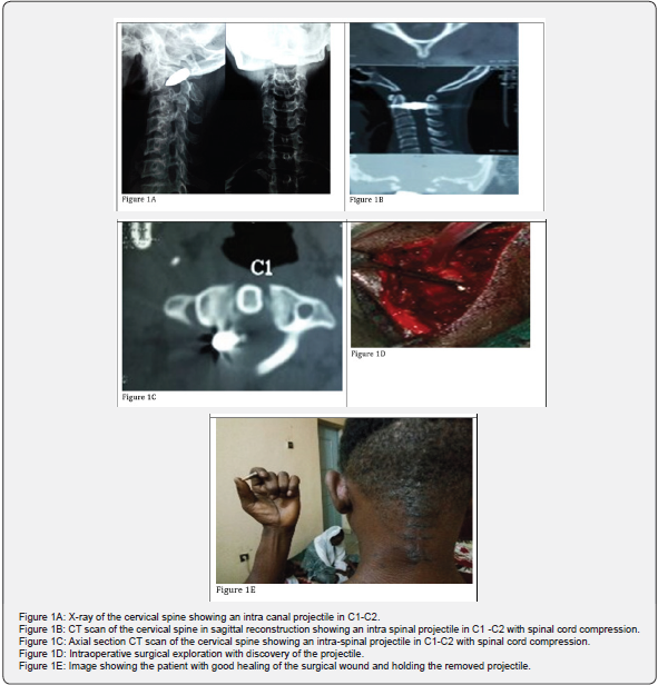
The 29-year-old patient was seen in a neurosurgery consultation for neck pain with right hemicorporeal heaviness that had been evolving for 03 months. The patient had a history of trauma with a cervical impact by firearm, 02 years ago, without associated neurological signs. The trauma had caused a single cervical wound of about 2 cm in length which had healed correctly. The clinical examination revealed a Brown-sequard syndrome consisting of a spinal syndrome, a right hemicorporeal pyramidal syndrome and a contralateral epicritic sensitivity disorder. Paraclinically, an X-ray of the cervical spine showed a C1-C2 intraductal projectile (Figure 1A) and a CT scan of the cervical spine showed a C1-C2 intra-spinal projectile with spinal cord compression (Figure 1B & 1C). The evolution before surgery was marked by neck pain and right hemiparesis. Surgical exploration (Figure 1D) revealed an intrarachid ball of which 1/3 was intramedullary and 2/3 was extra-medullary intraductal. The bullet was removed with a dural plasty. The postoperative course was simple with motor recovery from 2/5 to 4/5 in one month and good healing of the surgical wound (Figure 1E).
Discussion
Our study involved a case of traumatic spinal cord injury caused by a firearm. The clinical manifestation is sometimes late, and in our case it was hemiparesis two years later. An intramedullary bullet in C1 -C2 on CT and X-ray with possible slow migration of the projectile to the possible slow migration of the bullet into the spinal cord. Studies on vertebro-medullary wounds caused by knives and firearms are rare, but the increasing frequency of the latter in civilian practice makes them of interest [2]. Epidemiologically, there is a male predominance with an average age of 26 years with extremes ranging from 17 to 46 years. The diagnosis of spinal injury is easy when it is a spinal injury with a frank neurological syndrome. In other situations, spinal cord injury may go unnoticed, especially if the entry point of the wound is far from the spine and the clinical symptoms are discreet or difficult to interpret because of associated lesions. It is important to think of this type of lesion before any paraspinal wound and to carry out a meticulous neurological examination. Neurological deficits are frequent and polymorphic, and true Brown-Sequard syndromes have been reported in the literature [2]. This would explain the delay in diagnosis of a 2-year old ballistic spinal cord injury in our case [3].

The clinical manifestations depend on the level of injury and the quantity of preserved spinal cord. The loss of sympathetic tone, secondary to the SCI, disrupts the balance between sympathetic and parasympathetic tone and can cause arterial hypotension and extreme bradycardia, especially for lesions above T6. The neurogenic shock which results from this imbalance must be differentiated from spinal shock which is defined as a temporary clinical state of spinal cord sideration with impairment of motor functions (flaccid paralysis), sensory and autonomic nervous system, occurring a few hours after the installation of the lesion and which can last from a few days to a few weeks. Two particular clinical forms are to be known: the centromedullary syndrome and the Brown-Séquard syndrome [4].
In a traumatic context of the spine, MRI is currently the firstline examination in the presence of neurological signs [5]. This is not appropriate in our case because of the ballistic component of the trauma. Before the use of high-resolution CT scans, thinsection CT was very useful for highlighting bony abnormalities that may have been missed on standard radiographs. It also allows the evaluation of the extra spinal soft tissue [2-6]. Trauma to the upper cervical spine is associated with a predominance of odontoid fractures in 46.66% of cases [7] according to I. Tine et al. The management of the spinal cord injury must take place in a specialised centre. The aim is to avoid the development of secondary spinal injuries by continuing spinal immobilisation, ensuring sufficient spinal blood flow, adequate oxygenation and performing fixation or decompression surgery if necessary [4]. Surgical treatment of patients with spinal trauma aims to stabilise the injury, relieve spinal cord compression and facilitate resuscitation management, particularly during mobilisation. The optimal time to perform this surgery, if it is indicated, is not clearly defined in the literature. There is no randomised controlled trial evaluating this issue [4-8]. In our study, the evolution was marked by motor recovery from 2/5 to 4/5 in one month.
Conclusion
Firearm injuries are common and cause serious injuries, very often lethal or with significant sequelae, in a number of cases disabling. Spinal cord injuries may go undetected and only be diagnosed at the stage of complications [2] and the indication for MRI in ballistic spinal cord injuries must consider the nature of the projectile [1]. Hence the importance of a complete and systematic clinical and radiological assessment. Much progress remains to be made in the management of spinal cord injuries. Currently, their management involves many professionals and aims to limit the disability by avoiding aggravation or the occurrence of secondary injuries [4].
References
- A Daghfous , K Bouzaïdi , M Abdelkefi , S Rebai , A Zoghlemi, et al. (2015) Contribution of imaging in the initial management of ballistic trauma. Diagn Interv Imaging 96(1): 45-55.
- Yaqini K, Guartite A, Malki M, Louardi H, Abbassi O, et al. (2004) Vertebro-medullary wounds about 3 cases ©Masson, Paris, 2004 JEUR, 17, 37-41.
- P Pollion, K Senamaud-Dabadie, B Schnedecker, S Gonidec, T Musson, et al. (2014) Traumatisme crânien de type balistique chez un enfant de dix ans (Ballistic head injury in a ten-year-old child) La revue de médecine légale 5: 165-169
- Aurore Rodrigues (2018) Prise en charge des traumatisés médullaires, Le Praticien en anesthésie reanimation. PRATAN-778: 4.
- S Elrai, M Souei Mhiri, N Arifa Achour, K Mrad Daly, R Ben Hmida, et al. (2006) Contribution of magnetic resonance imaging in spinal cord injury J Radiol 87: 121-126.
- Jessica Pessayre (2015) Perioperative management of patients with chronic spinal cord injury, Le Praticien en anesthésie réanimation 19: 304-307.
- I Tine, ERB Atangana, PI Ndiaye, M Agbo-Panzo, AA Diop, M Faye Trauma of the spine at the Hôpital Principal de Dakar (HPD) : about 126 cases.
- YM Sakti, MA Saputra, T Rukmoyo, R Magetsari (2018) Spinal cord injury without radiological abnormality (SCIWORA) manifested as self-limited brown-SEQUARD syndrome. Trauma Case Rep 18: 28-30.


