




 JRANN Home


Abstract
Gene therapy is a novel method for treating genetic and oncological disorders. Conventional delivery vehicles like viral and non-viral physical vectors are facing serious limitations and drawbacks like the immunogenicity, cytotoxicity and the payload capacity. This review analyzes the nanoparticle-based gene delivery as an alternative method to overcome the problems from various common vectors by low mutagenesis, preservation of cell viability and the precision in molecular targeting. Also, the applications of nanocarrier in overcoming blood brain barrier, liver hepatocytes and some types of tumor microenvironments will be discussed. Despite there are lots of advantages of nanoparticle-based gene delivery, some points of view will be shared as the thought of the future directions by utilizing electrospun nanofiber as the support in order to transfer the advanced therapeutic to clinic.
Keywords: Nanotechnology; Nanoparticles; Gene therapy; Gene delivery
Introduction
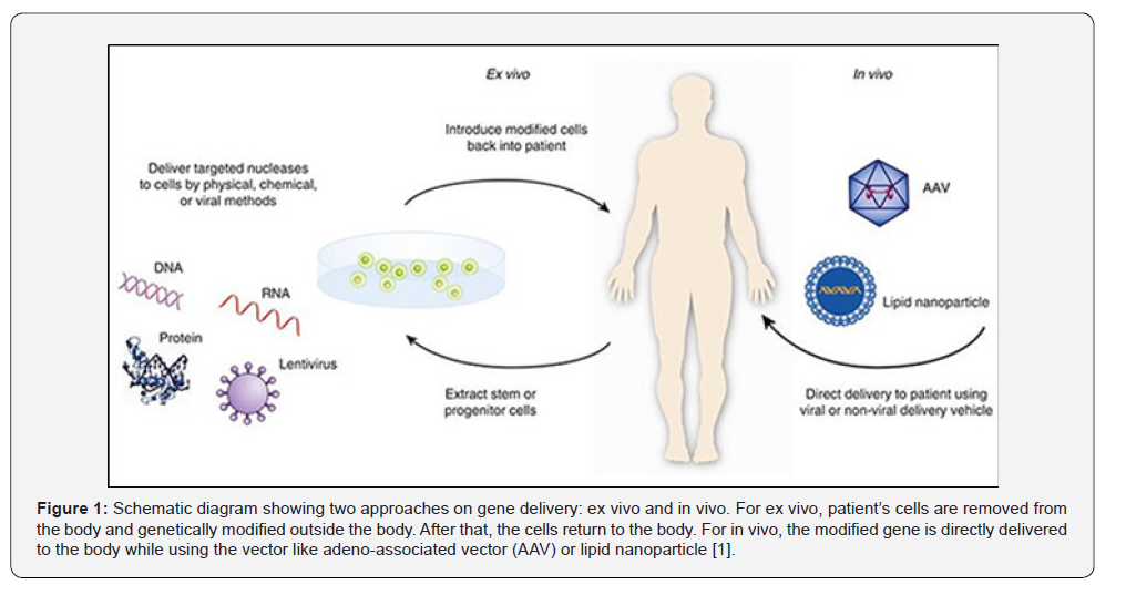
According to United States Food and Drug Administration (FDA), gene therapy is a technique that modifies a person’s genes to treat or cure disease which can be divided into replacing of disease-causing gene by a health copy of the gene, inactivating the malfunction disease-causing gene or introducing a new or modified gene for disease curing. The approach of gene therapy can be divided into ex vivo or in vivo in which the patients’ cells are removed and modified outside the body and reinfuse into body again or the therapeutic gene is directly delivered into patient’s body. Figure 1 shows these two therapeutic approaches and the Adeno-Associated Virus (AAV) or lipid nanoparticles as the examples of carrying vector. [1] Many diseases are caused by the single gene disorders where the mutations occurred in a single gene just like cystic fibrosis, hemochromatosis, etc. [2] In recent years, in order to improve the precision and specificity of disease treatment, gene therapy becomes an alternative of the convention therapeutic methods and there are several gene therapies (including viral and non-viral vectors) have been approved by FDA in recent years. For example, in 2024, Pfizer discovered an AAV based drug called Beqvez which focuses on Hemophilia B. Also, Rivfloza discovered by Novo Nordisk A/S in 2023 is a GalNAcbased gene therapy drug for primary hyperoxaluria [3]..
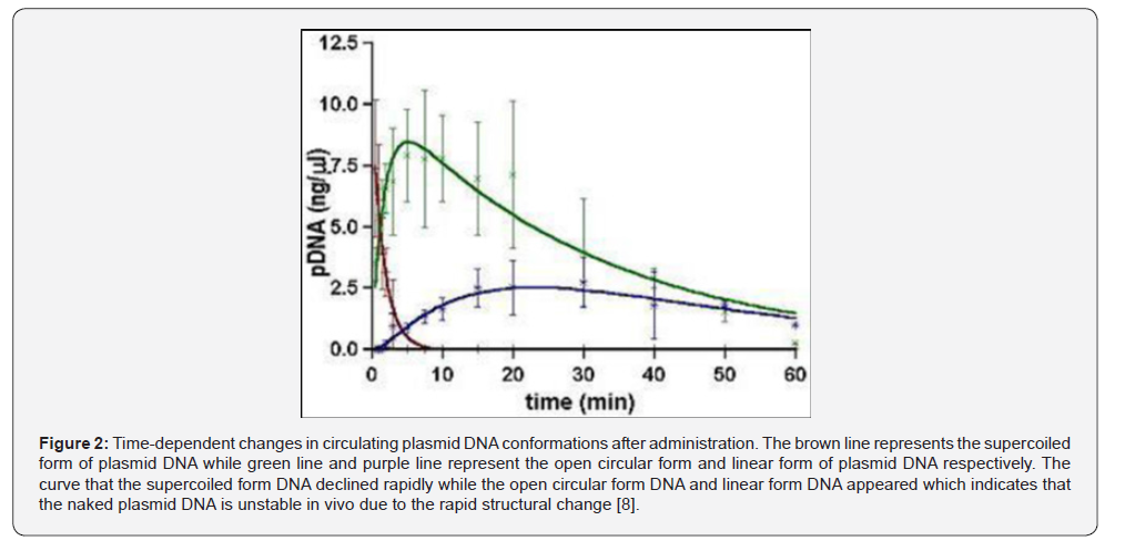
Even though gene therapy provides specific and efficient treatment for the disease, as mentioned above, conventional gene delivery is still facing lots of challenges. In terms of the immunogenicity and safety issue, gene therapy utilizes the viral vector as the cargo to deliver the genetic materials, Thomas et al. analyzed that when adeno viral vectors are delivered into the body, the immune responses would be triggered and then cause the risk of cytokine storms which is a serious safety issue for the gene delivery by the adeno viral vectors. [4] Also, Hecein-Bey- Abina et al. reported the first case that two patients with X-linked severe combined immunodeficiency (SCID-X1) treated by gene therapy and both of them got leukemia after three years. [5] As a result, the risk of cancer has also been taken into account for conventional gene therapy. With respect to limitations of such conventional non-viral physical methods as electroporation, microinjection or gene gun, Hardee et al. stated that these methods are highly invasive, especially electroporation, which may cause the permanent damage of the tissues. Moreover, the physical methods cannot deliver the genetic materials to whole body through the circulation system like viral vector resulting in low efficiency for metastatic diseases or multiple organ dysfunction syndrome. [6] On the other hand, the low stability of naked DNA or RNA is one of the major concerns for gene therapy. In early research by Kenji’s group, they utilized mice as the model the pharmacokinetics of plasmid DNA after intravenous injection and found that the degradation rate of naked plasmid DNA in circulation. It was suggested that the naked plasmid DNA was degraded by plasma deoxyribonuclease and the enzymes in other parts of body like the liver. [7] As shown in (Figure 2), the rapid drop of the supercoiled form of plasmid DNA (red line) indicates that the intact naked DNA is unstable in the circulation system which is not promising in gene therapy [8].
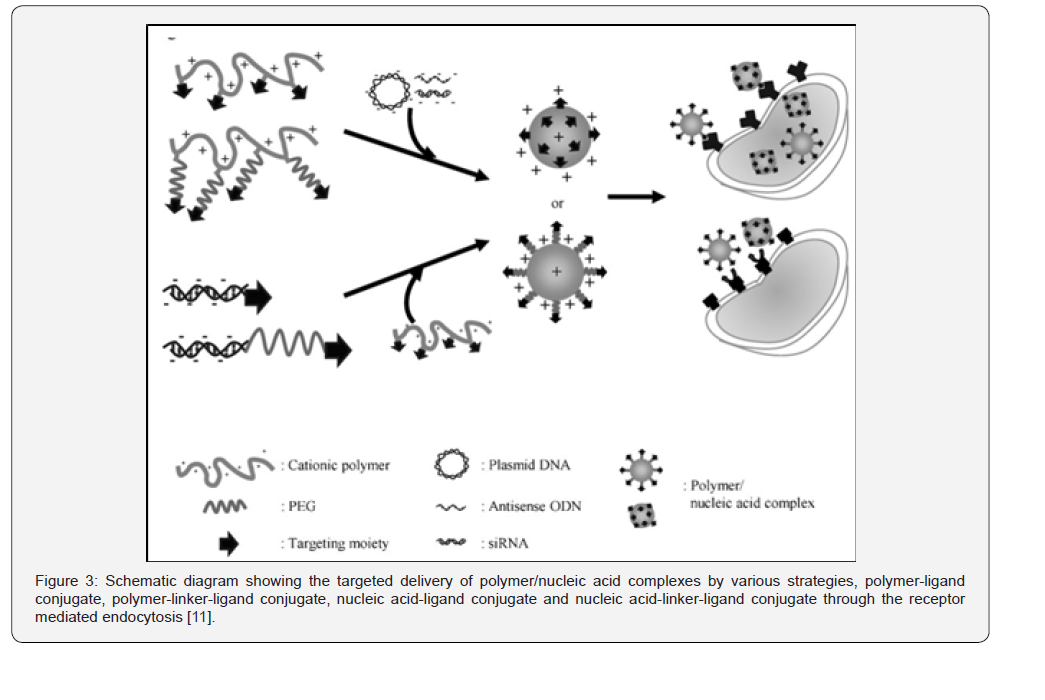
To deal with the aforementioned problems, nanoparticlebased gene delivery could be a solution for the gene therapy. According to Fabienne et al., they defined nanoparticles as “solid and spherical structure ranging around 100 nm in size and prepared from natural or synthetic polymers”. [9] In gene delivery, nanoparticles as the non-viral vector, the genetic materials such as DNA and RNA can be encapsulated or adsorbed inside the nanoparticles. With reference to Huayu et al. proposed mechanism, the electrostatic attraction between the cationic polymers and the negatively charged nucleic acid from the genetic materials causes the formation of stable nanocomplexes which protect the genetic materials from the degradation by the nucleases in the blood. The nanoparticles are then guided by surface ligands and enter the cell through endocytosis. Once inside the cell, the endosomal escape allows the nanocomplexes to leave the endosome and the genetic materials are then released to the cytoplasm. The genetic materials are transported to nucleus or cytosol for effective gene expression or silencing at the end [10]. In (Figure 3), the schematic diagram summarizes the general mechanism of polymeric nanoparticle gene delivery from the formation of the nanocomplex to the endocytosis.[11] Thanks to the mechanism described above, the nanoparticle gene delivery system achieves several merits comparing to the conventional gene delivery methods. For example, Kavanagh et al. [12] reported that the nanoparticle-based delivery is with lower risks of insertional mutagenesis and the reduction in the concern of immunogenicity compared to viral vectors. [12] Harris et al. demonstrated the change of peptide coating density shifted the in vivo gene delivery between different organs. They stated that the low coating density is for liver delivery and the delivery to the spleen and bone marrow will be the high coating density. By this finding, it can show that nanoparticle gene delivery having a tunable tissue specificity. [13] In this review, the advantages of nanoparticle gene delivery will be discussed as well as with their examples to illustrate the applications. At the end of the paper, my views will be discussed on how the nanoparticle-based gene therapy can be widely used in the future with the combination of nanofiber technology.
Review Section
Nanoparticles are as the carrier for the non-viral delivery of genetic materials such as DNA, RNA or genetically modified components to the cells in nanoscale. Various types of nanoparticles, including cationic compounds, inorganic nanoparticles and recombinant proteins, are used for incorporating with DNA or forming the complex with DNA. [10] Because of their characteristics just like the size or surface charge, nanoparticles become a promising carrier for gene delivery. In this section, the advantages of nanoparticles-based gene delivery together with their applications on specific cells will be discussed.
Advantages of using nanoparticles for gene delivery compared to viral vectors
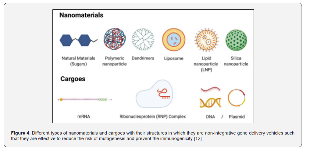
In terms of the safety concern, nanoparticles as genetic materials carrier perform lower risk in mutagenesis and reduce chance of the immunogenicity. According to Kavanagh et al., various types of nanomaterials are shown in (Figure 4), they serve as non-integrative gene delivery vehicles which means unlike the viral vectors that integrate the genetic materials for delivery. [12] Since the nanoparticles deliver the cargoes through cytosolic or episomal way which does not involve the process of integrating into DNA or other genetic materials. It is effective to minimize the risk of mutagenesis and prevent the disruption of the host gene. As some of the nanoparticles are made from natural or biocompatible materials, when comparing to viral vectors that may cause strong immune response because of the viral proteins, those nanoparticles would be less immunogenic.[12] Another study by Green et al. stated that nanoparticle as a non-viral vector could reduce immunogenicity in gene editing therapy. They demonstrated that the nanoparticles with surface modification by PEGylation could prolong circulation time and minimize immune detection. [14] As a result, this design of the nanoparticle could attenuate immune recognition such that alleviate immunogenicity during the in vivo transfer. Also, Yin et al. pointed out that since the nanocarriers, through the episomal delivery or direct mRNA translation in the cytoplasm, enable transient gene expression for the host cells, such that the risks of unintended genomic integration or mutagenesis can be avoided [15].
Regarding the scalability and the manufacturing cost, nonviral nanoparticles can be more scalable which can be a great advantage. Due to the extensive clinical and commercial adoption of gene therapy, the feasibility of mass production becomes a concern. For the viral vectors, with reference to Wright’s review, it requires large scale mammalian cell cultures, extensive purification processes for empty capsids removal as well as the straight quality control to ensure the batch-to-batch consistency [16]. By these manufacturing practices, the production of viral vectors for gene therapy cause the issue of excessive operation costs. However, when comparing to nanoparticle-based gene delivery method, the production of nanocarrier follows the standard chemical and physical processes instead of the complicated cellular machinery. Cullis et al. demonstrated the synthesis of lipid nanoparticles and polymeric vectors in which the process involved the scalable and continuous-flow technologies such as microfluidic mixing [17]. Their finding presented the microfluidic technology achieves high consistency and standardize manufacturing process while the nanocarrier is mass-producible. Also, the less complicate production operation could facilitate the scale-up which is crucial for lowering the manufacturing costs. On the other hand, another research focusing on Clustered Regularly Interspaced Short Palindromic Repeats (CRISPR) genome editing pointed out that nanoparticles as attractive platform due to the scale-up capability which means the nanoparticle gene delivery can be moved from laboratory scale to the reproducible production scale through the physicochemical manufacturing processes [18]. As a result, from previous studies, nanoparticle-based gene delivery is more advanced than the conventional viral vector gene delivery when it comes to the scalability as well as the manufacturing operation costs.
With respect to the payload capacity, nanoparticle-based gene delivery carrier does not be restricted by the payload. Based on the study by Victor et al., the payload capacity is the amount of therapeutic cargo in which a nanoparticle is able to carry, referring to the amount of nucleic acid molecules in each nanoparticle. [19] According to Wu et al., even though AAVs as the gold standard for many in vivo studies of gene therapy, their rigid protein capsids limit the maximum capacity for packaging to approximately 4.7 kilobases. The author pointed out that if the delivery involved the CRISPR system or even bulkier gene, the AAV vector would not be possible to do so due to the size limit of the AAV vector. [20] By contrast, since the structure of nanoparticlebased carrier is prepared by the electrostatic complexation or physical encapsulation which does not like the viral vector that is produced by biological packaging. Patil et al. in their study proved that polymeric or lipid nanoparticle carrier is able to encapsulate the plasmid DNA with large size or the multiple mRNA within a single nanoparticle. [21] Moreover, based on the finding by Kim et al., the combination of nanoparticles is uniquely suitable for the CRISPR system. They pointed out that nanoparticles not only can encapsulate the large plasmid DNA or mRNA, but also the ribonucleoprotein complexes. [22] Their findings can support that nanoparticles are highly adaptable system for gene therapy due to the broad cargo compatibility. As a result, for the payload capacity, the nanoparticle-based gene delivery is more advanced than the viral vector gene delivery.
Advantages of using nanoparticles for gene delivery compared to non-viral vectors other than nanoparticles
When it comes to the cytotoxicity and in vivo applicability, nanoparticles for gene delivery can minimize the cytotoxicity during in vivo applications. Electroporation as one of the nonviral physical methods, for example, it involves the use of electric pulse to transfect cells with DNA by the application of electric field to induce temporary pores in the cell membrane such that the DNA sequences can be taken up. [23] In the study of Gehl et al., the physical mechanism of electroporation was explained. They pointed out that the electroporation method operates within a narrow therapeutic window which indicates a reduced margin for error in dosing as well as the increase of risk of adverse effects. [24] Gehl et al. stated that the reason behind the narrow therapeutic window is that when the voltage is too low, the genetic materials cannot be entered the cell while for high voltage, it will cause the irreversible membrane rupture or even cellular necrosis and tissue trauma. [24] Also, Hardee et al. compared the physical non-viral gene delivery methods, including the electroporation, they pointed out that these technologies are with high cytotoxicity resulting in the low cell viability after the delivery. [6] Hence, the non-viral physical methods are not suitable to apply in vivo. However, nanoparticles gene delivery methods provide a solution for the problems mentioned above by the biomimetic approach and non-destructive way for delivering the genetic materials and entering the cells. Ramamoorth et al. [25] reported that the nanoparticles as the engineered carrier for genetic materials, these particles are with the highly modifiable physicochemical properties such as the controlled size and surface charge. By utilizing these properties, the nanocarrier can interact gently with the cell membrane without causing cell lysis. [25] Therefore, the cell viability can be preserved after the nanoparticle-based gene delivery while the therapeutic gene can be expressed due to the high survival rate of the cells. Another study by Whitehead et al. stated the nanoparticles, just like the lipid based and polymeric materials, are advantage in systemic administration. Due to the controlled particle size and the surface modification with PEG, the nanoparticle-based carrier can facilitate the absorption from the target tissue and inhibit the non-specific gene delivery. As a result, with the nanoparticle-based delivery, the genetic materials are allowed to be delivered through the bloodstream to the internal organs [26].
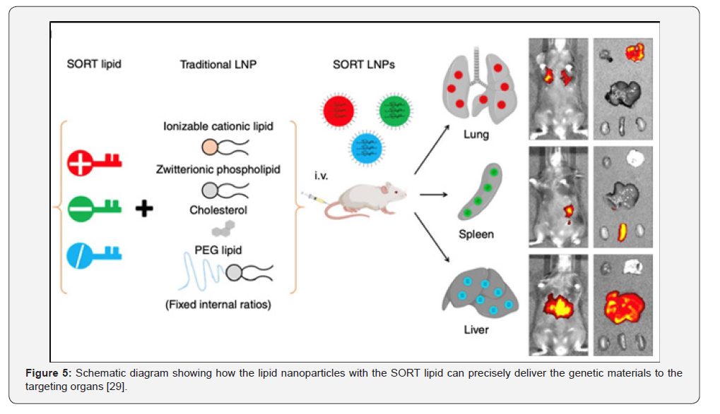
For the precision of gene delivery to the targeted tissues or organs, it would be one of the advantages of nanoparticles gene delivery over the conventional non-viral delivery. According to Yin et al. [15] traditional non-viral delivery just like the electroporation or gene gun requires the localized macroscopic force which forbids the systemic molecular targeting. [15] The preclusion of molecular targeting means the absent of relevant target which causes the gene therapy cannot be applied appropriately resulting in the reduction of applicability. [27] In contrast, Peer et al. [28] demonstrated that with the properties of nanoparticle-based gene carrier in which the surface modification with targeting ligands grafting to enable specific interactions with the receptors on the target cells. [28] Therefore, although the study from Peer et al. [28] is about cancer therapy, the principles of the nanocarrier targeting proposed by them is still able to prove the precision of nanoparticle-based gene delivery. Another important research from Cheng et al. suggested the Selective Organ Targeting (SORT) system which shows how the nanocarrier undergoes the precise delivery to the specific organs. With referring to (Figure 5), they utilized rat model to demonstrate the internal composition of lipid nanoparticles can redirect mRNA and CRISPR-Cas delivery preferentially to the organs just like lung, spleen or liver through the systemic administration which is a characteristic that cannot be achieved by the conventional physical method. [29] With the supporting research by Blanco et al. [30] when the nanoparticles reach the targeting site, the carriers can be internalized through endocytic pathways to support the intracellular delivery of the genetic materials cargo. [30] Based on the review of previous studies, in terms of the specific delivery of genetic materials, the nanocarriers can show an advantage over the conventional nonviral physical methods.
Examples of using nanoparticles for gene delivery to specific cells or tissues
For the brain neurons gene delivery, Blood-Brain Barrier (BBB) is the biggest limiting hurdle to Central Nervous System (CNS) due to its prevention of harmful reagents from entering the brain and the control of nutrients balance. [31] With the aid of nanoparticles, through the physical and biochemical ways, the problem by BBB can be overcame. Wong et al. analyzed how different nanocarrier systems including lipid-based formulations for surface modifications affect the penetration of the CNS. They emphasized the physicochemical properties of nanoparticles, such as small size, are important for the diffusion inside the brain parenchyma. [32] Yang et al. analyzed a number of nanoparticles platform such as lipid nanoparticles, polymeric nanoparticles and inorganic nanoparticles with targeting ligands surface modified. In terms of the administration route, they found that the intracerebroventricular, intrathecal and intranasal routes can access the CNS directly while bypassing the peripheral clearance. [33] Si et al. reviewed the engineering of the nanoparticles with surface ligands targeting receptors overexpressed to BBB endothelial cells for the transport into the brain parenchyma. The nanoparticles delivered nucleic acids including siRNA, miRNA and plasmid DNA to glioblastoma multiforme cells and the surrounded tumor microenvironment [34].
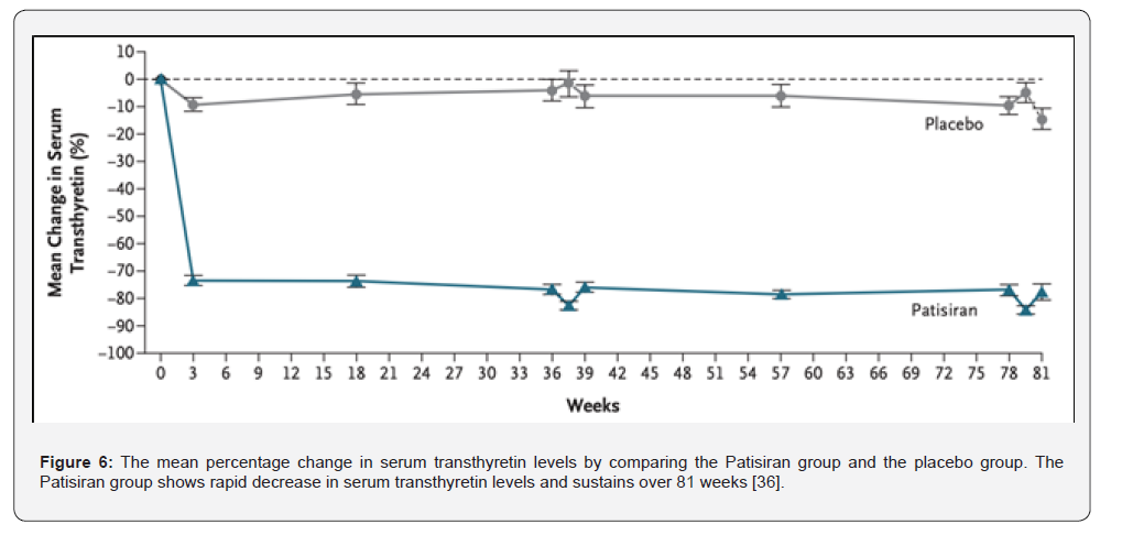
In terms of the liver hepatocytes, nanoparticles can deliver the mediated genetic materials effectively. Arjunan et al. reviewed the application of lipid nanoparticles mediated with nucleic acids to be delivered to hepatocytes for the therapy of hereditary liver disorders. They found that the lipid nanoparticles with optimized ionized formulations can deliver mRNA, siRNA or CRISPR systems for gene therapy which could be applied for liver disorders such as hereditary disease or alcoholic liver disease. [35] Adams et al. published their study regarding the Patisiran (Onpattro) which is the first lipid nanoparticle-based siRNA drug for effective and precise delivery and achieving gene silencing for the genetic fatal liver disease, Hereditary Transthyretin Amyloidosis (HATTR). The Patisiran was the first FDA approved therapeutic drug and the therapy can reduce serum transthyretin levels by 81% of the hATTR patient, as shown in (Figure 6), proving that the inhibition of the expression of the pathogenic proteins. [36] After the development of the Patisiran, Adams et al. performed a 5-year clinical research data to show the stability and safety using in human. [37] On the other hand, the long-term gene silencing could achieve the long-term effectiveness which is a main focusing point for the hereditary diseases. In the same research by Adams et al., they demonstrated the nanoparticle based delivery of siRNA can slow down the progression of neuropathy for the hATTR patients along the 5-year follow-up period, indicating that the nanoparticle based delivery is important for chronic disease management [37].
Another application of nanoparticle-based gene carrier would be the cancer cells as known as the tumor; by utilizing various types of nanoparticles, the therapeutic genetic materials provide the silencing of oncogenic targets within the tumor microenvironment. Schiffelers et al. [38] discovered a self-assembly based nanoplexes which integrates the Polyethyleneimine (PEI) and Polyethylene Glycol (PEG) as the sterically stabilized nanoparticles for delivering siRNA. The cationic PEI and negatively charged siRNA form a stable core which can protect the carried nucleic acid drug from degradation by nucleases in serum. Also, the surface conjugation of Arg-Gly-Asp (RGD) peptide ligands enables the targeting αvβ3 integrins which can improve the selective uptake efficiency of tumor cells due to the overexpression on tumor neovascular endothelial cells. [38] As a result, it causes the antiangiogenesis as well as inhibits the tumor growth. Instead of using the polymeric nanoparticles, Ferreira et al. reviewed the gold nanoparticle as the carrier for genetic materials for cancer therapy. Due to the high tunability, biocompatibility and the ease of surface modification, gold nanoparticles are considered as an ideal carrier for nucleic acid such as siRNA and plasmid DNA. Also, the photothermal effect of gold nanoparticles can combine the gene therapy and photothermal therapy for the anti-tumor effect. [39] Therefore, both the polymeric and the metallic based nanoparticles can also contribute to the gene therapy for cancer.
Nanoparticle based gene carrier can also be applied in Pancreatic Ductal Adenocarcinoma (PDAC) to overcome tumor’s dense stroma and deliver the nucleic acid. PDAC is a relatively uncommon cancer but it is expected to be the second highest mortality rate in 2030. [40] Won et al. reviewed the gene therapy using nanoparticle-based carrier to deliver RNA interference (RNAi) based therapeutics and CRISPR tools to overcome the cellular barrier in PDAC tumor for precise gene delivery to the tumor. [41] Wang et al. reviewed the previous studies about the applications of such different nanoparticles as lipid, micellar or gold nanoparticles for gene modulation and immunotherapy of pancreatic cancer therapy. They stated that nanoparticles are able to deliver immune and gene-based agents to target tissues and organs and thus inhibit the growth of tumor and metastasis due to the characteristics of nanoparticles including the high stability, permeability and low toxicity to nontarget tissues. [42] A recent study by Zhu et al. demonstrated the dual mRNA nanoparticle strategy for effective tumor neoantigen immunotherapy. The dual mRNA lipid polymeric based nanoparticles generate anti-PD-1 antibodies to overcome immunosuppression in PDAC tumors such that the growth of tumor can be inhibited [43].
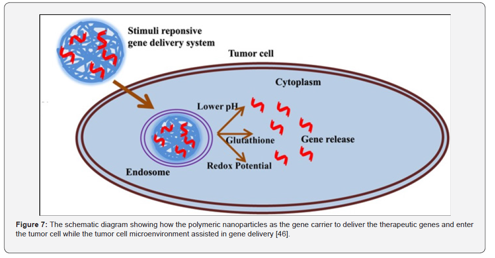
For the Non-Small Cell Lung Cancer (NSCLC) cells, previous studies showed promising results for using nanoparticles to deliver genetic materials for gene therapy of the non-small cell lung cancer. Chaudhary et al. [44] reviewed the strategic targeting of NSCLC utilizing gene delivery platforms. The biomolecule based nanoparticles deliver various types of nucleic acid just like plasmid DNA or mRNA to NSCLC cells. [44] Lee et al. reviewed the utilization of nanoparticles in the treatment of lung cancers in which the nanoparticles carry the gene molecules for transfection and targeting through systematic or localized administration. Due to the surface modification with targeting ligands, the tumor suppressor genes such as p53 can be delivered to the tumor cells precisely in order to inhibit the lung cancer cells growth which shows the advantage over the conventional drug with poor targeting and easy degradation issues. [45] Moreover, Narsireddy et al. [46] discussed how to utilize the polymeric nanoparticles as the alternative carrier of lipid or inorganic molecules to deliver nucleic acid therapeutics to the target tumor cells in order to overcome the limitation of the conventional chemotherapy. As shown in (Figure 7), they utilized polymeric nanoparticle as a stimuli responsive gene delivery system which is sensitive to the tumor microenvironment. After entering the cell, the genetic materials encapsulated by the polymeric nanoparticles were released for therapeutic use [46].
Discussion
Despite the nanoparticles-based gene carriers exhibit numerous advantages including the low toxicity, high cell viability or immunogenicity making it become a promising technology for gene therapy, some of the limitations should be taken into account before the clinical applications. For example, the nanoparticles are cleared rapidly and their poor retention at disease sites which cause the nanoparticles are hard for clinical translation of nonviral gene therapy. [47] As a result, combining the nanoparticles with another novel nanotechnology, electrospinning Nanofiber (NF) such that the nanocarrier for gene delivery can be more advanced by resolving the previous mentioned limitations. Also, in the last part of this section, some points of view of scaling up will be discussed.
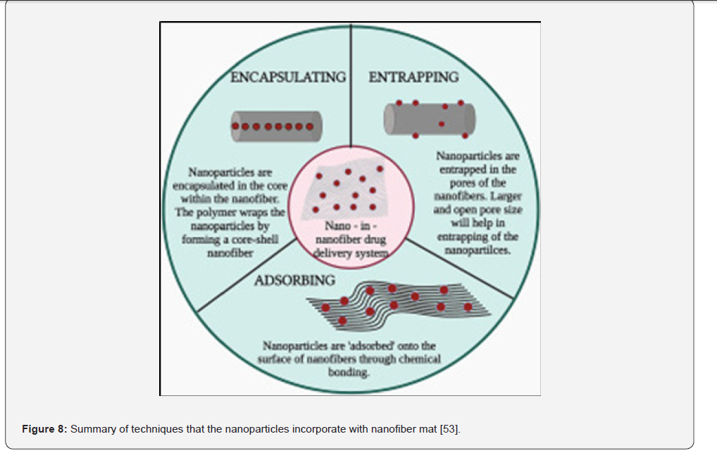
Electrospinning is a fiber fabrication method to produce ultrafine NFs with the application of high voltage with low current on the electroconductive polymer solution. [48] Previous studies discussed about some benefits of electrospun NFs drug delivery system including the safe release at target site as well as a costeffective procedure. [49-50] Also, previous study showed that the electrospun NFs can mimic the extracellular matrix and the NF membrane is biodegradable as well as the assistance in regeneration process. [51] Tong et al. demonstrated that the cells can be attached on the poly(hydroxybutyrate-co-hydroxyvalerate) (PHBV) NF mat by the cell culture experiments, while the cell spread and cell proliferation were found on the NF mat as well. [52] Since the polymer solution is the key for the electrospun NF, the nanoparticles can be added during the solution preparation process. After the preparation of the nanoparticles including the surface modification and encapsulation of genetic materials, as some of the nanoparticles can be dissolved in organic solvents, especially the polymeric based nanoparticles, they will be able to mix with the polymer electrospinning solution. After the electrospinning process, the nanoparticles can be incorporated with the NF mat by encapsulating, entrapping or adsorbing mode, as shown in (Figure 8). [53] On the other hand, Tong et al. [54] used PHBV to encapsulate Carbonated Hydroxyapatite (CHA) nanoparticles for electrospinning. The electrospun NF prepared by them can be applied in bone tissue engineering and showed promising results. [54] They also demonstrated the negatively charged scaffolds composed by poly (D, L-lactic acid) and CHA nanoparticles can enhance cell proliferation, alkaline phosphatase activity and mineralization while the structural integrity was maintained. [55] Hence, it is possible to prepare the materials for the electrospun NFs which could encapsulate the nanoparticles with genetic materials such that a double nano system could be created for genetic materials delivery and biological applications.
On the other hand, previous studies stated that the NF mat incorporated with nanoparticles. In terms of the targetability and the retention time of nanoparticles and drugs, the NF and nanoparticles composite can demonstrate an increase in these aspects. A study by Fatmanur et al. about vaginal infections performed the development of chitosan nanoparticles loaded with NF showed that the experimental set with the hybrid of nanoparticles and NFs can perform higher drug permeation through vaginal tissue thanks to the high surface area to volume ratio of the NFs as well as the porosity. Also, the polymer they utilized, polyvinylpyrrolidone (PVP), the NFs formed after electrospinning are with mucoadhesive properties which is suggested that causing an increase in interaction with vaginal mucosa and the release of drug within a required period of time. [56] Moreover, there was success case that performed by Chen et al. that they utilized the poly (d,l-lactic-co-glycolic acid) (PLGA) to encapsulate the nanoparticles consisted of chitosan and siRNA. The biodegradable NFs composite was able to release the siRNA at desired pH and the PLGA basd NFs acted as a protective barrier which prevented the siRNA from being hydrolyzed [57].
From the advantages and applications of the double nano system which includes the nanoparticles and NFs mentioned above, it sounds like the electrospun NF should be a promising candidate for the gene therapy. However, the electrospun NF is still not commercialized and transferred to clinical use. With reviewing the literatures published, the electrospinning setup is the needle type syringe which is still the laboratory scale. For the needle type electrospinning, the efficiency of fiber formation is low as the fiber can only be deposited on a rotating substrate. Also, the area of the NF mat is small for using the needle type setup which means the production capacity is low as well. The low production efficiency and low production capacity will contribute to high cost of the manufacturing resulting in extremely expensive of the final product. For the mass production of NF mat, it is worth to consider using the needleless electrospinning setup by transforming the production of NFs from laboratory scale to industrial scale. As reported by Lee et al., the application of needleless electrospinning can probably meet the large scale of manufacturing of NFs. [58] If large number of NFs can be manufactured within a short period of time, it may help the speed up of the NF based therapeutic products including the NF and nanoparticles gene delivery composite to be commercialized. Just like the product of NASK N95 respirators, FFP2 respirators, baby diapers, adult diapers and Nano AirPure flat media air filters commercial electrospun NF products, they are researched and manufactured in Hong Kong which are the successful cases showing that the electrospun NFs not only can be mass produced but also be commercialized from the laboratory bench to the market. [59] Nevertheless, the polymer solution formulations for various applications and the electrospinning parameters such as voltage, humidity or temperature are still to be adjusted and optimized.
Conclusion
In this review, the advantages of nanoparticle-based gene carrier over the conventional viral and non-viral vectors other than nanoparticles were discussed. The nanocarriers can mitigate the safety and immunogenicity concerns for the viral vectors while avoiding the high cytotoxicity. The highly tunable characteristic makes the nanocarriers to have high payload capacity, systemic stability and precise organ-specific targeting. In terms of the applications of the nanocarrier, the nanoparticle could successfully penetrate the blood-brain barrier as well as perform long term gene silencing in hereditary liver diseases. Also, the overcome of tumor stroma and the inhibition of tumor growth are also mentioned in this review. To share our point of view, the electrospun NF technology was discussed to offer a highly viable solution to the current hurdles just like the clinical transfer and commercialization. The double nano composite system not only mimics the extracellular matrix but also become a protective barrier to prevent enzyme degradation. It is hoped that the needleless electrospinning method can be all the more mature such that the technology can be scaled up production from laboratory to industrial level and these advanced therapeutic methods become a cost effective and accessible for gene therapy in the future.
Acknowledgement
The content of this review article is mainly based on that of a term paper in Department of Biomedical Engineering, The Chinese University of Hong Kong, Hong Kong Special Administrative Region, China. We would like to thank Ms. Sarinda Kwok, Chief Executive Officer from Profit Royal Pharmaceutical Limited, for the support of research and development activities. We would also like to thank Professor Kwok Pui Fung, Advisor, R&D Department from Profit Royal Pharmaceutical Limited for the guidance on the refinements of this review article.
References
- Administration USFaD (2026) What is Gene Therapy? U.S. Food and Drug Administration 2020.
- NCBI (2010) Single-Gene Disorders, in Understanding Genetics. 2010, National Center for Biotechnology Information (US): Bethesda.
- Geng G, Xu Y, Hu Z, Wang H, Chen X, et al. (2025) Viral and non-viral vectors in gene therapy: current state and clinical perspectives. eBioMedicine 118.
- Thomas CE, Ehrhardt A, Kay MA (2003) Progress and problems with the use of viral vectors for gene therapy. Nature reviews Genetics 4(5).
- Hacein-Bey-Abina S, Kalle CV, Schmidt M, Mccormack MP, Wulffraat N, et al. (2003) LMO2-Associated Clonal T Cell Proliferation in Two Patients after Gene Therapy for SCID-X1. Science 302(5644): 415-419.
- Hardee C, et al. (2017) Advances in Non-Viral DNA Vectors for Gene Therapy. Genes 8(2).
- Kawabata K, Takakura Y, Hashida M (1995) The Fate of Plasmid DNA After Intravenous Injection in Mice: Involvement of Scavenger Receptors in Its Hepatic Uptake. Pharmaceutical research 12(6).
- Houk BE, Hochhaus G, Hughes JA (1999) Kinetic modeling of plasmid DNA degradation in rat plasma. AAPS PharmSci 1(3).
- Danhier F, et al. (2012) PLGA-based nanoparticles: An overview of biomedical applications. Journal of controlled release 161(2): 502-522.
- Tian H, Chen J, Chen X (2013) Nanoparticles for Gene Delivery. Small (Weinheim an der Bergstrasse, Germany) 9(12).
- Mok H, Park TG (2009) Functional Polymers for Targeted Delivery of Nucleic Acid Drugs. Macromolecular bioscience 9(8).
- Kavanagh EW, Green JJ (2022) Toward Gene Transfer Nanoparticles as Therapeutics. Advanced healthcare materials 11(7).
- Harris TJ, et al. (2010) Tissue-specific gene delivery via nanoparticle coating. Biomaterials 31(5): 998-1006.
- Williford JM, et al. (2016) Critical length of PEG grafts on lPEI/DNA nanoparticles for efficient in vivo delivery. ACS biomaterials science & engineering 2(4).
- Yin H, et al. (2014) non-viral vectors for gene-based therapy. Nature reviews genetics 15(8): 541-555.
- Wright JF (2008) Manufacturing and characterizing AAV-based vectors for use in clinical studies. Gene therapy 15(11).
- Cullis PR, Hope MJ (2017) Lipid nanoparticle systems for enabling gene therapies. Molecular Therapy 25(7).
- Sioson VA, Kim M, Joo J (2021) Challenges in delivery systems for CRISPR-based genome editing and opportunities of nanomedicine. Biomedical Engineering Letters 11(3): 217-233.
- Li S, et al. (2022) Payload distribution and capacity of mRNA lipid nanoparticles. Nature communications 13(1).
- Wu Z, Asokan A, Samulski RJ (2006) Adeno-associated virus serotypes: vector toolkit for human gene therapy. Molecular therapy 14(3).
- Patil SD, Rhodes DG, Burgess DJ (2005) DNA-based therapeutics and DNA delivery systems: a comprehensive review. The AAPS journal 7(1).
- Kim M, et al. (2024) Advances in nanoparticles as non-viral vectors for efficient delivery of CRISPR/Cas9. Pharmaceutics 16(9).
- Matt C, et al. (2022) Chapter 11 - Gene Delivery Strategies. Elsevier Inc pp. 245-258.
- Gehl J (2003) Electroporation: theory and methods, perspectives for drug delivery, gene therapy and research. Acta physiologica Scandinavica 177(4).
- Ramamoorth M, Narvekar A (2015) Non-viral vectors in gene therapy- an overview. Journal of clinical and diagnostic research: JCDR 9(1).
- Whitehead KA, Langer R, Anderson DG (2009) Knocking down barriers: advances in siRNA delivery. Nature reviews Drug discovery 8(2).
- Min H-Y, H-Y. L (2022) Molecular targeted therapy for anticancer treatment. Experimental & molecular medicine 54(10).
- Peer D, et al. (2007) Nanocarriers as an emerging platform for cancer therapy. Nature nanotechnology 2(12).
- Cheng Q., et al. (2020) Selective organ targeting (SORT) nanoparticles for tissue-specific mRNA delivery and CRISPR–Cas gene editing. Nature nanotechnology 15(4).
- Blanco E, Shen H, Ferrari M (2015) Principles of nanoparticle design for overcoming biological barriers to drug delivery. Nature biotechnology 33(9).
- Kimura S, Harashima H (2020) Current status and challenges associated with CNS-targeted gene delivery across the BBB. Pharmaceutics 12(12).
- Wong HL, Wu XY, Bendayan R (2012) Nanotechnological advances for the delivery of CNS therapeutics. Advanced drug delivery reviews 64(7).
- Yang J, Luly KM, Green JJ (2023) Nonviral nanoparticle gene delivery into the CNS for neurological disorders and brain cancer applications. Wiley Interdisciplinary Reviews: Nanomedicine and Nanobiotechnology 15(2).
- Si J-X, et al. (2025) Nanoparticle-based delivery systems for targeted therapy in brain tumors: Progress, challenges and perspectives. International Journal of Oncology 67(4).
- Arjunan P, et al. (2024) Lipid-nanoparticle-enabled nucleic acid therapeutics for liver disorders. Acta Pharmaceutica Sinica B 14(7).
- Adams D, et al. (2018) Patisiran, an RNAi therapeutic, for hereditary transthyretin amyloidosis. New england journal of medicine 379(1).
- Adams D, et al. (2025) Five-year results with patisiran for hereditary transthyretin amyloidosis with polyneuropathy: a randomized clinical trial with open-label extension. JAMA neurology 82(3).
- Schiffelers RM, et al. (2004) Cancer siRNA therapy by tumor selective delivery with ligand-targeted sterically stabilized nanoparticle. Nucleic acids research 32(19).
- Ferreira D, et al. (2020) Gold nanoparticles for vectorization of nucleic acids for cancer therapeutics. Molecules 25(15).
- Park W, Chawla A, O’Reilly EM (2021) Pancreatic Cancer: A Review. JAMA: the journal of the American Medical Association 326(9).
- Won E-J, et al. (2022) Gene Therapy Using Nanocarriers for Pancreatic Ductal Adenocarcinoma: Applications and Challenges in Cancer Therapeutics. Pharmaceutics 14(1).
- Wang X, et al. (2024) Novel insight and perspectives of nanoparticle-mediated gene delivery and immune-modulating therapies for pancreatic cancer. Journal of nanobiotechnology 22(1).
- Zhu Q, et al. (2025) Dual mRNA nanoparticles strategy for enhanced pancreatic cancer treatment and β-elemene combination therapy. Proceedings of the National Academy of Sciences - PNAS 122(11).
- Chaudhary S, et al. (2021) Strategic targeting of non‐small‐cell lung cancer utilizing genetic material‐based delivery platforms of nanotechnology. Journal of biochemical and molecular toxicology 35(7).
- Lee HY, Mohammed KA, Nasreen N (2016) Nanoparticle-based targeted gene therapy for lung cancer. American journal of cancer research 6(5).
- Amreddy N, et al. (2018) Polymeric Nanoparticle-Mediated Gene Delivery for Lung Cancer Treatment. Springer Nature: CHAM. pp. 233–255.
- Gonzalez JC, et al. (2025) Nano Approaches to Nucleic Acid Delivery: Barriers, Solutions, and Current Landscape. Wiley interdisciplinary reviews Nanomedicine and nanobiotechnology 17(2).
- Elsadek NE, et al. (2022) Electrospun Nanofibers Revisited: An Update on the Emerging Applications in Nanomedicine. Materials 15(5).
- Hu X, et al. (2014) Electrospinning of polymeric nanofibers for drug delivery applications. Journal of controlled release 185(1).
- Venugopal J, et al. (2008) Nanotechnology for Nanomedicine and Delivery of Drugs. Current pharmaceutical design 14(22).
- Miguel SP, et al. (2018) Electrospun polymeric nanofibres as wound dressings: A review. Colloids and surfaces B Biointerfaces 169: 60-71.
- Tong H-W, Wang M, Lu WE (2011) Electrospun poly (hydroxybutyrate-co-hydroxyvalerate) fibrous membranes consisting of parallel-aligned fibers or cross-aligned fibers: Characterization and biological evaluation. Journal of Biomaterials Science, Polymer Edition 22(18).
- Shetty K, Bhandari A, Yadav KS (2022) Nanoparticles incorporated in nanofibers using electrospinning: A novel nano-in-nano delivery system. Journal of controlled release 350: 424-434.
- Tong H-W, et al. (2010) Electrospinning, characterization and in vitro biological evaluation of nanocomposite fibers containing carbonated hydroxyapatite nanoparticles. Biomedical Materials 5(5).
- Tong H-W, Wang M, Lu WW (2013) Enhancing the biological performance of osteoconductive nanocomposite scaffolds through negative voltage electrospinning. Nanomedicine 8(4): 577-89.
- Tuğcu-Demiröz F, et al. (2021) Development and characterization of chitosan nanoparticles loaded nanofiber hybrid system for vaginal controlled release of benzydamine. European journal of pharmaceutical sciences 161.
- Chen M, et al. (2012) Chitosan/siRNA Nanoparticles Encapsulated in PLGA Nanofibers for siRNA Delivery. ACS nano 6(6): 4835-44.
- Lee J, et al. (2023) Recent progress in preparing nonwoven nanofibers via needleless electrospinning. Macromolecular Materials and Engineering 308(9).
- (2026) NASK Nanofiber Technology PRODUCTS NASK Nanofiber Technology 2025.

























