Nitroglycerine and Furosemide in Hypertensive Pulmonary Edema with Wavy Triple Sign (Yasser’s Sign), Tee-Pee Sign, and Camel-Hump T-Wave in Suspected COVID Pneumonia-Interpretations and Prognostication
Yasser Mohammed Hassanain Elsayed*
Critical Care Unit, Kafr El-Bateekh Central Hospital, Damietta Health Affairs, Egyptian Ministry of Health (MOH), Damietta, Egypt
Submission: April 14, 2023; Published: April 24, 2023
*Corresponding author: Yasser Mohammed Hassanain Elsayed, Critical Care Unit, Kafr El-Bateekh Central Hospital, Damietta Health Affairs, Egyptian Ministry of Health (MOH), Damietta, Egypt
How to cite this article: Yasser Mohammed Hassanain Elsayed*. Nitroglycerine and Furosemide in Hypertensive Pulmonary Edema with Wavy Triple Sign (Yasser’s Sign), Tee-Pee Sign, and Camel-Hump T-Wave in Suspected COVID Pneumonia-Interpretations and Prognostication. J of Pharmacol & Clin Res. 2023; 9(2): 555761. DOI: 10.19080/JPCR.2023.09.555761
Abstract
Rationale: Hypertensive pulmonary edema is a lethal sequence of hypertensive crises. It represents a remarkable presentation in the critical care department for hypertensive patients. ECG abnormalities are frequently identified complications in COVID-19 patients. Wavy triple an electrocardiographic sign (Yasser’s sign) is a new specific diagnostic sign seen in the cases of hypocalcemia. There is a strong link between COVID-19 infection and the Wavy triple ECG sign (Yasser’s sign). Tee-Pee sign and Camel-Hump T-wave are strongly relevant to electrolyte disorders. Nitroglycerine is a strong vasodilator and antihypertensive drug. Furosemide is the strongest diuretic drug.
Patient concerns: A 67-year-old, Farmer, married Egyptian male patient was presented to the intensive care unit with hypertensive pulmonary edema, various new ECG signs, and suspected COVID-19 pneumonia.
Diagnosis: Hypertensive pulmonary edema with Wavy triple sign (Yasser’s sign), Tee-Pee sign, and Camel-Hump T-wave in suspected COVID pneumonia.
Interventions: Chest CT, electrocardiography, and oxygenation.
Outcomes: Good outcomes with dramatic responses in the presence of several remarkable critical risk factors were the results.
Lessons: The association of COVID pneumonia with hypertensive crises, pulmonary edema, bilateral pleural effusion, Wavy triple ECG sign (Yasser’s sign), Tee-Pee sign, and Camel-Hump T-wave in the elderly patient is highly interesting and remarkable. An elder age, male, diabetes, hypertensive crises, pulmonary edema, hypocalcemia, hypokalemia, hypernatremia, pleural effusion, and COVID-19 pneumonia are constellation serious risk factors. Wavy triple ECG sign (Yasser’s sign), Tee-Pee sign, and Camel-Hump T-wave may be transient. Nitroglycerine and furosemide are highly effective in hypertensive pulmonary edema with suspected COVID pneumonia.
Keywords: Nitrates; Diuretics; COVID-19 pneumonia; Camel-Hump T-Wave; Tee-Pee sign; Hypertensive crises; Wavy Triple (Yasser’s Sign); Pulmonary edema; Hypocalcemia
Abbreviations: COVID-19: Coronavirus disease 2019; ECG: Electrocardiogram; ICU: Intensive care unit; IHD: Ischemic Heart Disease; O2: Oxygen; PVCs: Premature ventricular contractions; SGOT: Serum glutamic-oxaloacetic transaminase; SGPT: Serum glutamic-pyruvic transaminase; VR: Ventricular rate
Introduction
Hypertensive crises with acute elevations in blood pressure that are associated with end-organ damage such as acute myocardial infarction, cerebrovascular accident, acute pulmonary edema, or acute renal failure are called is defined as a hypertensive emergency. Hypertensive crises represent the most immediate danger to those afflicted and the most dramatic proof of the lifesaving potential of antihypertensive therapy. Hypertensive crises are present when markedly elevated blood pressure or > 180/120 mmHg are common issues in the emergency department. Prompt diagnosis, based primarily on signs and symptoms is essential. Appropriately aggressive therapy will often result in a satisfactory outcome [1]. Pulmonary edema is one of the most common acute respiratory disorders. Its diagnosis and treatment of the disease are still clinical problems [2]. COVID-19 mortality is primarily driven by abnormal alveolar fluid metabolism of the lung, leading to fluid accumulation in the alveolar airspace. This condition is generally referred to as pulmonary edema and is a direct consequence of severe acute respiratory syndrome coronavirus 2 (SARS-CoV-2) infection. There are multiple potential mechanisms leading to pulmonary edema in severe Coronavirus Disease (COVID-19) patients [3]. Nitroglycerin is a vasodilatory medication. It is currently off-label and non-FDA-approved uses include acute management of hypertensive crises [4]. Indeed, nitroglycerin has an arterial and venous vasodilatory effect. But the more desired effects primarily are caused by venodilation [5]. Venodilation causes blood pooling inside the venous system, reducing cardiac preload, decreasing cardiac work, and reducing anginal symptoms [6].
Furosemide is one of the most powerful diuretics but has the lowest toxicity. It acts as a diuretic by binding to the potassium sodium cotransporter on the thick ascending limb of the loop of Henle. Furosemide caused a significant decrease in blood pressure, with no remarkable effect on heart rate. It improves the symptoms of pulmonary edema by reducing excess fluid in the lungs. The reduced liquids will improvement of pulmonary symptoms [2]. The Wavy triple ECG sign (Yasser Sign) is a recently novel diagnostic sign innovated in hypocalcemia [7]. The analysis for this sign in the author’s interpretations is based on the following
a) Different successive three beats in the same lead are affected.
b) All ECG leads can be implicated.
c) An associated elevated beat is seen with the first of the successive three beats, a depressing beat with the second beat, and an isoelectric ST-segment in the third one.
d) The elevated beat is either accompanied by ST-segment elevation or just an elevated beat above the isoelectric line.
e) Also, the depressed beat is either associated with STsegment depression or just a depressing beat below the isoelectric line.
f) The configuration for depressions, elevations, and isoelectricities of the ST-segment for the subsequent three beats are variable from case to case. So, this arrangement is non-conditional.
g) Mostly, there is no participation among the involved leads.
The author intended that is not conditionally included in a special coronary artery for the affected leads [7]. The Tee-Pee sign is a new sign like a shape of a traditional Native American Indian’s home. It is found in hyperkalemia, hypocalcemia, and hypomagnesemia. It causes precordial QRS-complexes with peaked T-waves prominent U-waves, and elongation of the descending limb of the T-wave [8]. Camel hump T-waves is also an innovative sign appointing to T-waves that have a double-peak. Two causes are involved in a camel-hump T-waves: Prominent U waves fused to the end of the T-wave are identified in severe potassium depletion. It is more frequently seen with sinus tachycardia and heart block. Indeed, these T-wave abnormalities may be seen in hypothermia and severe brain damage. So, it is a non-specific sign [8].
Case Presentation
A 67-year-old, Farmer, married heavy smoker Egyptian male patient was presented to the intensive care unit (ICU) with tachypnea, dyspnea, palpitations, and chest pain. Generalized body aches, cough, fatigue, anorexia, and loss of smell were associated symptoms. The patient started to complain of fever 9 days ago. He has direct contact with a confirmed case of COVID-19 pneumonia 10 days ago. Otherwise diabetes on insulin mixture (60 units daily; BID, 40-20) and hypertension on captopril tablets (25mg, OD) the patient denied a history of other relevant diseases, drugs, or other special habits. Informed consent was taken. Upon general physical examination, generally, the patient appeared anxious, diaphoretic, centrally cyanosed, irritable, orthopneic, tachypneic, and distressed with a regular rapid pulse rate of VR; 110 bpm, blood pressure (BP) of 220/140 mmHg, respiratory rate of 34 bpm, a temperature of 38 °C, and pulse oximeter of oxygen (O2) saturation of 88%. Coarse generalized chest crepitations were heard on chest auscultations. Currently, the patient was admitted to ICU for Hypertensive pulmonary edema with Wavy triple sign (Yasser’s sign), Tee-Pee sign, and Camel-Hump T-wave in suspected COVID pneumonia. Initially, the patient was treated with O2 inhalation by O2 system line (100%, by nasal cannula, 8L/ min) one sublingual isosorbide dinitrate tablets (5 mg, as needed), 3 frusemide IV 3 amps (40 mg), sublingual captopril tablet (25 mg, as needed), and continue nitroglycerine IVI (10 mg/50 ml solvent, 5 ug/min, and titrated according to BP) were given. The patient was maintained treated with cefotaxime; (1000 mg IV TID), azithromycin tablets (500 mg, OD), oseltamivir capsules (75 mg, BID only for 5 days), hydrocortisone sodium succinate (100 mg IV BID), and paracetamol (500 mg IV TID as needed).


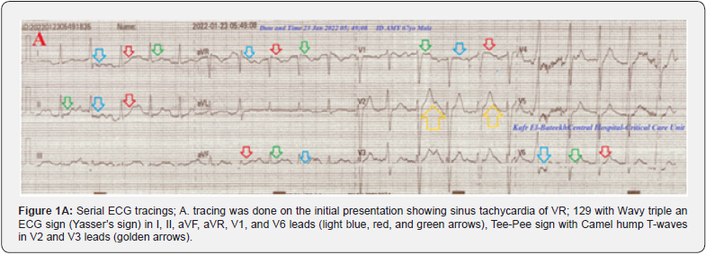
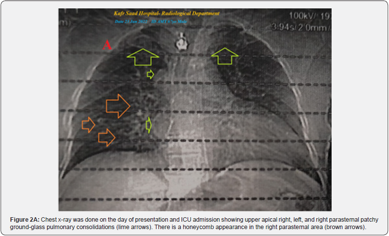
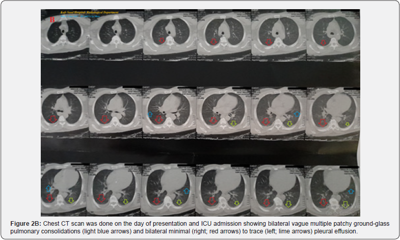
After controlling the BP, SC enoxaparin 80 mg, BID), captopril tablets (25 mg; BID), aspirin tablets (75 mg, OD), frusemide IV amp (40 mg IV TDS), and nitroglycerin retard capsules (2.5mg, BID) were added. The patient was daily monitored for temperature, pulse, blood pressure, ECG, and O2 saturation. The initial ECG tracing was done on the initial presentation showed sinus tachycardia of VR; 129 with Wavy triple an ECG sign (Yasser’s sign) in I, II, aVF, aVR, V1, and V6, Tee-Pee sign with Camel hump T-waves in V2 and V3 (Figure 1A). The second ECG tracing was taken within 1 minute of the above ECG tracing sinus tachycardia of VR; 130 with Wavy triple an ECG sign (Yasser’s sign) in V4-V6 leads, there are three premature ventricular contractions (PVCs) in V1-3 leads and two premature atrial contractions (PACs) in I, and II leads (Figure 1B). The third ECG tracing was taken within 10 hours of the initial ECG and treatment showing NSR of VR of 86 with the disappearance of all the above abnormalities. There is evidence of tremor artifacts (Figure 1C). The plain chest-XR film was done on the day of presentation and ICU admission showing upper apical right, left, and right parasternal patchy ground-glass pulmonary consolidations. There is a honeycomb appearance in the right parasternal area (Figure 2A). The chest CT was done on the day of presentation and ICU admission showing bilateral vague multiple patchy ground-glass pulmonary consolidations and bilateral minimal (right) to trace (left) pleural effusion (Figure 2B). The initial complete blood count (CBC); Hb was 13.6 g/dl, RBCs; 5.65*103/mm3, WBCs; 9.9*103/mm3 (Neutrophils; 76 %, Lymphocytes: 19%, Monocytes; 4%, Eosinophils; 1% and Basophils 0%), and Platelets; 214*103/mm3. CRP was high (24g/dl). SGPT (22 U/L) and SGOT were normal (14U/L). Serum albumen was normal (4.0gm/dl).
Serum creatinine (1.3mg/dl) and uric acid were normal (5.9 mg/dl). RBS was slightly high (257mg/dl). Plasma sodium was high (161mmol/L). Serum potassium was slightly low (3.3mmol/L). Ionized calcium was low (0.7mmol/L). The troponin test was negative. D-dimer was normal (0.25 mg/dl) ABG showed partially compensated respiratory alkalosis. Hypertensive pulmonary edema with Wavy triple sign (Yasser’s sign), Tee-Pee sign, and Camel-Hump T-wave in suspected COVID pneumonia was the most probable diagnosis. Within 10 hours of the above management, the patient finally showed nearly dramatic clinical and mostly electrocardiographic improvement. The patient was discharged after clinical stabilizations and continued on aspirin tablets (75 mg, OD), clopidogrel tablets (75 mg, OD), captopril tablets (25 mg; BID), frusemide tablets (40 mg OD), and oral nitroglycerine capsule (2.5 mg, twice daily). Further recommended cardiac and chest follow-up was advised.
Discussion
Overview
i. A 67-year-old, Farmer, married Egyptian male patient was presented to the intensive care unit (ICU) with hypertensive pulmonary edema, various new ECG signs, and suspected COVID-19 pneumonia.
ii. The primary objective for my case study was the presence of hypertensive pulmonary edema, bilateral pleural effusion with Wavy triple sign (Yasser’s sign), Tee-Pee sign, and Camel-Hump T-wave in suspected COVID-19 pneumonia in ICU.
iii. The secondary objective for my case study was the question; how would you manage this case in the ICU?
iv. Interestingly, the presence of a positive history of contact with a confirmed COVID-19 case, bilateral vague ground-glass consolidation, and some laboratory COVID-19 suspicion on top of clinical COVID-19 presentation with fever, dry cough, generalized body aches, anorexia, and loss of smell will strengthen the higher suspicion of COVID-19 diagnosis.
v. The existence of respiratory alkalosis is the indicator for the current Wavy triple sign or (Yasser’s sign) of hypocalcemia. vi. The T-wave-like shape of a traditional Native American Indian’s home with hypocalcemia, precordial QRS-complexes with peaked T-waves prominent U-waves and elongation of the descending limb of the T-wave supporting the diagnosis of Tee-Pee sign [8]. But there is hypokalemia and not hyperkalemia.
vii. The presence of T-waves that have a double-peak with prominent U waves fused to the end of the T-wave with hypokalemia and sinus tachycardia suggests the diagnosis of Camel hump T-waves [8]. But the hypokalemia is mild.
viii. The change of leads of Wavy triple sign (Yasser’s sign) from I, II, aVF, aVR, V1, and V6 to V4-V6 leads within one minute indicating the diagnosis of Movable-weaning off an electrocardiographic phenomenon in hypocalcemia (changeable phenomenon or Yasser’s phenomenon of hypocalcemia) [9].
ix. Wavy triple sign (Yasser’s sign), Tee-Pee sign, and Camel- Hump T-wave disappear without specific treatment.
x. There is dramatic ECG and clinical improvement after IV nitroglycerine and furosemide.
xi. Acute pulmonary embolism was the most probable differential diagnosis for the current case study.
xii. I can’t compare the current case with similar conditions. There are no similar or known cases with the same management for near comparison.
xiii. The only limitation of the current study was the unavailability of echocardiography.
Conclusion and Recommendations
a) The association of COVID pneumonia with hypertensive crises, pulmonary edema, bilateral pleural effusion, Wavy triple ECG sign (Yasser’s sign), Tee-Pee sign, and Camel- Hump T-wave in the elderly patient is highly interesting and remarkable.
b) An elder age, male, diabetes, hypertensive crises, pulmonary edema, hypocalcemia, hypokalemia, hypernatremia, pleural effusion, and COVID-19 pneumonia are constellation serious risk factors.
c) Wavy triple ECG sign (Yasser’s sign), Tee-Pee sign, and Camel-Hump T-wave may be transient.
d) Nitroglycerine and furosemide are highly effective in hypertensive pulmonary edema with suspected COVID pneumonia.
- Case Report
- Abstract
- Introduction
- Case Presentation
- Discussion
- Conclusion and Recommendations
- References
References
- Elsayed YMH (2019) Nitroglycerin in Suboptimal Versus the Remaining Doses in Hypertensive Crises Regards Propylene Glycol; Efficacy and Safety (Nitroglycerin on Trace Study); Retrospective Observational Study. Ann Med & Surg Case Rep 3: 1-12.
- Barzegari H, Khavanin A, Delirrooyfard A, Shaabani S (2021) Intravenous furosemide vs nebulized furosemide in patients with pulmonary edema: A randomized controlled trial. Health Sci Rep 4(1): e235.
- Cui X, Chen W, Zhou H, Gong Y, Zhu B, et al. (2021) Pulmonary Edema in COVID-19 Patients: Mechanisms and Treatment Potential. Front Pharmacol 12: 664349.
- Kim KH, Kerndt CC, Adnan G, Derek JS (2023) Nitroglycerin. In: StatPearls. Treasure Island (FL): StatPearls.
- Arnold WP, Mittal CK, Katsuki S, Murad F (1977) Nitric oxide activates guanylate cyclase and increases guanosine 3':5'-cyclic monophosphate levels in various tissue preparations. Proc Natl Acad Sci U S A 74(8): 3203-3207.
- Thadani U, Lipicky RJ (1994) Short and long-acting oral nitrates for stable angina pectoris. Cardiovasc Drugs Ther 8(4): 611-23.
- Elsayed YMH (2019) Wavy Triple an Electrocardiographic Sign (Yasser Sign) in Hypocalcemia. A Novel Diagnostic Sign; Retrospective Observational Study. EC Emergency Medicine and Critical Care 3(2): 1-2.
- Elsayed YMH (2020) Hypocalcemia-induced Camel-hump T-wave, Tee-Pee sign, and bradycardia in a car-painter of a complex dilemma: A case report. Cardiac 2(1): 1-5.
- Elsayed YMH (2021) Movable-Weaning off an Electrocardiographic Phenomenon in Hypocalcemia (Changeable Phenomenon or Yasser’s Phenomenon of Hypocalcemia)-Retrospective-Observational Study. CPQ Medicine 11(1): 1-35.


