The Close-mouthed Causality - A Case of Calcified Canal
Geon Pauly*, Roopashri Rajesh Kashyap, Raghavendra Kini, Prasanna Kumar Rao, Gowri P Bhandarkar and Meghana HC
Department of Oral Medicine and Radiology, AJ Institute of Dental Sciences, India
Submission: March13, 2018; Published: April 09, 2018
*Corresponding author: Geon Pauly N, Postgraduate student, Department of Oral Medicine and Radiology, AJ Institute of Dental Sciences, Kuntikana, NH-66. Mangaluru, PIN- 575004 Karnataka, India, Tel: +918905102696; Email: geonpauly@gmail.com
How to cite this article: Geon P, Roopashri R K, Raghavendra K, Prasanna K R, Gowri P B, Meghana HC. The Close-mouthed Causality - A Case of Calcified Canal. JOJ scin. 2018; 1(1): 555553. DOI: 10.19080/JOJS.2018.01.555553
Abstract
The dental pulp can be aptly considered 'the heart' of the tooth. It is the part in the centre of a tooth, which is made up of living connective tissue and cells. It remains protected in a space within a tooth known as the pulp chamber. Thus, the pulp chamber is considered a very important and integral part of the tooth. However, the pulp chamber undergoes different types of morphological and pathological alterations. Calcified canals are one amid the most commonly found pathological alterations.
Keywords: Dental pulp; Dental pulp cavity; Physiologic calcification
Clinical Image
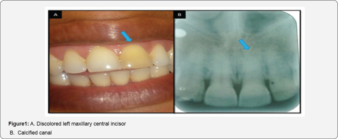
A 37-year-old medically fit female patient visited our department with a chief complaint of a discolored tooth in upper front teeth region since 5 years. She had no associated discomfort or pain and gave a history of a fall 5-6 years prior to date. Her past medical, dental and family histories were non-contributory. Intraoral examination revealed that the maxillary central incisor of the second quadrant had a generalized yellowish discoloration (Figure 1A). No tenderness elicited on palpation and percussion. A provisional diagnosis of Ellis Class IV fracture was documented. Electric pulp testing yielded a negative response. Intraoral periapical radiograph revealed calcification of the entire pulp canal, suggestive of trauma induced calcification (Figure 1B). As the patient was completely asymptomatic, she was referred for prosthodontic evaluation to improve the esthetics of the tooth.

Tooth calcification is considered to be the pulpal sequelae to trauma and aging phenomenon in an individual. The exact mechanism of pulp canal calcification is not entirely understood [1]. It has been suggested that if pulp remains vital after pulpal injury, the resultant blood clot and haemorrhage formed would act as nidus for calcification [2]. Calcification of the root canal (also referred to as pulp canal obliteration) is a common sequel following luxation injuries to permanent teeth. Abbott and Yu had discussed the terminology regarding this condition and had recommended the use of the term “calcification” as it more accurately describes the nature of changes seen [3].
The two chief morphologic forms of pulp calcifications are discrete pulp stones and diffuse calcifications. Pulp calcifications have been seen associated with genetic or systemic conditions such as dentin dysplasia, dentinogenesis imperfecta, Vander Woude syndrome [4]. On the treatment front, completely calcified canals can be left untreated as they totally asymptomatic in most cases. But such patients must be kept on constant observation, for it is a causality which spreads silently underneath, and if discoloration of the coronal aspect of the teeth is noted, prosthetic rehabilitation becomes mandatory. In case of partially calcified canals management includes; orifice recognition, biomechanical preparation and use of chelating agents like EDTA can be adjudged [5].
References
- Akpata ES (1969) Traumatized anterior teeth in Lagos school children. J Niger Med Assoc 6: 40-45.
- Sardhara Y, Dhanak M, Parmar G (2016) Management of Maxillary Central Incisor with Calcified Canal: Case Report. IOSR Journal of Dental and Medical Sciences 15(1): 24-27.
- Raghuvanshi S, Jain P, Kapadia H (2015) Endodontic Management of Traumatized Permanent Anterior Teeth with Radiographically Calcified Root Canal - Report of Two Cases. IJOCR 3(3): 76-80.
- Pauly G, Kashyap RR, Kini R, Rao PK, Bhandarkar GP, et al. (2017) The Pulpless Tooth: A Case of Calcified Pulp Canal. J Trauma Treat 6: i105.
- Nayak V, Kini R, Rao PK, Baliga A, Bhandarkar GP, et al. (2017) Trauma induced calcification: An Enigma. Pacific J Med Sci 17: 66-69.


