The Application of Non-Destructive Analytical Techniques for the Characterization of Modern Artistic Materials: A Case Study on the Works of N G Pentzikis
Nick Tachmazidis1,2*, Georgios Karagiannis2, Stamatios Amanatiadis2 and Eleni Pavlidou1
1Aristotle University of Thessaloniki, Department of Physics, Greece
2Ormylia Foundation, Art Diagnosis Centre, Greece
Submitted: March 02, 2023; Published: March 16, 2023
*Corresponding author: Nick Tachmazidis, Aristotle University of Thessaloniki, Department of Physics, Greece, Ormylia Foundation, Art Diagnosis Centre, Greece
How to cite this article: Nick Tachmazidis, Georgios Karagiannis, Stamatios Amanatiadis and Eleni Pavlidou. The Application of Non-Destructive Analytical Techniques for the Characterization of Modern Artistic Materials: A Case Study on the Works of N G Pentzikis. JOJ Material Sci. 2023; 7(4): 555720. DOI:10.19080/JOJMS.2023.07.555720
Abstract
This study aims to provide a detailed analysis of the materials and technique used in the creation of three paintings by the Greek artist N. G. Pentzikis. Non-destructive techniques, including Raman, Fourier Transform Infrared (FTIR) and X-ray fluorescence (XRF) spectroscopy, and imaging techniques such as Infrared Reflectography (IRR) and ultraviolet fluorescence (UVF) were employed to investigate the pigments used in the multilayered painted surface of his artworks as well as the unique “psifarithmisi” technique invented by the artist. The results provided valuable insights into the composition of the pigments used. The non-destructive methodology utilized in this study can have broader applications for the analysis of other multilayered artistic techniques and can serve as an important tool in preventing art forgery.
Keywords: Non-destructive analysis; Multilayered painted surface; Spectroscopy; Pigments; Psifarithmisi; Imaging technique
Introduction
Nondestructive analysis techniques are essential in the field of cultural heritage science, as they allow for the investigation of materials and techniques used in artwork without damaging the object. Techniques such as Raman and Fourier Transform Infrared (FTIR) spectroscopy provide a wealth of information about the molecular structures and compositions of painting elements, including binders, pigments, and varnishes, and have an extensive database reference for both inorganic and organic materials [1-4]. These methods can be used in conjunction with other analytical techniques such as X-ray Fluorescence (XRF), which is used for elemental analysis [5], to create a comprehensive understanding of the materials used in an artwork. The identification of materials is critical for the documentation of an artist’s palette, which is valuable for authentication purposes. Additionally, analyzing the technique of an artist is necessary to prevent mistakes during conservation processes and to identify art forgeries. Imaging techniques such as Ultraviolet Fluorescence (UVF) and Infrared Reflectography (IRR) can also be utilized to document the artist’s technique without damaging the artwork.
This study specifically focuses on the nondestructive analysis of three paintings by N G Pentzikis (1908-1993), a Greek writer and painter who used a unique multilayer painting technique known as psifarithmisi (Greek: ψηφαρίθμηση; /psi̱faríthmi̱si̱/), which is similar to pointillism. The name psifarithmisi originates from the Greek words “ψηφίδα” (mosaic tile) and “αρίθμηση” (numbering) and is based on an algorithmic system where texts from various sources are dissected into words, then letters, and each letter is translated to a number. Each number is then matched to a specific color and meaning using a wide variety of different shapes, such as simple point brush strokes, “<”, “>”, “O”, “O” with a dot, lines, rhomboid, trapezoid, and wavy shapes. This results in multilayered paintings, with estimates suggesting that some paintings could contain up to 28 layers of 7500 brushstrokes [6].
Hands, 1975, tempera on paper, 31.5 x 23.5 cm
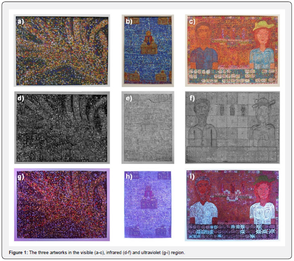
The artwork entitled “Hands” (Figure 1a), which was created in 1975, featuring two hands rendered in a pointillistic style using a large variety of brushstrokes in blue, red, green, orange, yellow, and purple color against a green-brown background. Though the artwork is a deviation from the artist’s usual religious themes, it serves as a study of his psifarithmisi technique. The yellow tones used to depict the inner parts of the hands in some parts combine with red and orange brushstrokes, and the outlines of the hands were created using white and blue brushstrokes. The brushstrokes are characterized by two main kinds, the circular or dotted shape and that with wide and straight strokes. The fingertips of the hands display a circular pattern indicating fingernails, while the right-hand features broad strokes of yellow and white color suggesting the phalanges of the fingers.
Transfer of the relics of Saint Bartholomew and other Martyrs, 1969, tempera on cardboard, 24 × 17cm
The 1969 painting “Transfer of the relics of Saint Bartholomew and other Martyrs” (Figure 1b) showcases Pentzikis’ unique artistic technique characterized by the use of various brushstroke shapes such as crosses, lines, semicircles, squares, rectangles, “Λ”- shaped peaks, and sinusoidal curves. The painting consists of light blue, blue, yellow colors with the addition of yellow-green, yellow- brown, white, brown, red-brown, and purple in some areas.
Pentzikis masterfully creates a spatial dynamic in the painting between the central figure of Saint Bartholomew and the surrounding figures representing the martyrs. The blue tones used in the painting represent the metaphysical realm where immaterial beings interact with the material world. The blue tones also symbolize the serenity and vastness of the metaphysical realm, as well as the turbulence of the material world due to human problems and sins. The contrasting colors of browns, reds, and whites complement the blue tones, creating a harmonious interplay between the foreground and background colors.
The central figure of Saint Bartholomew is emphasized by the less dense brushstrokes surrounding it and stands on a unique reliquary with two domes, compared to the one dome of the other figures’ reliquaries. However, all of the reliquaries share the feature of the three crosses. The robes of the figures differ, with the central figure’s robe being blue, the upper right figure’s robe being yellow-green, and the rest of the figures having less defined shapes in their robes with varying shades of red and yellow. Overall, the painting showcases Pentzikis’ artistic style and his ability to create a spatial dynamic and harmonious interplay between colors and shapes.
Couple, 1975, tempera on cardboard, 26.5 × 36cm
The artwork titled “Couple” (Figure 1c), a typical example of the psifarithmisi technique, is characterized by orange-colored shades dominating two-thirds of the painting and blue-colored shades in the remaining portion. The artist employs a variety of symbolic shapes mainly with red-blue shades on an orange background. The blue and white colors are seen mainly in circular patterns of shapes. The colors in the painting are thin and have multiple layers, giving them a unique texture.
While the central theme of the painting appears to be a couple, three figures in the center of the painting that resemble saints can be distinguished. The man of the couple on the left, blends with his surroundings, but subtle brown brushstrokes in his hair and beard, as well as his shirt with blue colored brushstrokes resembling trattegio, distinguish him. The woman on the right has a stronger contrast with a bright yellow hat, orange-red hair, and white and yellow-brown tones in her skin and blouse. The bottom and perimeter of the painting feature various decorative symbols consisting of squares with various shapes.
According to the artist, the orange color represents “unbuilt light” and he used it mainly for temples [7]. In addition to that, the artist’s choice of celebratory shapes and colors, suggests that the couple is experiencing joy, perhaps in the form of an engagement or wedding.
Research Aim
The aim of this research is to analyze the modern artistic materials in multilayered painted surfaces, specifically studying the stratigraphy, pigments used, and the unique technique utilized by the artist, through nondestructive techniques such as Raman, FTIR, and XRF spectroscopy, as well as IRR and UVF imaging techniques.
Materials and Methods Used
The present study was carried out at the Teloglion Fine Arts Foundation in Thessaloniki, Greece, employing the state-of-theart iTomography infrastructure of the “ORMYLIA” Foundation. The primary aim of this investigation was to obtain significant insights into the materials and techniques utilized in the examined paintings through the analysis of data obtained from a diverse range of modalities, including Raman spectroscopy, Fourier Transform Infrared (FTIR) spectroscopy, X-ray Fluorescence (XRF) spectroscopy, Infrared (IR) camera imaging, and ultraviolet filter-based photography [8-10].
i-Tomography
The infrastructure employed in this study was designed with a high degree of flexibility, enabling the precise positioning of various measurement modalities to facilitate the analysis of cultural heritage objects. The infrastructure incorporated different focal lengths and geometries to achieve accurate and detailed measurements. Two levels of positioning were implemented to ensure precise measurements of the artwork. The first level, coarse positioning, involved clamping the artwork with soft foam padding between two rails, which were manually positioned in the Y axis using four half-turn brake handles. The artwork was then mounted on a moving dolly on rails that allowed for X and Z movement. The second level, fine positioning, utilized three linear stages with an accuracy of ±1μm and two rotation stages with 3 arcmin accuracy and 12 arcsec repeatability to provide yaw and pitch rotations. The platform on which the measurement modalities were mounted was placed on the pitch rotary stage, while the yaw was performed by the stage on which the artwork was placed. Overall, this infrastructure allowed for precise and flexible positioning of the measurement modalities to achieve accurate and detailed analysis of cultural heritage objects [8].
Fourier transform Infrared (FTIR) spectroscopy
The infrared spectra of the point measurements were obtained through reflectance mode, utilizing the ALPHATM FTIR Spectrometer of Brucker TM, equipped with the Quick Snap TM External reflection module. This module featured a custom-made 1mm diameter, and the measurements were conducted with 64 scans, 2cm-1 resolution, and a range between 365-7500cm-1. The module was placed in front of the spectrometer, and the spectra were collected accordingly. To ensure accurate results, the obtained spectra were compared with the reference spectra available in the Infrared and Raman Users’ Group database (IRUG, www.irug.org) [1].
Raman spectroscopy
The Raman spectroscopy analysis was conducted using the i-RamanTM EX spectrometer manufactured by BWTEKTM. The laser employed in this analysis had an operating wavelength of 1064nm and was focused directly on the painted surface. The number of scans and the milliwatts (mW) used during the analysis were adjusted based on the requirements of each measurement. The obtained spectra were compared with the reference spectra available in various databases, including the Infrared and Raman Users’ Group database (IRUG, www.irug.org), the SOP spectral library (soprano.kikirpa.be), and the Cultural Heritage Science Open source (chsopensource.org/pigments-checker) [1,2,11]. This approach facilitated the identification of the pigments and materials used in the analyzed artwork and enabled the characterization of the molecular structures present in the painted layers.
X-ray fluorescence (XRF) spectroscopy
The portable Niton XL3t GOLDD+XRF Analyzer manufactured by Thermo Scientific was employed in this study for elemental analysis of the inorganic pigments found in the paintings. This instrument is compact and lightweight with dimensions of 244 × 230 × 95.5 mm3 and weight of less than 1.3kg. The XRF analyzer is equipped with a fast detector that has an energy resolution of less than 185 eV at 60,000 cps, with 4 μsec shaping time. The beam focusing area of the instrument is 3mm, which provided high spatial resolution. The Niton XL3t GOLDD+XRF Analyzer was used as a supplementary technique for its ability to rapidly identify and quantify elements present in the sample.
Infrared Reflectography (IRR)
The image mosaic of the infrared reflectography (IRR) was created using the XMID-FPA-640 IR camera manufactured by XenICsTM, which operates in the 1-5μm spectral range. The camera was mounted on a moving/robotic stage and captured a series of 640 × 512 pixel infrared images, which were algorithmically stitched together to reveal a detailed image of the artwork in the mid-infrared region of the electromagnetic spectrum. The camera features an efficient, thermoelectrically cooled detector with control and communication electronics, enabling high-quality image acquisition.
Ultraviolet Fluorescence (UVF) Photography
Similar to the IRR, the UVF technique employed a Canon EOS- 1Ds Mark II digital camera with a filter covering the spectral range of 400-700 nm. The camera was set at a 4-second exposure, f/4.0 aperture, ISO 640 sensitivity, and a 50mm lens. The light source used was a Phillips Mercury Fluorescent Blacklight HPW 125W TS lamp, which emitted at a wavelength of 380nm. This setup allowed for the capture of images in the ultraviolet range of the electromagnetic spectrum.
Results and Discussion
Spectroscopic techniques
For the investigation of the pigments of the colored brushstrokes, the FTIR, Raman, and XRF spectroscopic techniques were utilized. The use of a multi-layer technique and a wide range of colors in the artwork resulted in increased noise in the spectra, which posed a challenge for interpretation. A tabular summary of the findings has been included in table 1 for ease of reference.
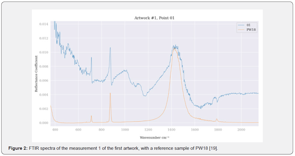
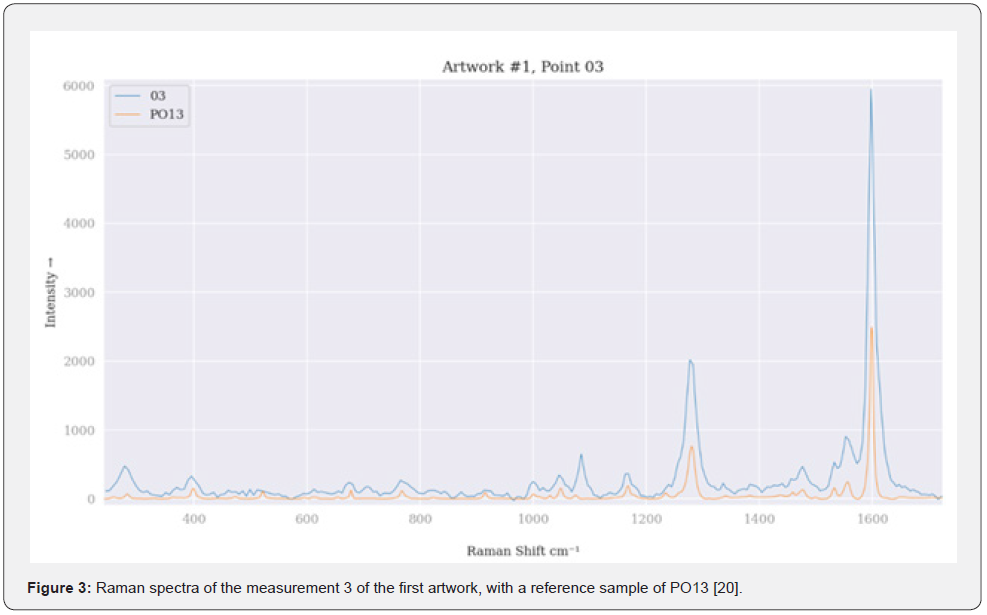
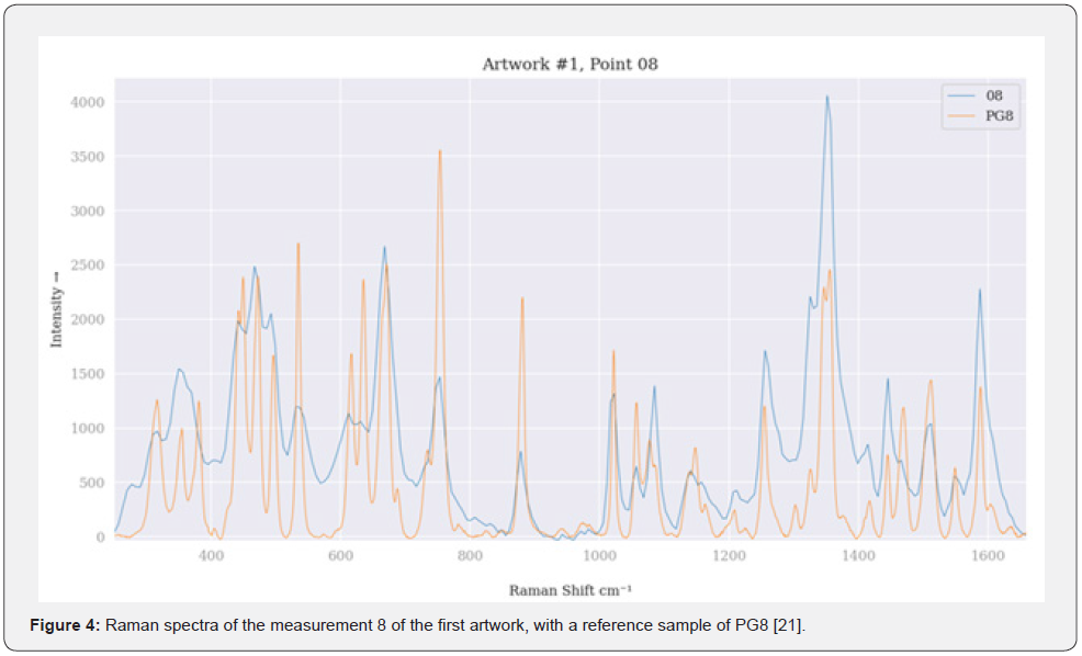
Hands: In the first artwork, the inorganic pigment, Chalk (PW18, CaCO3, C.I. 77220), was identified in all the IR and Raman spectra (Figure 2) as well as the XRF technique which revealed high levels of Calcium (Ca), indicating its use as a filler to enhance opacity [12-14]. The blue brushstrokes contained Ultramarine Blue (PB29, Al6Na8O24S3Si6, C.I. 77007) in its synthetic form, due to no differences in Ca or Magnesium (Mg) [15], while the orange brushstrokes contained Benzidine Orange (PO13, C32H24Cl2N8O2, C.I. 21110) (Figure 3). The white brushstrokes contained Titanium White (PW6, TiO2, C.I. 77891) in the tetragonal crystal structure of Anatase. The green brushstrokes contained Nitroso Green (PG8, C30H18FeN3NaO6, C.I. 10006) where the Raman bands showed some differences due to resolution (Figure 4), but XRF showed increased levels of Iron (Fe) which confirms the result. The yellow brushstrokes contained Hansa Yellow (PY1, C17H16N4O4, C.I. 11680) or Pigment Yellow 1:1 (PY1:1) which is a variant of the first.
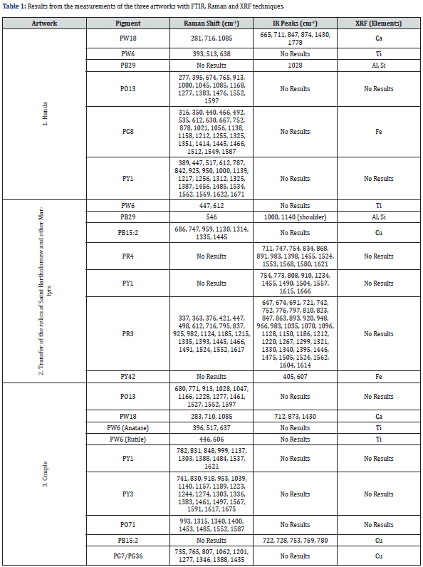
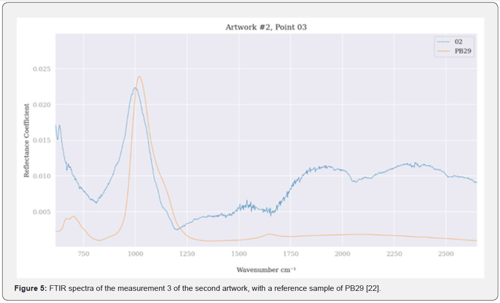
Transfer of the relics of saint Bartholomew and other martyrs: In the second artwork, the XRF revealed high levels of Titanium (Ti) and Ca, as well as moderate levels of Zinc (Zn), which suggest the presence of inorganic white pigments, such as Titanium White (PW6) and Chalk (PW18), and Zinc Oxide White (PW4, ZnO, C.I. 77947) used as paint additives. In blue-colored areas of the painting, the organic Phthalocyanine Blue (PB15:2) was identified along with the inorganic Ultramarine Blue (PB29) (Figure 5) and Titanium White (PW6), in the tetragonal crystal form of Rutile. The FTIR technique also showed two peaks at 609 and 634 cm-1, that can be assigned to the color with red-orange hue. This might be a mixture of Permanent Red R (PR4, C.I. 12085) and PY1 which is used in commercial colors [16,17], but the result could not be fully interpreted due to spectral noise. In the red mantle, the organic Toluidine Red (PR3, C16H10ClN3O3, C.I. 12120) was identified based on its characteristic peaks in the Raman spectrum.
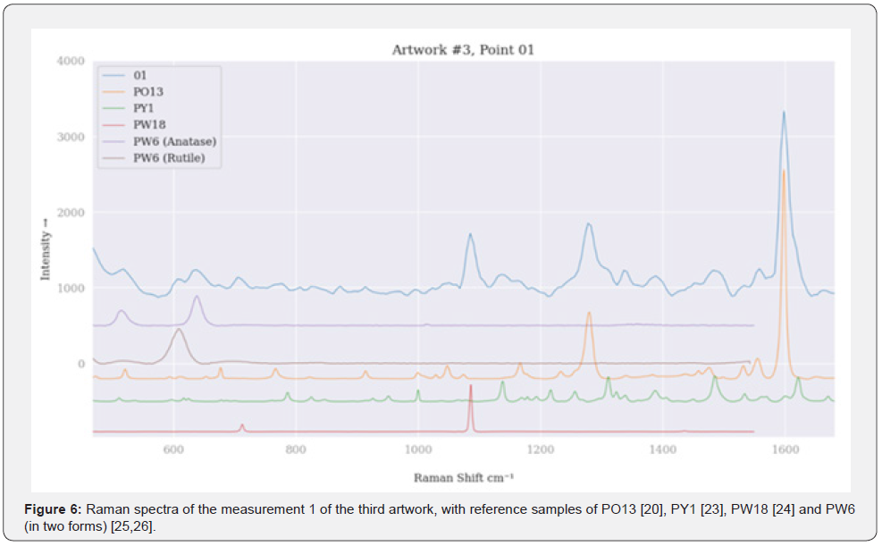
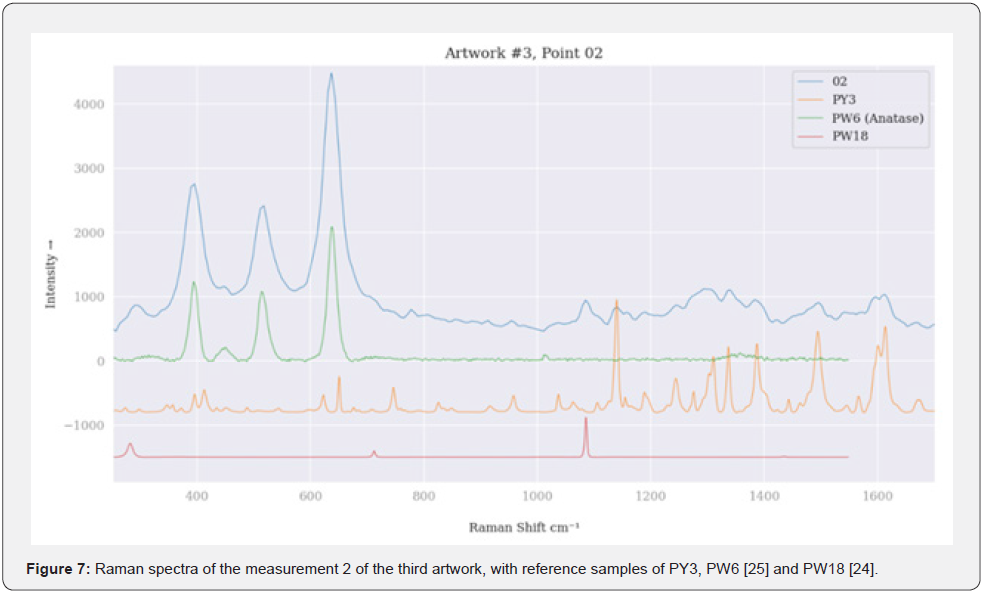
Couple: In the third artwork, the acquired results from XRF analysis are consistent with those obtained from the examination of the first artwork, Hands. Specifically, the highest proportions of Ca were detected, while Ti displayed a high degree of absorption. These findings suggest that these elements were an additive to the pigments utilized by the artist, potentially extending to the paper substrate. Additionally, some degree of Fe absorption was observed, which may be attributed to background colors. The organic PO13 was detected in the yellow hat with orange brushstrokes of the female figure, while the inorganic PW18 was identified in all Raman spectra. PW6, was found in two crystal forms, Anatase and Rutile. In the yellow brushstrokes there were two pigments which were identified as PY1 (Figure 6) and Hansa Yellow 10G (PY3, C16H12Cl2N4O4, C.I. 11710) (Figure 7). The orange shades were attributed to organic Pyrrole Orange (PO71, C20H10N4O2, C.I. 56120), while the purple tones were likely produced by Alizarin Crimson (PR83, C14H8O4, C.I. 58000) but couldn’t be confirmed. The blue pigment in the woman’s blouse was identified as the PB15:2. The green colors seemed to consist of another pigment that is in the phthalocyanine class, that can be attributed to either the Phthalocyanine Green BS (PG7, C32Cl16CuN8, C.I. 74260) or Phthalocyanine Green YS (PG36, C32Br6Cl10CuN8, C.I. 74265) [18], but the results were inconclusive. The yellow-brown pigment in the halo was identified as the earth inorganic pigment, Yellow Ochre (PY42, Fe2O3 • H2O, C.I. 77492) which is some of the most widespread pigments used since ancient times [12]. The FTIR showed some alterations from the reference spectrum, but the XRF showed high levels of Fe, confirming the result. Other clay pigments variations such us, red ochres, siennas or umbers may have been used due to elevated levels of Fe and Manganese (Mn) showed by the XRF technique, but the other techniques did not show clear results to identify the above pigments.
Imaging techniques
The utilization of Ultraviolet Fluorescence (UVF) in art analysis enables the discernment of multiple colors that may not be visible to the naked eye. This technique allows for a more comprehensive understanding of an artwork’s chromatic composition, contributing to the examination of the artist’s style and choices. Conversely, the application of Infrared Reflectography (IRR) permits insight into the painting’s early stages, as it enables the visualization of initial layers that may have been subsequently obscured or altered during the creation of the artwork. By utilizing both UVF and IRR techniques, a more comprehensive analysis of an artwork can be achieved, providing valuable insights into the artist’s creative process and stylistic preferences. An analytic overview of the behavior of colors in the paintings can be seen in table 2.
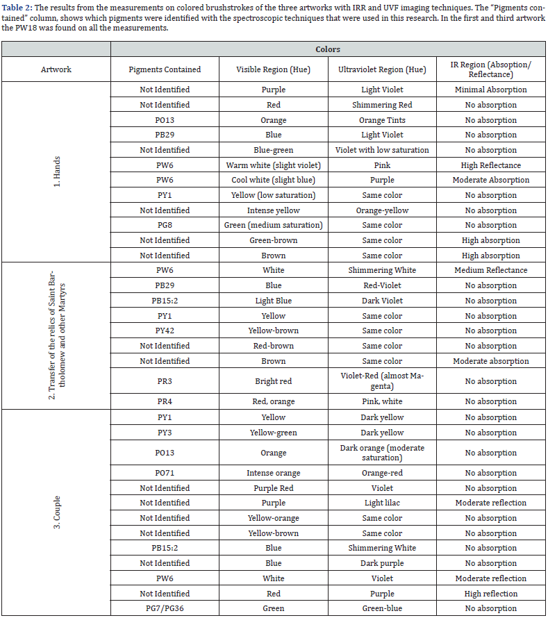
Hands: In the first artwork, the absence of blended brushstrokes suggests that the painter used very little solvent to dilute the colors in most of the painting. However, some areas display evidence of solvent use and mixture, which could be accidental rather than intentional. Some colors that exhibit fluorescence in the UV light are purple, red, orange, blue and yellow that have minimal to no absorption in the IR region (Figures 1d & 1g). Additionally, two distinguishable shades of white can be seen, which show completely different behaviors under UV and IR spectra. Infrared reflectography revealed hidden details of the painting, such as shapes and patterns. The brown color of the signature in the upper right corner showed the highest absorption in the IR spectrum, followed by the green-brown color that can be seen throughout the painting.
Transfer of the relics of Saint Bartholomew and other Martyrs: In the second artwork, the white pigment, PW6, exhibits fluorescence as it appears as a shimmering, intense white color, whereas in the IR region it shows reflection. The blue colors made with the PB29 exhibit fluorescence and show a red-violet hue. In sparse amounts, however, the strokes are barely distinguishable, revealing clearer background shapes in the infrared spectrum. Another blue pigment, PB15, fluoresces in the UV region, showing dark violet colors. This results in the painting appearing to have inverted colors (Figure 1h). Interestingly, the red pigment, PR4, also fluoresces in the UV region, were its red hue changes to a violet, magenta-like hue. In the IR region (Figure 1e), the painting showed clear patterns of the psifarithmisi technique, as almost none of the pigments used in the upper layers showed any particular absorption. This resulted in many rectangular and square shapes with a cross shape inside, along the entire length of the panel becoming visible. The shapes have fine lines, indicating the use of a thin brush for the first brushstrokes of the psifarithmisi, while a thicker brush was used in the upper layers.
Couple: In the third artwork, the predominant colors in the ultraviolet spectrum are red-violet, contrasted with white colors in blouses and dark purple in the lower part of the painting, as can be seen in figure 1i. The yellow pigments in the hat shift to dark green tones in the infrared spectrum, with no absorption observed in the IR region. The PO13 and PO71 pigments exhibit distinct fluorescent properties in the UV spectrum, with the former displaying a darker orange tone of moderate saturation and the latter producing more intense orange-red tones. In the infrared spectrum, neither pigment shows any absorption. The blue pigment in the couple’s shirts fluoresces and appears as a shimmering white with a slight blue tint, possibly due to the presence of white pigment. The white color present in the women’s blouse in the UV spectrum appears with violet tones due to the fluorescent color of the background, while it appears as gray in areas where white is in greater concentration, also affected by the color of the background. The purple pigments also exhibit fluorescence and shift towards violet and lilac tones in the UV spectrum, while the red pigment utilized in the patterns exhibits a purplish hue. The green pigment used in the patterns exhibits relatively lower saturation and takes on a greenish-blue tint in the UV spectrum. As with the previous painting (section 4.2.2) the IR region showed early stages of psifarithmisi technique where the painter used a pencil to create a 6 x 9 matrix made up of squares, most of which have either the Greek letters Delta (“Δ”) or Epsilon (“E”) in the center, apart from the three central squares in the middle of the second row that contain three figures (Figure 1f). Moreover, it is evident that the artist used a pencil to draw the outline of the figures, which show thick lines. That suggests that the artist may have used the pencil sideways. Another use case of the pencil can be seen in the outline of the decorative pattern at the bottom of the painting.
Conclusion
The current study showcases the efficacious use of non-invasive methodologies for the analysis of modern artistic materials in a multilayered painted surface. The i-Tomography infrastructure, coupled with Raman, FTIR, and XRF spectrometers, and IRR and UVF imaging techniques, furnished insightful results on the stratigraphy of the three paintings, “Hands”, “Transfer of the relics of Saint Bartholomew and other Martyrs” and “Couple”, the pigments employed, and the unique psifarithmisi technique utilized by Pentzikis. As discussed in section 6.1.3 of the present analysis, the first and the third artworks evinced substantial levels of Ca alongside the detection of the PW18 pigment in all recorded measurements. It is posited that this outcome may be ascribed to the application of an identical color brand, given that both works were executed within the same year, specifically in 1975. Additionally, the first artwork did not manifest the square configurations observable in the succeeding two artworks. In contrast, the second artwork evinced several diminutive squares, while the third artwork featured sizable squares containing a center aligned letter [19-23].
Moreover, the imaging techniques used in this study proved to be promising in revealing the underlying layers of the psifarithmisi technique, consequently verifying the artist’s claims on his other painting, “Moni Simonos Petra”, wherein he employed a series of squares on the painting’s surface to denote various phonetic components of speech [7]. Furthermore, the non-destructive approach employed in this study may have broader applications for investigating other multilayered techniques. Ultimately, documenting the materials and techniques used by artists can be a significant tool in preventing the misuse of materials during conservation and art forgery [24-26].
Acknowledgment
This research is a component of the PhD thesis of Nick Tachmazidis in the Physics Department at Aristotle University of Thessaloniki. The authors express their gratitude to the administration of the Teloglion Fine Arts Foundation in Thessaloniki, Greece, for allowing the analysis of N. G. Pentzikis’ works. The measurements were performed at the Teloglion Fine Arts Museum. Nick Tachmazidis acknowledges the support received from the “ORMYLIA” Foundation, the Art Diagnosis Center, and Dr. G. Karagiannis for facilitating access to the necessary equipment.
References
- Pretzel PBAB, Lomax SQ (2022) IRUG - Infrared and Raman Users Group Spectral Database 2007. 1-2.
- Wim F, Saverwyns S (2012) Identification of synthetic organic pigments: the role of a comprehensive digital Raman spectral library. Journal of Raman spectroscopy 43(11): 1536-1544.
- Burgio L, Clark R (2001) Library of FT-Raman spectra of pigments, minerals, pigment media and varnishes, and supplement to existing library of Raman spectra of pigments with visible excitation. Spectr Acta A 57(7): 1491-1521.
- Signe V, Virro K, Leito I (2005) Web-based infrared spectral databases relevant to conservation. CAC Journal p. 10-17.
- Sawczak M, Kamińska A, Rabczuk G, Ferretti M, Jendrzejewski R, et al. (2009) Complementary use of the Raman and XRF techniques for non-destructive analysis of historical paint layers. Applied surface science 255(10): 5542-5545.
- Pentzikis NG (1990) Water Overflow, Paratiritis.
- Bakolas N (1984) Painters of Thessaloniki: The first generations, Regos, Fotakis, Lefakis, Paralis, Pentzikis, Svoronos, Sachinis, Natsis (Catalog). Thessaloniki: Northern Greece Publishing SA.
- Karagiannis G, Karamanos T, Athanasopoulos E, Panayiotou K, Amanatiadis S, et al. (2018) Development of an i-Tomography infrastructure for non-destructive documentation of cultural heritage objects. IEEE International Conference on Imaging Systems and Technologies, Krakow, Poland.
- Amanatiadis S, Apostolidis G, Karagiannis G (2018) Infrared hyperspectral spectroscopic mapping imaging from 800 to 5000 nm. A step forward in the field of infrared imaging in 1st International Conference TMM_CH Transdisciplinary Multispectral Modelling and Cooperation for the Preservation of Cultural Heritage, Eugenides Foundation, Athens, Greece.
- Karagiannis G (2016) High resolution spectroscopic mapping imaging applied in situ to multilayer structures for stratigraphic identification of painted art objects. Optics, Photonics and Digital Technologies for Imaging Applications 9896. SPIE, pp. 343-347.
- Cristina CM, Cosentino A, Mangone A (2016) Pigments Checker version 3.0, a handy set for conservation scientists: A free online Raman spectra database. Microchemical Journal 129: 123-132.
- Eastaugh N, Walsh V, Chaplin T, Siddall R (2008) Pigment Compedium: A Dictionary and Optical Microscopy of Historical Pigments, Elsevier.
- Virgil E (2021) Why Some Paints Are Transparent and Others Opaque. Natural Pigments, Inc.
- (2006) Getty Conservation Institute, Tate, the National Gallery of Art, "Modern Paints Uncovered," in Modern Paints Uncovered Symposium, Los Angeles.
- Miliani C, Daveri A, Brunetti BG, Sgamellotti A (2008) CO2 entrapment in natural ultramarine blue. Chemical Physics Letters 466(4-6): 148-151.
- Giménez P, Linares A, Sessa C, Bagán H, García JF (2022) Capability of Far-Infrared for the Selective Identification of Red and Black Pigments in Paint Layers. Spectrochimica Acta Part A: Molecular and Biomolecular Spectroscopy 266: 120411.
- Sennelier (2021) Sennelier - A History in Color, Sennelier.
- Winsor, Newton (2021) Spotlight on: Phthalo Green, Winsor & Newton.
- (2021) University of Barcelona, IMP00480, Chalk, primarily calcium carbonate, PW18, Kremer, BBAA-UB, tran, Infrared and Raman Users Group (IRUG) Spectral Database.
- (2021) Bern University of the Arts (BUA), Art Technological Laboratory, ROD00061 PO13, Disazopyrazol, CI21110, Clariant, 104886, 785, HKB, scat, Infrared and Raman Users Group (IRUG) Spectral Database.
- Wim F (2021) PG8, Raman 785. Royal Institute for Cultural Heritage (KIK/IRPA).
- (2021) Straus Center for Conservation, Harvard University Art Museums, IMP00324 Ultramarine blue, synthetic, PB29, Weber, Forbes, 81, SCC, tran, Infrared and Raman Users Group (IRUG) Spectral Database.
- (2021) Bern University of the Arts (BUA), Art Technological Laboratory, ROD00036, PY1, Monoazo Yellow, CI11680, Heubach, 100100, 785, HKB, scat. Infrared and Raman Users Group (IRUG) Spectral Database.
- Museum of Fine Arts, Boston (2021) RMP00052, Calcite marble, Calcium carbonate, 785, MFAB, scat, Infrared and Raman Users Group (IRUG) Spectral Database.
- Museum of Fine Arts, Boston (2021) RMP00074 Titanium dioxide, anatase, E&A, Forbes, MFA538, 785; scat, Infrared and Raman Users Group (IRUG) Spectral Database.
- (2021) Philadelphia Museum of Art, RMP00146 Titanium white, rutile form, PW6, F&S, Forbes, PMA; scat, Infrared and Raman Users Group (IRUG) Spectral Database.


