Analysis of Mineral Pigments from the Gnishikadzor Area, Southeastern Armenia
Yeghis Keheyan1*, Alessandro Latini2, Daniella Ferro2, Stella Nunziante Cesaro1, Dmitri Arakelyan3, Artur Petrosyan4 and Boris Gasparyan5
1Scientific Methodologies Applied to Cultural Heritage (SMATCH), Italy
2Department of Chemistry, Sapienza University of Rome, Italy
3Institute of Geological Sciences, Republic of Armenia
4Institute of Archaeology and Ethnography, Republic of Armenia
5Department of Archaeology and Ethnography, Yerevan State University, Republic of Armenia
Submitted: August 10, 2022; Published: November 7, 2022
*Corresponding author: Yeghis Keheyan, Scientific Methodologies Applied to Cultural Heritage, Rome, Italy
How to cite this article: Yeghis K, Alessandro L, Daniella F, Stella N C, Dmitri A, et al. Analysis of Mineral Pigments from the Gnishikadzor Area, Southeastern Armenia. JOJ Material Sci. 2022; 7(2): 555709. DOI:10.19080/JOJMS.2022.07.555709
Abstract
The territory of the Republic of Armenia is very rich with ores and different types of deposits, including resources of natural mineral pigments. They differ by large variation of colours and are represented by painted ores, clays, and earths, among which the most significant is the group of paints with yellow, red and brown shades (ochre). Vayots Dzor Province in South-Eastern Armenia is among the rich areas where painted earths are widely spread. Presence of red and brown ochre are very well visible in the south-western part of the province, in the gorge of the Gnishik River, which is also known as the Noravank Gorge, due to the monastic complex of Noravank located here. Red colour rocks in the area of the Noravank Gorge (Gnishikazdor) represented by the sedimentary strata of the Upper Devonian and are determined by the Famennian Stage (375-359 million years). The samples analysed were taken from the foothills of the Noravank Monastery and analysed by different techniques: Scanning electron microscopy (SEM) with energy dispersive spectroscopy (EDS); FT-IR spectroscopy; XRD diffraction analysis, which allow to indicate the presence of different elements trough contrast variations (atomic number contrast), to determine spectral ranges where absorption peaks were detected, as well as to perform phase identifications. The results show that the concretion is a hard, compact mass of matter formed by the precipitation of mineral cement within the spaces between particles, and is found in sedimentary rock or soil. It is composed of a carbonate mineral such as calcite; an amorphous or microcrystalline form of silica such as chert, flint, jasper or an iron oxide or hydroxide such as goethite and hematite. Implementation of such kind of study is valuable for the future comparison of similar finds from the nearby prehistoric archaeological contexts, where inhabitants exploited red ochre as a pig.
Keywords: Mineral paints; Red ochre; Areni-1 cave; Vayots Dzor Province; Republic of Armenia
Introduction
The mountains of Armenia conceal deposits of ores. Alaverdi (Northern Armenia) and Kapan (Southern Armenia) localities are rich of copper deposits, molybdenum was found in the southeast (Dastakert deposit), in the central and southeastern areas are iron ore deposits (Hrazdan, Abovyan and Svarants deposits). Besides, there are industrial stocks of aluminium-nepheline-syenites, as well as barite with admixture of gold and silver, the deposits of lead, zinc, manganese, gold, platinum, antimony, mercury, and arsenic. There are also rare earth metals: bismuth, gallium, indium, selenium, thallium, tellurium, rhenium. Tuffs (red, orange, yellow, pink, and black), marble, travertines, limestones, are great as building and finishing materials. Semiprecious and ornamental stones are represented by agates, jaspers, amethysts, beryls, rubies, obsidians, onyxes, turquoise.
The area of the country is also rich with resources of natural mineral pigments, where 17 deposits were registered and studied. They differ by large variation of colours and are represented by painted ores, clays and earths, among which the most significant is the group of paints with yellow, red and brown shades (ochre) [1,2].
The colour shade of ochre depends on the type of the iron oxide chromophore. The red ochre contains mainly haematite (Fe2O3), while the yellowish one is rich in hydrated iron oxide goethite, FeO·OH), [3]. The presence of other minerals, such as clay minerals or some metal oxides, can also influence the colour of the ochre. The classification of ochre can be also made according to the matrix composition of kaolinite (Al2SiO5) (OH)4 and/or gypsum (CaSO4·2H2O), and/or sulphate, [4]). Green earth is a clay pigment consisting of hydrated iron, magnesium, and aluminium potassium silicates. Colour varies from a dark, greyish blue green to a dark, dull yellowish green. The colour of green earth is derived from the presence of the following minerals: glauconite or celadonite. As the yellow and red ochre, the green earth or “terreverte” has been used as a pigment all over the world since ancient times [4,5]. They have been found in artworks all over the world and in any historical period, probably due to their availability, high coloring capacities and stability to the light and to the different weather conditions.
Armenian mineral pigments were also used since the dawn of the human civilization and their exploitation by the local inhabitants continued until 1940, after which they were processed on industrial level [2,6].
Vayots Dzor Province in South-Eastern Armenia is among the rich areas where painted earths are widely spread and are represented by large deposits in Agarakadzor and Yeghegnadzor [6]. Meanwhile presence of red and brown ochre are very well visible in the south-western part of the province, in the gorge of the Gnishik River (Gnishikadzor, “dzor” in Armenian means gorge), which is also known as the Noravank Gorge, due to the monastic complex of Noravank (means New Monastery in Armenian) located here The samples analysed in this article was taken from the foothills of the Noravank Monastery, left from the road, where section of red coloured sediment is exposed during the construction of the road (Figure 1). Red colour rocks in the area of the Noravank Gorge (Gnishikazdor) are represented by the sedimentary strata of the Upper Devonian and are determined by the Famennian Stage (375-359 million years). In this area, they are exposed in the core of the so-called Gnishik anticline, spread in the basin of the middle reaches the Gnishik River. The entire stratum of Devonian deposits here is 385 m thick and is represented by ferruginous dark gray and fractured organogenic limestones, which then turn into sandy limestones with a phosphorite content. Ferruginous quartzites with large impregnations of iron oxides are also exposed in ferruginous sandy limestones, shales with carbonate nodules and rich brachiopod fauna: Productella capetatiformis Abrahamian, Plicatifera meisteri, Cyrtospirifer verneuili, Camarotoechia baitaversis, etc (Figure 2).


First evidence of exploitation of similar red coloured ochre from the area was recorded in Late Chalcolithic horizons of Areni-1 cave, located 7km north from the exposure, 2km northeast from the village of Areni, on the left bank of the Arpa River, near the point of its confluence with the tributary Gnishik and at an elevation of 1070m above the sea level. Areni-1 is a threechambered karstic cave. The excavations here began in 2007 and the major significance of this archaeological site was abundantly clear during the initial excavations when very well preserved Chalcolithic (4,300 – 3,400 BCE) and Medieval (4th –18th centuries CE) occupations were exposed. Areni-1 exhibits a transitional culture between Chalcolithic and EBA, which sheds light on the formation and the early stages of the Kura-Araxes culture in the region. Chalcolithic finds from the first gallery of the cave include numerous large storage vessels, some of which contained human skulls – of two adolescent males and a female. Grape remains and vessels typically used for wine storage, together with the results of chemical analyses of the contents, point to Chalcolithic wine production at the site. The cave had been used for different purposes since the end of the 5th millennium BCE: it was a shelter, a storeroom for food; it was used for wine production and for ritual purposes, including burial. All the data indicate clear social complexity and a ritual/productive area. Its strategic location, suitable climate of the Vayots Dzor Province compared to the surrounding mountainous area, and its numerous watercourses and highly fertile soils, make this area especially suitable for human settlement and agricultural development. Indeed, the oldest leather shoe in Eurasia and one of the oldest pieces of evidence for wine production was discovered in the Areni-1 Cave, dated to the Late Chalcolithic – and wine is still today one of the area’s main products [7-15].
Local red ochre was used by the Chalcolithic inhabitants of the cave in different purposes, i.e., for rock-paintings, in symbolic behavior (for coloring the inner parts of the ritual vessels and clay constructions, the compacted floors, as red ochre was the symbol of blood and revival as well as for decorating basketry and pottery [11,13] (Figure 2).
Experimental
The samples from Gnishikadzor or the Noravank were analysed by different techniques. Below are reported the techniques applied to characterize completely these fabulous stones.
Techniques
Scanning electron microscopy (SEM) with energy dispersive spectroscopy (EDS): SEM-EDS micro-morphological and chemical investigations were carried out by a LEO 1450 VP -INCA 300 scanning electron microscope coupled with a electronic probe for X-ray microanalysis, resolution of 3,5nm with the possibility to analyze nonconductive sample by operating in novacuum conditions. The interfacing with EDS gives the possibility to have qualitative and quantitative composition of elements into area observed. For quantitative analysis this method is not sensible under 0.1% in weight. Electron beam energy is 20keV to allow the detection most of the chemical elements starting from boron. Under these experimental conditions the ancient samples have been analysed without any treatment, by using the apparatus in low vacuum. The observations in backscattered electron allow suggest the presence of different elements trough contrast variations (atomic number contrast).
FT-IR spectroscopy: The FTIR microspectra were collected with a Bruker Optic Alpha-R portable interferometer with an external reflectance head covering a circular area of about 5mm in diameter. The samples were placed directly in front of the objective and spots were selected for analysis. The recorded spectral range was 7500-375cm-1 acquired with 200 scans or more, with a resolution of 4cm-1. Spectra reported in the text, however, show only the spectral range where absorption bands were observed (4000-375cm-1). This analysis is non-destructive and non-invasive. The spectra of powdered samples were obtained using the diffuse reflectance infrared Fourier transform (DRIFT) module. In addition, very small amounts of samples were dispersed in potassium bromide (KBr, FTIR grade purity, Fluka) at different concentrations (sample/ KBr 1/100 to sample/KBr 1/1000). These were studied by collecting 200 scans or more in the same spectral range and resolution. Fourier-Transform infrared (FTIR) spectra were recorded using an Alpha FT-IR spectrometer (Bruker) equipped with the Diffuse Reflection Infrared Fourier Transform (DRIFT) module in the spectral range 7500-375cm-1 at a resolution of 2cm-1 cumulating at least 200 scans. The powdered samples were dispersed in potassium bromide (KBr), FT-IR grade of purity, Fluka) in excess. Figures reported spectral ranges where absorption peaks were detected
XRD diffraction analysis: The X-ray powder diffraction analysis has been performed in the angular range 10-90° in 2θ with a Panalytical X’Pert Pro MPD diffractometer (Cu Kα radiation, λ=1.54184 Å) equipped with X’Celerator ultrafast RTMS detector. The angular resolution (in 2θ) was 0.001°. A 0.04 rad soller slit, a 1° divergence slit, and a 20mm mask have been used on the incident beam path, while a 6.6mm anti-scatter slit and a 0.04 rad collimator have been used on the diffracted beam path. Phase identification has been performed with the Panalytical High Score Plus software.
Results and Discussion
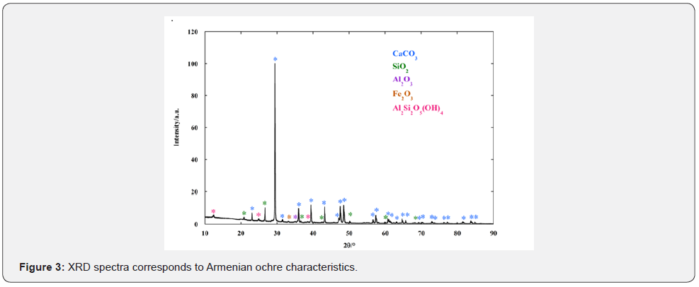
XRD spectra (Figure 3) show prevalently the presence of ochre. From phase analysis the following chemical composition was evidenced; CaCO3, SiO2, Al2O3, Fe2O3, Al2S2(OH)4.


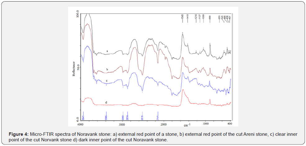
Different stones from the same locality have been cut off. The stone has been cut and analyzed in all parts by FTIR. It was very hard to cut it. Internal part was grey and white. The spectra obtained is reported and compared in figure 4. At 536cm-1 is hematite band and at 473cm-1 is iron oxide band. Quartz band is at 799cm-1. Infrared spectroscopy was employed to analyze the exterior red surface of the stone samples. In addition, a small stone was cut in order to examine the inner side, which appeared grey and white.
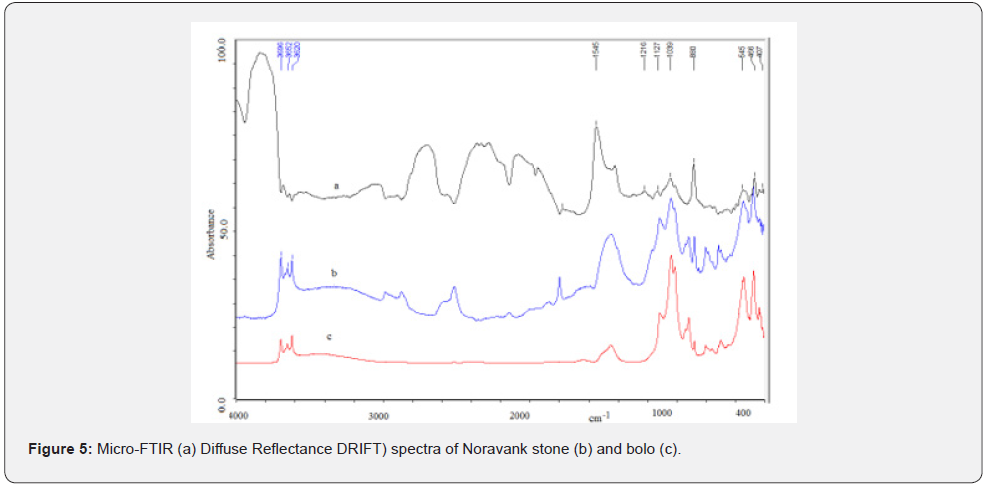
Specular reflectance produces derivative- shaped peaks in the region below 1200cm-1 because of the restrahlen effect [16]. All spectra show intense band with peaks at 1545 and 1418cm-1 assigned to the C-O stretching mode of calcite (sparitic limestone). The features at 880 and 2515cm-1 also belong to calcite and respectively assigned to O-C-O bending and combination mode. The features in the 1200-1000 cm-1 interval confidently suggest the presence of silicates, probably kaolin, characterizes by the peaks at about 3690 and 3620 cm-1 assigned to C-H stretching modes [17]. At the lower frequency range of the spectra reported in figures 5a & 5b, bands are also observed at 545 and 466cm-1, indicating the existence of iron oxides molecules in the samples [18].


The proposed assignment seems supported by the absence of mentioned features in the spectra of clear and dark points of the samples. In the last case infrared analysis shows the presence of calcium carbonate as unique component of the stone. Compares micro-FTIR (a) and DRIFT spectra (b) of a red point of the Noravank stone. Spectral differences observed can be attributed only to the different techniques employed. In fact, DRIFT spectrum confirms the components individuated in reflectance analysis suggesting only a minor content of calcium carbonate with respect to the silicates content. In addition, the DRIFT spectrum of a sample of Armenian bole is also reported (c).
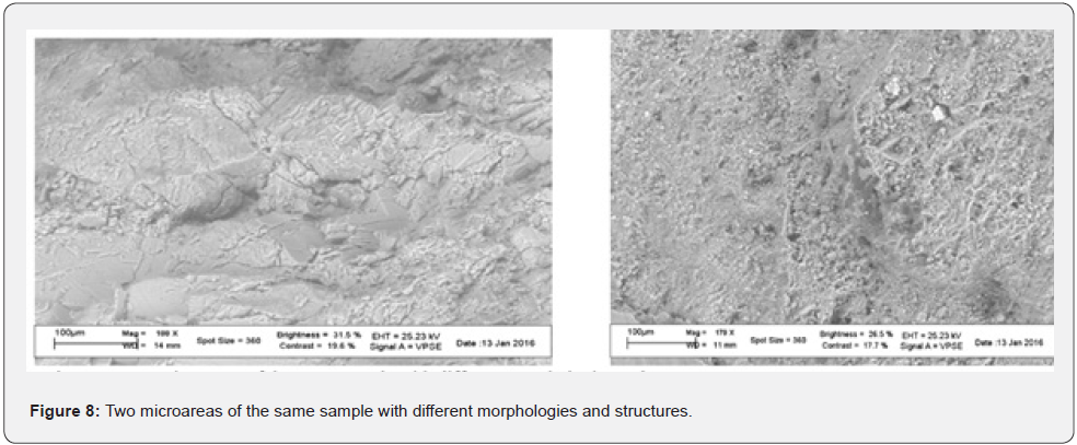
SEM- EDS. The micromorphological analysis, using SEM the image detector with secondary electron resolution in non-in-air conditioning has obtained a very well-defined aspect compared to the petrographic material, with clear crystalline formations of a solid structure is observable figure 6.
Other parts of the same stone were analyzed by SEM to identify the different structures, as tested with the other techniques used figure 7 (Table 1). Various points in the area shown in figure 7 and are analyzed in EDS as reported in table 2. Several EDS analyses were carried out on several areas of the sample, the more significative are reported in table 2.


What was observed at the SEM-EDS is in line with the other types of investigations, while not providing data on the molecular formulations, but only on the composition of elements, it may be useful to consider the morphological and microstructural aspect, where, for example, it is never found its trigonal crystalline habit, but the observation of powdery material, figure 7, could be associated with its presence in conjunction with quartz and other minerals, which would explain the red color felt when handling the stone.
Hematite gave following oxide compositions; FeO 29.8%, Fe2O3 15, MgO 2,6, Al2O3 8.1, CaO 16.55, SiO2 26.7, K2O 0.55, TiO2 0.4%. The concretions with following oxide composition have been detected FeO 5.63, Fe2O3 2.8, MgO 1.55, Al2O3 11.42, CaO 33.34, SiO2 44.4.
A concretion is a hard, compact mass of matter formed by the precipitation of mineral cement within the spaces between particles and is found in sedimentary rock or soil. Concretions form within layers of sedimentary strata that have already been deposited. They usually form early in the burial history of the sediment before the rest of the sediment is hardened into rock. This concretionary cement often makes the concretion harder and more resistant to weathering than the host stratum. They are commonly composed of a carbonate mineral such as calcite; an amorphous or microcrystalline form of silica such as chert, flint, or jasper; or an iron oxide or hydroxide such as goethite and hematite. They can also be composed of other minerals that include dolomite, ankerite, siderite, pyrite, marcasite, barite, and gypsum. Although concretions often consist of a single dominant mineral, other minerals can be present depending on the environmental conditions, which created them. For example, carbonate concretions, which form in response to the reduction of sulfates by bacteria, often contain minor percentages of pyrite. Other concretions formed as a result of microbial sulfate reduction, consist a mixture of calcite, barite, and pyrite.
Conclusion
Implementation of different techniques applied to characterize completely the mineral pigment sample from Noravank is а valuable data, showing a need to conduct similar analyses for the other deposits in the Vayots Dzor Province and all Armenia. Such a study can help to create a reliable database of the mineral pigments of the country and to compare the results with the similar studies of the samples discovered from the archaeological contexts. The mineral pigments, especially, ochre, have been intensively used by prehistoric and historic populations for different purposes, especially in creation of rock-paintings, decorating the pottery and basketry, as well as in rituals. Exploitation of pigments by ancient societies will shed new light on the questions of utilization of mineral resources in the territory of Armenia and the raw-material circulation in the landscape, as well as aspects of symbolic behaviour during the complex ritual games, which took place inside the caves and other sacral spaces. This also can be significant example of benefit achieved by the combination of different scientific disciplines and tools regarding deeper study of the ancient past (Figure 8).

References
- Geology of the Armenian SSR (1966) Non-metallic minerals. In: Mkrtchyan SS (Edt.), Armenian SSR Academy of Sciences Press, Yerevan, Russia.
- Tarayan NP (1949) Mineral paints. In: Mineral resources of the Armenian SSR. Non-metallic minerals, Armenian SSR Academy of Sciences Press, Yerevan, Russia 2: 400-410.
- Cornell RM, Schwertmann U (1996) Iron Oxides: Structure, Properties, Reactions, Occurrences and Uses. In: VCH, New York, USA.
- Hradil D, Grygar T, Hradilová J, Bezdicka P (2003) Clay and iron oxide pigments in the history of painting. Appl Clay Sci 22: 223-236.
- Casellato U, Vigato PA, Russo U, Matteini M (2000) A Mössbauer approach to the physico-chemical characterization of iron-containing pigments for historical wall paintings. J Cultural Heritage 1(3): 217-232.
- Geology of the Armenian SSR (1967) Metallic minerals. In: Mkrtchyan SS (Edt.), Armenian SSR Academy of Sciences Press, Yerevan, Russia.
- Pinhasi R, Gasparian B, Areshyan G, Zardaryan D, Smith A, et al. (2010) First direct evidence of Chalcolithic footwear from the Near Eastern Highlands. Plos One 5(6).
- Barnard H, Dooley AN, Areshian G, Gasparyan B, Faull KF (2011) Chemical evidence for wine production around 4000 BCE in the Late Chalcolithic Near Eastern Highlands. Journal of Archaeological Science 38(5): 377-384.
- Areshian GE, Gasparyan B, Avetisyan PS, Pinhasi R, Wilkinson K, et al. (2012) The Chalcolithic of the Near East and south-eastern Europe: discoveries and new perspectives from the cave complex Areni-1., Armenia. Antiquity 86(331): 115-130.
- Wilkinson K, Gasparyan B, Pinhasi R, Avetisyan P, Hovsepyan R, et al. (2012) Areni-1 cave, Armenia: A Chalcolithic-Early Bronze Age settlement and ritual site in Southern Caucasus. Journal of Filed Archaeology 37(1): 20-33.
- Zardaryan D (2014) About some types of decorations on the Chalcolithic pottery of the Southern Caucasus. In: Gasparyan B, Arimura M (Edt.), Stone Age of Armenia, A Guidebook to the Stone Age Archaeology in the Republic of Armenia, Monograph of the JSPS-Bilateral Joint Research Project, Kanazawa University Press, Printed in Japan, pp. 207-218.
- Smith A, Bagoyan T, Gabrielyan I, Pinhasi R, Gasparyan B (2014) Late Chalcolithic and Medieval archaeobotanical remains from Areni-1 (Birds’ Cave), Armenia. In: Gasparyan B, Arimura M (Eds.), Stone Age of Armenia. A Guidebook to the Stone Age Archaeology in the Republic of Armenia, Monograph of the JSPS-Bilateral Joint Research Project, Kanazawa University Press, Printed in Japan, pp. 233-260.
- Stapleton L, Margaryan L, Areshian GE, Pinhasi R, Gasparyan B (2014) Weaving the ancient past: Chalcolithic basket and textile technology at the Areni-1 cave, Armenia, In: Gasparyan B, Arimura M (Eds.), Stone Age of Armenia. A Guidebook to the Stone Age Archaeology in the Republic of Armenia, Monograph of the JSPS-Bilateral Joint Research Project, Kanazawa University Press, Printed in Japan, pp. 219-232.
- Bobokhyan A, Meliksetyan Kh, Gasparyan B, Avetisyan P, Chataigner C, et al. (2014) Transition to extractive metallurgy and social transformation in Armenia at the end of the Stone Age. In: Gasparyan B, Arimura M (Eds.), Stone Age of Armenia, A Guidebook to the Stone Age Archaeology in the Republic of Armenia, Monograph of the JSPS-Bilateral Joint Research Project, Kanazawa University Press, Printed in Japan, pp. 283-313.
- Gasparyan B, Dan R, Petrosyan A, Vitolo P, Haydosyan H, et al. (2020) The Vayots Dzor Project (VDP): a preliminary overview of the first three years’ activities (2016-2018). Aramazd, Armenian Journal of Near Eastern Studies 10(1-2): 143-183.
- Jana M, Peter K (2001) Baseline studies of the clay minerals society source CLAYS: infrared methods. Clays and Clay Minerals 49(5): 410-432.
- Tironi A, Trezza MA, Irassar EF, Scian AN (2012) Thermal treatment of kaolin: effect on the pozzolanic activity. Procedia Materials. Science 1: 343-350.
- Ying SL, Jeffrey SC, Andrea LW (2012) Infrared and Raman spectroscopic studies on iron oxide magnetic nanoparticles and their surface modifications. Journal of Magnetism and Magnetic Materials 324(8): 1543-1550.


