




 JOJCS Home



Abstract
Background: Collagen plays a central role in maintaining skin structure, elasticity, and hydration. Age-related decline in collagen synthesis contributes to visible signs of skin aging, including wrinkles, dryness, uneven pigmentation, and loss of radiance. Oral hydrolyzed collagen supplements have emerged as a promising nutritional strategy to support skin health. However, the biological activity and efficacy of collagen peptides may vary depending on their source and manufacturing process.
Aim: This pilot study aimed to evaluate the efficacy and safety of a fish-derived collagen peptide supplement (Naticol® x3Peptide), containing at least 25% di- and tripeptides, on several clinical signs of skin aging in women aged 55-70 years.
Methods: A single-arm, open-label pilot trial was conducted in 44 women aged between 55 and 70 years. Participants received a daily oral dose of 2.5g of Naticol® x3Peptide for 8 weeks. Clinical evaluations were performed at baseline, after 3 weeks, and after 8 weeks of supplementation. The assessed parameters included crow’s feet wrinkle severity, skin hydration, facial dark spots, skin color, and complexion radiance. Safety and tolerability were also monitored throughout the study.
Results: Daily supplementation with Naticol® x3Peptide resulted in significant improvements in several markers of skin aging compared with baseline measurements. Participants showed a reduction in crow’s feet wrinkle severity and a significant increase in skin hydration after 8 weeks of supplementation. In addition, facial complexion radiance improved, while facial dark spots became lighter and less visible over time. The supplement was well tolerated, with no major safety concerns reported during the study period.
Conclusion: The results of this open-label pilot study support the efficacy and safety of oral supplementation with fish-derived hydrolyzed collagen peptides with enhanced di- and tripeptide content. Daily intake of 2.5g of Naticol® x3Peptide significantly improved wrinkle appearance, skin hydration, complexion radiance, and pigmentation parameters in women with aging skin.
Keywords: Collagen peptides; Dietary supplement; Skin rejuvenation; Radiance; Hydration; Wrinkles; Lightening
Abbreviations: AEs: Adverse Events; AGEs: Advanced Glycated End Products; b*: Yellow–Blue Color Component of the Skin Tone; BMI: Body Mass Index; C.L.B.T: Color–Luminosity–Brightness–Transparency; ECM: Extracellular Matrix; EEMCO: European Group on Efficacy Measurement of Cosmetics; Gly: Glycine; HACCP: Hazard Analysis Critical Control Point; Hyp: Hydroxyproline; IL: Interleukine; ITA°: Individual Typology Angle; L*: Luminance; MMP: Matrix Metalloproteinase; MW: Molecular Weight; NMFs: Natural Moisturizing Factors; PRIMOS: Phase-Shift Rapid In Vivo Measurement of Skin; Pro: Proline; TGF-β: Transforming Growth Factor; TNF-⍺: Tumor Necrosis Factor; UV: Ultraviolet
Introduction
The dermis is the thickest inner layer of skin and is the place where several major functions of the skin take place. Blood vessels which are absent from the most superficial cutaneous layer ensure nutrition. By detecting pressure, temperature, and pain and transmitting signals to the brain, nerve endings ensure sensory perception [1-3]. Amongst the various components of the dermis, fibroblasts play a major role in skin biology. They secrete a gel-like fundamental substance and fibers such as elastin and collagen that ensure mechanical protection. The dermal fibers confer suppleness, elasticity, and tensile strength to the skin, while the principal macromolecules of the fundamental substance, such as proteoglycans and glycosaminoglycans, exert a hydrating and lubricating effect. As the skin ages, the most significant changes occur in the dermis, for example, a reduction in blood microcirculation and a degradation of collagen and elastin fibers leading to a decrease in skin radiance, firmness and elasticity as well as the development of wrinkles. With aging, the number and size of fibroblasts decrease resulting in a diminution of collagen biosynthesis. In parallel, metalloprotease enzyme activity increases, leading to the degradation of collagen fibers. As a result of the harmful effects of this dual process, collagen fibers decrease in number and become more fragmented and fragile [3,4].
Collagens constitute a large family of proteins. In the human body and in the dermis in particular, type I collagen is the most common protein, providing structural support to the extracellular matrix of connective tissues [5-7]. As previously mentioned, ageing has many physiological and functional consequences which originate at the molecular level.
Post-translational modifications of collagen, mainly glycosylation and glycation, occur after collagen synthesis. Due to collagen’s long half-life of about 15 years, these modifications gradually accumulate over time [8]. Advanced Glycation End products (AGEs) also contribute significantly to skin aging by cross-linking collagen fibers, which alters their mechanical properties and affects the function of the extracellular matrix (ECM) [9].
Given the importance and crucial role of collagen fibers in maintaining healthy and functional skin, and the established detrimental effect of their gradual decline with age, supplying the human body with collagen peptides able to promote endogenous collagen synthesis appears to be a logical approach. Globally, over the past few years, skin-beneficial functional foods have gained significant attention. More specifically, collagen-containing oral supplements are now among the most popular products to address signs of ageing. The advantages of taking oral hydrolyzed collagen supplements for skin hydration and elasticity are now very well established [10,11]. The anti-wrinkles effect of these supplements has also been demonstrated in several studies [12]. Recent human studies have also suggested that the oral administration of collagen peptides could improve the radiance of the complexion and the overall beauty/healthy appearance of the facial skin [13-15].
While several in vitro and in silico studies suggest that collagen peptide hydrolysates may influence cutaneous pigmentation via tyrosinase inhibition [16-18], clinical evidence demonstrating a direct pigmentation-lightening effect of oral collagen in humans is scarce. On this basis and given the importance of pigmented disorders with aging [19], we also wanted to examine the potential effects of our fish collagen peptide supplement on facial dark spots.
Importantly, the amino acid composition of collagen, and more specifically the peptide sequences that constitute it, along with their associated biological activities, are strongly influenced by both the species of origin and the industrial extraction method [20]. This makes it necessary to evaluate each new oral collagen formulation individually.
Our study aimed to assess the potential of a new specific fish collagen peptide supplement (Naticol® x3Peptide) with a minimum 25% di and tripeptide content to improve several parameters related to skin aging.
Materials and Methods
Ethics and trial design
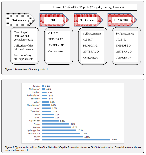
The protocol, CRFS, and informed consent documents of this single-arm, open-label study, were reviewed and approved by the Human Research Ethics Committee of Thammasat University (Faculty of Medicine, Bangkok, Thailand). A certificate of approval 241/2023 was granted to the study (project MTU-EC-IM-5-192/66). Because Thailand currently has no legal or regulatory requirement for clinical trial registration in a public registry, the study was not registered in the Thai Clinical Trials Registry. The ethical principles of the Declaration of Helsinki, Good Clinical Practices and regulations from the Personal Data Protection Act B.E. 2562 (2019) of Thailand were respected. Prior to any study-related procedures, potential subjects received oral and written explanations about the aims, possible cosmetic benefits, constraints and potential risks, and details of the study; those who gave their signed informed consent, and authorizations to use their pictures for publication, were then included in this study. The study duration was 8 weeks and comprised 3 visits (baseline before the first intake of the investigational product, T+3 weeks and T+8 weeks). An overview of the study protocol is presented in Figure 1.
Study population and inclusion and exclusion criteria
Forty-five (45) healthy Asian females, aged between 55 and 70 years old, were enrolled on the basis of the following inclusion criteria: having skin phototype III or IV according to Fitzpatrick’s scale, having any skin nature, having crow’s feet wrinkles of grade ≥3 (according to the scale of the “Skin Aging Atlas - Vol 2. Asian type” [21], having a dull complexion according to the C.L.B.T. scale [19], having one or several dark spots on the cheeks with a diameter ≥2.5mm, not having ingested oral supplements for at least 1 month. Excluded were those subjects who had a diagnosed or highly probable allergy to fish; had taken food supplements in the 3 months prior to the beginning of the trial; had an unstable dietary pattern; reported excessive alcohol, caffeine-based product, or tobacco consumption; had medical conditions or received anti-aging, anti-wrinkle, or pigmentary disorder therapies that could confound the evaluations (such as medicinal, aesthetic, or cosmetic treatments); or had a Body Mass Index (BMI) of ≥27.5kg.m-2. Detailed inclusion/exclusion criteria are provided in Supplementary Table S2.. Participants were instructed to not modify their nutrition and cosmetic habits, to avoid starting any whitening/anti-wrinkle/anti-aging treatments, and to avoid taking any food supplements other than the product under investigation during the study period. The full list of proscriptions can also be found in Supplementary Supplementary Table S2.
Because this pilot study was exploratory, no formal sample size calculation was performed. The study’s primary goal was to gather preliminary data on the effect of the test product on complexion radiance prior to planning a larger trial. Nevertheless, previous studies using collagen-based oral supplements and similar assessment methods (such as corneometry and wrinkles evaluation) have achieved meaningful results with populations of 20 to 40 subjects [22,23]. Therefore, we decided to include 45 subjects. Considering 10% potential dropout cases or exclusions due to protocol violations, the objective was to complete the study with at least 40 subjects.
Trial intervention
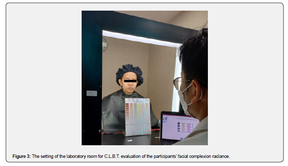
The investigational product (Naticol® x3Peptide) was a nutritional supplement containing 100% fish collagen peptides with a mean Mw of 1200 Da, comprising >25% di- tripeptide. The typical amino acid composition of the formulation is presented in Figure 2. It was presented in 250 grams sachets with a spoon allowing a dose of 2.5g. The product had to be taken orally, once daily at 2.5g per serving, 20 minutes before a meal in the morning, for a total duration of 8 weeks. All dietary supplements were manufactured by Weishardt International (Graulhet, France). Control and quality assurance measures were performed in accordance with the requirements of Good Manufacturing Practices, based on, among other thing, batch traceability and HACCP (Hazard Analysis Critical Control Point) analysis. During the trial period, the subjects were instructed to maintain their regular diet. To ensure the subjects’ proper compliance (i.e., that the appropriate amounts of test product were being used), the test products were weighed at T0, T+3 weeks and T+8 weeks. A declaration form was also distributed to each subject together with the sachets during the initial visit, checked after 3 weeks, and collected back at the final visit to monitor the regularity of consumption of the investigational product.
Evaluation methods for treatment efficacy
All measurements and evaluations were performed after a 20-minute acclimatization period of rest in controlled rooms (20-24°C, 40-60% RH). or any given subject, measurements were always performed at the same time for all appointments to avoid bias due to natural circadian variations. Each evaluation method was consistently handled by the same technician(s) at all kinetic stages to prevent operator-dependent biases.
For each subject, wrinkles of one crow’s feet area (left or right randomly selected) were investigated by using the PRIMOS system (Canfield Scientific Europe, Utrecht, Netherlands), a gold standard skin surface relief analysis technique. Phase-shift Rapid In vivo Measurement of Skin (PRIMOS) is an optical three-dimensional imaging technique allowing a non-invasive profilometric analysis of the skin surface. It is based on the principle of projection of a pattern of parallel light lines onto the skin, with this pattern being shifted in phase over time. A digital camera captures multiple successive images of the studied skin surface. Deformations of the pattern in the sequence of images caused by the cutaneous relief allow us to reconstruct a three-dimensional map of the surface of the skin, i.e. of wrinkles in our study [24,25]. By matching the same skin areas at the three evaluation times, PRIMOS 5.7 software allowed for the determination of the average wrinkles depth and length, the mean and maximum depth of the biggest wrinkle (µm), the wrinkles count, and their average area (mm2) and volume (mm3).
The facial skin complexion was evaluated based on a visual assessment of six descriptors by three trained judges, according to a set of specific scales of the C.L.B.T. method developed by Musnier et al. [15], and slightly adapted to take into consideration the specificities of Asian skin [19]. The C.L.B.T. acronym stands for the six descriptors to be evaluated: the colors (olive, yellow and pink hues), luminosity, brightness and transparency of the skin.
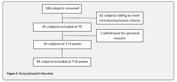
The evaluation was conducted in a laboratory equipped with a dedicated scoring table to ensure consistent lighting conditions and maintain fixed judge-subject relative positioning. Subjects were seated between two symmetrically positioned “daylight” lamps and wore black caps and gowns to cover their clothing and avoid the potential influence of extrinsic colors (Figure 3). Evaluations were carried out by 3 trained judges who were neither allowed to communicate with one another nor allowed to check the results from the previous evaluation time; their scores were then averaged to increase the reliability of the assessment. The color evaluation was based on structured visual scales presenting percentages of saturation of olive, yellow and pink hues (0-100%). The luminosity, brightness and transparency descriptors evaluations were carried out using analogical scales ranging from 0 to 10 (e.g., 0 for no luminosity at all and 10 for maximal luminosity). The scores were captured by the evaluator using in-house software to move a cursor along the different scales. The interpretation of C.L.B.T. results is based on significant variations in the descriptors taken separately. A positive change in skin radiance is characterized by increases in the pink saturation percentage and the luminosity, brightness and transparency scores, along with decreases in the yellow and olive saturations percentages. If the majority (or all) of the descriptors change in the anticipated way as outlined above, the effectiveness of the test product in enhancing skin complexion radiance can be verified. Methodological details can also be found in a paper from Dumoulin et al. [26].
To assess hydration of the outer layers of the skin, a Corneometer® MPA 580 (Courage+Khazaka electronic, Köln, Germany) was used according to EEMCO guidelines [27]. Measurements of the skin’s electrical capacitance (in arbitrary units, a.u.) were performed in triplicate on the left or right cheek (randomly selected), then averaged. To ensure consistency, the locations of the measurement sites were marked at the baseline visit.
Other measurements involved the use of the Antera 3D® camera (Miravex Limited, Dublin, Ireland). In this method, the skin is illuminated with multi-directional light emitting diodes of different wavelengths spanning over the entire visible spectrum; then, the camera collects the reflected light, and the reflection angle is used to extract high-resolution topographic features of the skin. From the reconstructed skin surface image, color analysis is also possible [28,29]. In our study, we therefore used the Antera 3D® device to track the changes in the color of facial dark spots over time. Images of the cheeks with dark spots were acquired for each subject at all stages of kinetics using an Antera 3D® camera. To ensure reproducible positioning of the camera at each visit, individual transparent mappings of the skin surface were performed. To assess color changes in the dark spots throughout the study, the CIE-L*a*b* values were determined by using the device’s software. Additionally, the individual typological angle was calculated using the formula ITA°=Arctg [(L*-50)/b*].(180/π). L* and ITA° are the most effective indicators of a whitening or lightening effect [30], where higher L* and ITA° values indicate lighter skin.
After 3 and 8 weeks, the subjects were required to fill out questionnaires to evaluate their overall opinion and their attitude regarding the effectiveness of the product being tested. The questionnaires were filled out at the testing facilities, in front of a mirror with standardized lighting, without any extrinsic influences. In particular, self-assessment questionnaires were filled out by the subjects before any measurements were carried out to avoid influencing the subjects’ judgment about the test product. For each item, the possible answers were “Completely agree”, “Somewhat agree”, “Somewhat disagree” and “Completely disagree”.
Safety assessment
Subjects were asked to call the Investigator to report any possible symptoms of discomfort, related or not to the tested product, immediately. A follow-up form was given to all subjects at the beginning of the study. This form allowed the participants to record adverse events (AEs) on a daily basis and document the nature, intensity and duration of the symptom(s). Interviews of the subjects were also conducted at T+3 weeks and T+8 weeks to find out if they had experienced any adverse events during the course of the study.
Statistical analysis
All statistical analyses were performed using SigmaStat 3.5 (Systat software Inc., Delaware, USA) and Past 1.37 (Palaeontological Association) softwares. Quantitative variables are described as mean values with their respective standard deviations. Qualitative variables are reported as numbers and percentages. The normality of distributions was verified using the Shapiro-Wilk test, with the threshold set at 1%. Depending on the normality of the data, the two-tailed paired Student’s t-test or non-parametric Wilcoxon test was used for comparisons versus baseline. The threshold of significance was set to p<0.05.
With regard to the self-assessment questionnaires, the analysis involved calculating the frequencies of the different possible answers (given as percentages) to each qualitative question. To evaluate the efficacy and the appreciation of the product for each item, two percentages, Z1 and Z2, were calculated as follows: Z1 = favorable opinion (Ex: “Completely agree” + “Somewhat agree”) and Z2 = unfavorable opinion (Ex: “Completely disagree” + “Somewhat disagree”). The statistical difference in frequencies (%) between favorable and unfavorable opinions was evaluated using the Chi2 test at 5%.
Results
Participant characteristics
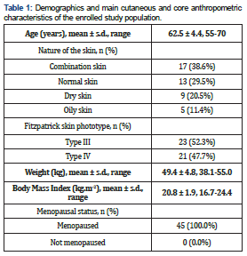
A total of 106 potential participants were screened; of these, 45 subjects were enrolled in the study. A total of 44 subjects completed the 8-week- trial (Figure 4). Therefore, the objective to retain at least 40 assessable subjects at the end of the study period was achieved. The demographics and main characteristics of the study panel are presented in the Table 1.
Subject compliance
With a recommended daily dose of 2.5g, the observed usage of the test product was below the expected usage. Of note, a few subjects ingested just slightly more than 50% of the instructed amount over the study period. Overall, the actual average daily dose of Naticol® x3Peptide used was approximately 1.8g rather than the instructed 2.5g. This lower-than-planned intake is a factor important to consider in relation to the observed effects described thereafter, and will be discussed at the end of this paper.
Treatment efficacy
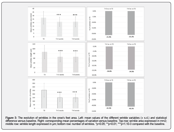
Figure 5 summarizes the results of the profilometric assessment of crow’s feet area wrinkles using the PRIMOS system. After 3 weeks of oral administration of the Naticol® x3Peptide, very significant decreases in crow’s feet wrinkles were observed compared with the baseline for their average area (from 26.98 ± 8.98mm2 down to 21.21 ± 6.59mm2, i.e., -21.4%; p<1.10-3) and average length (86.48 ± 28.63μm down to 65.73 ± 20.84μm, i.e., -24.0%; p<1.10-3). A very important reduction in the number of wrinkles was also observed after 3 weeks (-41.5%; p<1.10-3). The results after 8 weeks of intake confirmed those observed at T+3 weeks. Compared with the baseline, significant decreases were observed for the average area (from 26.98 ± 8.98mm2 down to 21.27 ± 7.12mm2, i.e., -21.2%; p<1.10-3) and average length (86.48 ± 28.63μm down to 65.45 ± 20.64μm, i.e., -24.3%; p<1.10-3) of wrinkles in the crow’s feet area. The average number of wrinkles was reduced by -42.2% compared with that at T0. Interestingly, the anti-wrinkle effect reached its peak after 3 weeks and was maintained until the end of the study period while pursuing the cure.
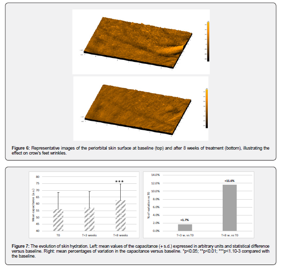
To visually illustrate the observed anti-wrinkle effect, Figure 6 thereafter shows representative 3D reconstructions of one “average” subject’s crow’s feet area, taken at baseline and after 8 weeks of the product intake.
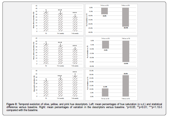
After 8 weeks of use, Naticol® x3Peptide was able to significantly improve the hydration of the outer layers of skin (Figure 7). Indeed, a significant increase in the mean capacitance, measured via corneometry, was found: from 56.1 ± 13.7a.u. at baseline up to 62.6 ±12.2a.u. after 8 weeks of daily intake (i.e. +11.6%; p<1.10-3). After 3 weeks, a slight increase, not reaching the level of statistical significance, was observed (+1.7%).
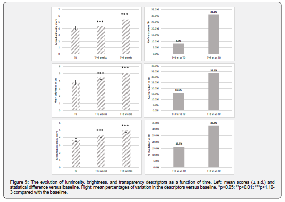
In the visual evaluation of facial skin radiance using the C.L.B.T. method, highly significant reductions in mean olive and yellow saturations were observed at both T+3 weeks and T+8 weeks compared with baseline (Figure 8).
From a value of 35.6 ± 2.5% before use of the tested oral supplement, the mean olive saturation percentage progressively decreased to 32.7 ± 2.7% (i.e., -8.2%; p<1.10-3) and 27.3 ± 2.3% (i.e., -23.3%; p<1.10-3) after 3 and 8 weeks, respectively. Similarly, a significant decrease in the mean yellow saturation percentage was achieved at T+3 weeks compared with the baseline (from 36.3 ± 2.6% down to 32.9 ± 2.5%, i.e. -9.3%; p<1.10-3). The decrease was even more important after 8 weeks (-24.9%; p<1.10-3). Considering the pink component of the complexion significant increases in the mean saturation percentage were found after 3 weeks (from 25.5 ± 1.9% at T0 up to 28.3 ± 2.3%, i.e., +11.0%; p<1.10-3) and 8 weeks of supplement intake (from 25.5 ± 1.9% at T0 up to 32.8 ± 2.5, i.e., +29.0%; p<1.10-3) (Figure 8). The significant decreases in olive and yellow hues, together with the significant increase in pink color, represent improved skin radiance thus highlighting the beneficial effect of Naticol® x3Peptide after only 3 weeks [14,15].
Results achieved for physical descriptors of skin radiance were also remarkable (Figure 9). From a mean grade of 4.1 ± 0.3 at baseline, luminosity significantly increased to 4.4 ± 0.3 at T+3 weeks (i.e., +8.3%; p<1.10-3), and to 5.4 ± 0.4 at T+8 weeks (i.e., +31.1%; p<1.10-3). For the brightness indicator, the mean grade increased from 3.8 ± 0.3 at T0 up to 4.5 ± 0.3 at T+3 weeks (i.e., +16.1%; p<1.10-3), and reached +5.1 ± 0.4 (i.e. +33.6%; p<1.10-3) at the end of the follow-up period. Finally, very significant increases were observed for skin transparency. From a mean grade of 3.7 ± 0.2 at baseline, the transparency mean grade progressively increased to 4.3 ± 0.3 (i.e., +16.5%; p<1.10-3) at 3 weeks and 5.0 ± 0.3 (i.e., +32.8%; p<1.10-3) after 8 weeks of treatment. These variations can be interpreted as a more intense reflection of light by the skin, a more even-looking complexion, and a fairer and younger appearance of the facial skin [14]. Thus, the C.L.B.T. clinical evaluations showed a general and highly significant improvement in all descriptors of cutaneous radiance after both 3 and 8 weeks of supplementation.
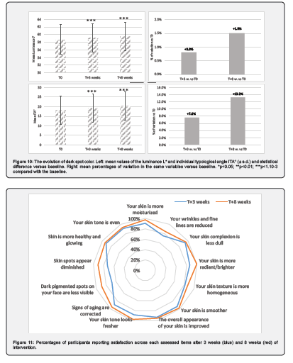
The color measurements of pigmented spots performed via Antera 3D® image analysis are graphically summarized in Figure 10. Continuous increases in the mean L* and ITA° values were observed throughout the study. The luminance L* significantly increased from 58.69 ± 3.87 at baseline to 59.16 ± 3.71 at T+3 weeks (i.e., + 0.8%;p<1.10-3) and 59.55 ± 3.71 at T+12 weeks (i.e. + 1.5%; p<1.10-3). Similarly, from 18.03 ± 7.42 prior to oral supplementation, the mean ITA° significantly increased to 19.39 ± 7.49 (i.e. + 7.6%; p<1.10-3) after 3 weeks and to 20.40 ± 7.36 (i.e., +13.2%; p<1.10-3) after 8 weeks. This indicated that significant lightening of the facial dark spots occurred gradually after 3 and 8 weeks of oral supplementation with Naticol® x3Peptide.
The tested oral supplement Naticol® x3Peptide received highly positive evaluations from the study participants, with all claims being significantly supported by the results. After 3 weeks of treatment, the percentages of favorable opinions ranged between 73% and 95%. At the end of the 8-week study, all the tested items were even more appreciated since the favorable opinions generally progressed and ranged between 84% and 100% (Figure 11). In line with the results, the question « Would you say that your skin is more radiant/brighter» received 100% positive opinions which is totally in line with the improvement in complexion radiance demonstrated by the C.L.B.T. clinical evaluations. The detailed results from the self-assessment questionnaires are provided in Supplementary Material Table S1.
Safety assessment
No functional or clinical sign imputable to the tested oral supplement was reported or observed during the 8-week trial period. Therefore, the tolerance of Naticol® x3Peptide formulation was considered to be excellent under the conditions of this study.
Discussion
The growing use of dietary supplements to enhance skin appearance reflects the link between skin and overall health, a trend amplified by the rapid global aging, with the population aged 60 and over expected to exceed 2 billion by 2050 [31,32].
To exert a biological effect upon the skin, orally administered collagen peptides must reach the dermis. This ability of collagen hydrolysates to reach their target after ingestion was precisely demonstrated by Watanabe-Kamiyama et al. [33] who investigated the absorption mechanism of low-molecular-weight collagen hydrolysates labeled with 14C in rats [33].
Collagen-derived products are hydrolyzed into small peptides in the gastrointestinal tract before being absorbed into the bloodstream, from which they are transported to the skin. The literature reports the presence of free amino acids, di- and tri-peptides released into plasma after collagen ingestion [34].
Naticol® x3Peptide, the hydrolyzed collagen peptides evaluated in our study, are sourced from fish skins and thereby consists primarily of type I collagen. Naticol® x3Peptide, with a mean molecular weight of 1.2kDa has a rich di-tripeptide content. As compared with larger collagen peptides, the smaller molecular size of hydrolyzed collagen peptides allows for better absorption in the body. Moreover, after ingestion, collagen hydrolysates are further cleaved during gastrointestinal passage, giving rise to elevated levels of new di- and tri-peptides and free amino acids in the blood circulation [35]. This enhanced bioavailability ensures that the collagen is more effectively utilized by the body, leading to improved outcomes in skin health. Mechanistically, di- and tri-peptides act as bioactive signaling molecules to activate dermal fibroblasts proliferation, upregulate collagen elastin and hyaluronic acid synthesis [36]. It has also been shown that collagen peptides intake can preserve the ECM by reducing matrix metalloproteinase 2 activity [37-39]. This dual activity, promoting synthesis and decreasing the biodegradation of collagen fibers, explains the interest in collagen hydrolysates for anti-aging applications.
In line with this, previous in vitro experiments on human fibroblasts have confirmed that Naticol® x3Peptide significantly inhibits the expression of pro-inflammatory cytokines such as IL-12, IL-1β, and TNFα while enhancing the expression of anti-inflammatory mediators such as IL-10 and TGF-β (data under publication).
In terms of safety, overall tolerability of Naticol® x3Peptide was confirmed by 100% of participants, with no adverse events reported during the 8-week study. The fish collagen peptides were derived from gelatin via enzymatic hydrolysis, produced in compliance with regulatory standards for agri-food products intended for human consumption. Hydrolyzed collagen, such as hydrolyzed gelatin, is classified as ‘Generally Recognized As Safe’ (GRAS) by the FDA, and its immunogenicity is considered low, especially for low-molecular-weight collagen hydrolysates [44].
This pilot trial confirmed the anti-wrinkle effect of collagen-based oral supplements. Indeed, this study revealed significant improvement in the appearance of the crow’s feet wrinkle area after 3 and 8 weeks of oral administration of a 2.5 g daily dose of Naticol® x3Peptide compared with the baseline.
These findings are consistent with previous studies showing that collagen supplementation improves facial wrinkles by stimulating dermal fibroblasts, enhancing Type I collagen, elastin, and hyaluronic acid production, and supplying key amino acids for collagen synthesis. Additionally, its anti-inflammatory and antioxidant properties help reduce oxidative stress, down-regulate pro-inflammatory cytokines, and inhibit collagen-degrading enzymes, collectively supporting extracellular matrix integrity and reducing wrinkle formation [40-42].
Another key benefit of the treatment was increased skin hydration after 8 weeks, consistent with previous studies and recent meta-analyses [10,11]. Collagen peptides enhance skin moisture by stimulating filaggrin production and increasing ceramides, lipids, and hyaluronic acid, thereby reducing transepidermal water loss [10].
Skin radiance, an uncommon focus in collagen peptide research and the most original finding of this study, showed promising results. The C.L.B.T. clinical visual evaluation, carried out by experienced and carefully trained experts, demonstrated a general and highly significant improvement in all the descriptors of skin radiance after 3 and 8 weeks of intervention.
Finally, the tested oral supplement was also found to be able to significantly lighten the participants’ facial dark spots after 3 and 8 weeks of ingestion, as demonstrated via the colorimetric analysis of digital photographs of the cheeks taken with an Antera 3D® camera. The mechanism of action of the tested supplement on pigmented spots has not been established. However, some oligopeptides are known to exhibit depigmenting activity. Notably, collagen-derived peptides reported to inhibit melanin biosynthesis predominantly originate from marine sources, particularly fish [17,18,43].
Regarding the disparity in kinetics, we observed wrinkle improvement was maximal at week 3 and subsequently plateaued, whereas the other parameters under investigation (hydration, C.L.B.T. descriptors of the complexion radiance, color of dark spots) continued to improve between weeks 3 and 8. One possible explanation could be that wrinkle reduction and skin hydration/optical parameters may involve distinct physiological mechanisms that progress at different rates. Wrinkle appearance is influenced by early changes in dermal hydration and biomechanical properties, which may respond relatively rapidly to collagen peptide supplementation, resulting in an initial improvement followed by an apparent plateau. In contrast, parameters such as skin hydration, luminance, brightness, and color may be more closely associated with the gradual modulation of skin physiology, including changes in dermal matrix organization and epidermal renewal (8 weeks corresponding approximately to two skin renewal cycles). In our experience, skin color changes usually require more than 1 month, i.e. more than one full cycle to become significant. The same rule should also apply to the olive; yellow and pink descriptors used for the evaluation of the radiance of the complexion.
The strengths of this pilot study rely on the use of objective skin measurement techniques for a robust and quantifiable outcome assessment (wrinkle assessment using the PRIMOS system, skin hydration measurement via corneometry, and facial dark spot color analysis with the Antera 3D® camera). In addition, the use of a dedicated clinical evaluation method for skin radiance (C.L.B.T.) allowed us to clearly evidence a marked enhancement of the participants’ skin complexion since all (six) descriptors of radiance exhibited very significant improvements after 3 and 8 weeks of supplement intake. This supports our initial hypothesis that the oral administration of collagen peptides could improve the complexion radiance and overall beauty/healthy appearance of facial skin, as suggested by a few recent studies [45].
The highly positive results from both instrumental and clinical measurement methods are mirrored by the high percentages of favorable opinions expressed by the participants in self-assessment questionnaires regarding the tested oral treatment after 3 and 8 weeks of use (Figure 8).
Finally, although participants often consumed less than the recommended 2.5g/day, beneficial effects of the oral supplement were still observed suggesting that adherence to the full recommended dose could potentially yield even greater results. While the study’s open-label design and reliance on baseline comparisons represent limitations, these promising results provide a strong rationale for confirmation in a randomized, placebo-controlled trial.
Conclusion
Taken together, the experimental results of this 8-week clinical trial substantiated Naticol® x3Peptide as a safe dietary supplement capable of attenuating several cutaneous signs of aging such as wrinkles and pigmentary dark spots, which are a major concern for mature women, as well as significantly improving skin hydration. Notably, facial complexion radiance also showed a highly significant improvement. Considering the overall results, Naticol® x3Peptide appears to be a potent ingredient for attenuating the visible signs of skin aging.
References
- Lotfollahi Z (2024) The anatomy, physiology and function of all skin layers and the impact of ageing on the skin. Wound Prac Res 32: 6-10.
- Brito S, Baek M, Bin BH (2024) Skin structure, physiology, and pathology in topical and transdermal drug delivery. Pharmaceutics 16(11): 1403.
- Abdo JM, Sopko NA, Milner SM (2020) The applied anatomy of human skin: a model for regeneration. Wound Med 28: 100179.
- Haydont V, Bernard BA, Fortunel NO (2019) Age-related evolution of the dermis: clinical signs, fibroblast and extracellular matrix dynamics. Mech Ageing Dev 177: 150-156.
- Salo AM, Myllyharju J (2021) Prolyl and lysyl hydroxylases in collagen synthesis. Exp Dermatol 30(1): 38-49.
- León-López A, Morales-Peñaloza A, Martínez-Juárez VM, Vargas-Torres A, Zeugolis DI, et al. (2019) Hydrolyzed collagen – Sources and applications. Molecules 24(22):
- Shenoy M, Abdul NS, Qamar Z, Al Bahri BM, Al Ghalayini KZK, et al. (2022) Collagen structure, synthesis, and its applications: a systematic review. Cureus 14(5): e24856.
- Verzijl N, DeGroot J, Thorpe SR, Bank RA, Shaw JN, et al. (2000) Effect of collagen turnover on the accumulation of advanced glycation end products. J Biol Chem 275(50): 39027-39031.
- Avery NC, Bailey AJ (2006) The effects of the Maillard reaction on the physical properties and cell interactions of collagen. Pathol Biol (Paris) 54(7): 387-395.
- Jung K, Kim SH, Joo KM, Lim SH, Shin JH, et al. (2021) Oral intake of enzymatically decomposed AP collagen peptides improves skin moisture and ceramide and Natural Moisturizing Factor contents in the stratum corneum. Nutrients 13(12): 4372.
- Pu SY, Huang YL, Pu CM, Kang YN, Hoang KD, et al. (2023) Effects of oral collagen for skin anti-aging: a systematic review and meta-analysis. Nutrients 15(9): 2080.
- Duteil L, Queille-Roussel C, Maubert Y, Esdaile J, Bruno-Bonnet C, et al. (2016) Specific natural bioactive type 1 collagen peptides oral intake reverse skin aging signs in mature women. J Aging Res Clin Practice 5: 84-92.
- Evans M, Lewis AD, Zakaria N, Pelipyagina T, Guthrie NA (2021) Randomized, triple-blind, placebo-controlled, parallel study to evaluate the efficacy of a freshwater marine collagen on skin wrinkles and elasticity. J Cosmet Dermatol 20(3): 825-834.
- Trehan A, Anand R, Chaudhary G, Garg H, Verma MK (2024) Efficacy and safety of skin radiance collagen on skin and hair matrix: a placebo-controlled clinical trial in healthy human subjects. Clin Cosmet Invest Dermatol 17: 581-591.
- Musnier C, Piquemal P, Beau P, Pittet JC (2004) Visual evaluation in vivo of complexion radiance using the C.L.B.T. sensory methodology. Skin Res Technol 10(1): 50-56.
- Zheng J, Xu D, Li T (2025) The inhibitory effect of peptide hydrolysate of Type I collagen derived from pig skin on melanogenesis in B16F10 melanoma cells. Biomolecules 15(2): 220.
- Zu XY, Li MJ, Xiong GQ, Cai J, Liao T, et al. (2023) Silver carp (Hypophthalmichthys molitrix) Scales Collagen Peptides (SCPs): preparation, whitening activity screening and characterization. Foods 12(7): 1552.
- Xue W, Liu X, Zhao W, Yu Z (2022) Identification and molecular mechanism of novel tyrosinase inhibitory peptides from collagen. J Food Sci 87(6): 2744-2756.
- Perin F (2012) New method for evaluation of Asian skin radiance. Personal Care Asia Pacific 3: 35-37.
- Cadar E, Perterau AM, Prasacu I, Ionescu AM, Pascale C, et al. (2024) Marine antioxidants from marine collagen and collagen peptides with nutraceuticals applications: a review. Antioxidants 13(8): 919.
- Bazin R, Flament F (2010) Skin ageing atlas. Volume 2, Asian type, Editions Med’Com: Paris, France, pp. 40-41.
- Aguirre-Cruz G, León-López A, Cruz-Gómez V, Jiménez-Alvarado R, Águirre-Alvarez G (2020) Collagen hydrolysates for skin protection: oral administration and topical formulation. Antioxidants 9(2): 181.
- Asserin J, Lati E, Shioya T, Prawitt J (2015) The effect of oral collagen peptide supplementation on skin moisture and the dermal collagen network: evidence form an ex vivo model and randomized, placebo-controlled clinical trials. J Cosmet Dermatol 14(4): 291-301.
- Jaspers S, Hopermann H, Suaermann, G, Hoppe U, Lunderstädt R, et al. (1999) Rapid in vivo measurement of the topography of human skin by active image triangulation using a digital micromirror device. Skin Res Technol 5(3): 195-207.
- Jacobi U, Chen M, Frankowski G, Sinkgraven R, Hund M, et al. (2004) In vivo determination of skin surface topography using an optical 3D device. Skin Res Technol 10(4): 207-214.
- Dumoulin M, Gaudout D, Lemaire B (2016) Clinical effects of an oral supplement rich in antioxidants on skin radiance in women. Clin Cosmet Invest Dermatol 9: 315-324.
- Berardesca E, Loden M, Seru J, Masson P, Rodrigues LM (2018) The revised EEMCO guidance for the in vivo measurement of water in the skin. Skin Res Technol 24(3): 351-358.
- Linming F, Wei H, Angi L, Yuanyu C, Hens X, et al. (2018) Comparison of two skin imaging analysis instruments: the VISIA® from Canfield vs the ANTERA 3D® CS from Miravex. Skin Res Technol 24(1): 3-8.
- Messaraa C, Metois A, Walsh M, Hurley S, Doyle L, et al. (2018) Wrinkles and roughness measurement by the Antera 3D and its application for evaluation of cosmetic products. Skin Res Technol 24(3): 359-366.
- Petit L, Pierard GE (2003) Skin-lightening products revisited. Int J Cosmet Sci 25(4): 169-181.
- Draelos ZD (2010) Nutrition and enhancing youthful-appearing skin. Clin Dermatol 28(4): 400-408.
- World Population Prospects 2024: Summary of results.
- Watanabe-Kamiyama M, Shimizu M, Kamiyama S, Taguchi Y, Sone H, et al. (2010) Absorption and effectiveness of orally administered low molecular weight collagen hydrolysate in rats. J Agric Food Chem 58(2): 835-841.
- Kawaguchi T, Nanbu PN, Kurokawa M (2012) Biodistribution of prolylhydroxyproline and its metabolites after oral administration in rats. Biol Pharm Bull 35(3): 422-427.
- Virgilio N, Schön C, Mödinger Y, van der Steen B, Vlemicks S, et al. (2024) Absorption of bioactive peptides following collagen hydrolysate intake: a randomized double-blind crossover study in healthy individuals. Front Nutr 11: 1416643.
- Jariwala N, Ozols M, Bell M, Bradley E, Gilmore A, et al. (2022) Matrikines as mediators of tissue remodelling. Adv Drug Deliv Rev 185: 114240.
- Zague V, de Freitas V, da Costa RM, de Castro GA, Jaeger RG, et al. (2011) Collagen hydrolysate intake increases skin collagen expression and suppresses matrix metalloproteinase 2 activity. J Med Food 14(6): 618-624.
- Ivaskiene T, Viskelis J, Streimikyte P, Savickaitè M, Mobasheri A, et al. (2025) Collagen supplementation and regenerative health: advances in biomarker detection and smart material integration. Front Nutr 12: 1716166.
- Cao C, Xiao Z, Tong H, Liu Y, Wu Y, et al. (2022) Oral intake of chicken bone collagen peptides anti-skin aging in mice by regulating collagen degradation and synthesis inhibiting inflammation and activating lysosomes. Nutrients 14(8): 1622.
- Proksch E, Schunck M, Zague V, Segger D, Degwert J, et al. (2014) Oral intake of specific bioactive collagen peptides reduces skin wrinkles and increases dermal matrix synthesis. Skin Pharmacol Physiol 27(3): 113-119.
- Kim DU, Chung HC, Choi J, Sakai Y, Lee BY (2018) Oral intake of low-molecular-weight collagen peptide improves hydration elasticity and wrinkling in human skin: a randomized double-blind placebo-controlled study. Nutrients 10(7):
- Maia Campos PMBG, Franco RSB, Kakuda L, Cadioli GF, Costa GMD, et al. (2021) Oral supplementation with hydrolyzed fish cartilage improves the morphological and structural characteristics of the skin: a double-blind placebo-controlled clinical study. Molecules 26(16): 4880.
- Dymek MT, Warszycki D, Podlewska S, Sikora E (2024) Novel tripeptides as tyrosinase inhibitors: in silico and in vitro Int J Mol Sci 25(24): 13509.
- Avila Rodriguez MI, Rodríguez Barroso LG, Sánchez ML (2018) Collagen: a review on its sources and potential cosmetic applications. J Cosmet Dermatol 17(1): 20-26.
- Choi FD, Sung CT, Juhasz MLW, Mesinkovsk NA (2019) Oral collagen supplementation: a systematic review of dermatological applications. J Drugs Dermatol 18(1): 9-16.

























