




 JOCCT Home



Abstract
Introduction: Myocardial infarction is one of the leading causes of death in the world with 8.7 million fatalities in 2015 and is generally considered a disease of the elderly. Recent data, however, shows that the disease among young adults though is not common accounts for 1 in 20 of all AMI cases. The management of this patient cohort has not been well categorised. This study, therefore, audited the investigation and treatment of patients aged ≤45 years of age presenting with acute MI to a tertiary cardiac centre over a 15-year period. The aim was to answer the following questions 1) Did all patients undergo invasive investigation with appropriate coronary revascularisation? 2) Did all patients receive appropriate secondary prevention measures including pharmacological therapy in appropriate dosages? 3) Was systematic investigation undertaken to detect fewer common causes of AMI in this young patient group?
Method: A secondary data analysis method has been employed, using the data stored on the MINAP database for 7455 patients presenting with an AMI to The Royal Sussex County hospital between May 2013 and May 2018. Those patients presenting with a first AMI were divided into two Groups: Group-1 ≤45 years of age and Group-2 ≥46 years of age. Full demographics, risk factors, investigations and treatment were analysed for both groups. In addition, more detailed analysis was performed in the last 50 consecutive patients in Group- 1 with review of angiographic films, echocardiographic images and medication dosage on discharge.
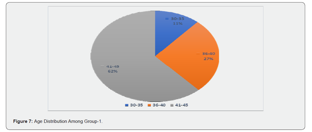
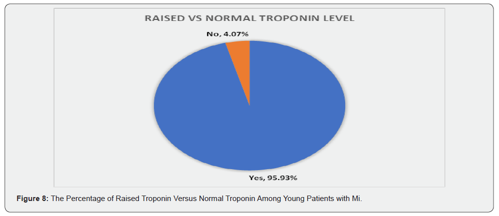
Results: 376 patients (5%) of all patients admitted with MI were ≤45 years of age (mean age 39.68 ± 4.07, 86.2% male) of whom 62% were 41-45 years old, 27% were 36-40 years old and 11% were 30-35 years old. Eighty percent had a smoking history, 47.1% had a positive family history of MI and the mean BMI was 29.27 ± 13.07. The most common previous medical conditions were hypertension (21.3%), hypercholesterolemia (17%), asthma (11.4%) and diabetes (7.7%). The troponin level was raised in 95.93% and 75.5% had ST segment elevation on presentation. The highest prevalence culprit lesion was LAD (50%), and 56% had single vessel disease. At angiography 60% had TIMI grade 0 at presentation, while after PCI 92% had TIMI grade 3 and 8% TIMI grade 2. The mean LVEF before PCI was 46.9% ± 9.5% and on average improved by 10% post PCI. Only one patient was screened for PFO using an echocardiogram with bubble study. Eighty-four percent had not been screened for any clotting abnormalities. On discharge, 72% of the patients were discharged on all appropriate medication. Twelve percent had not been given to any antiplatelet agent, whereas the most common antiplatelet regimen was Aspirin with Clopidogrel (68%). The usual prescribed ACEI/ARB was Ramipril, 60% received either Ramipril 1.25mg or 2.5mg. Bisoprolol was the typical prescribed Beta blocker, 54% were prescribed Bisoprolol 1.25mg or 2.5mg. Eighty four percent were administered a statin with most common prescription (70%) being Atorvastatin 80mg.
Conclusion: Our results show that majority of the young patients with MI underwent appropriate invasive investigations and revascularisation with 92% having TIMI 3 flow in the culprit coronary lesion post intervention. Almost all young patients post MI diagnosis received all appropriate secondary prevention drugs, but contrary to the NICE guidelines not all patients received all four types of preventive drugs which are antiplatelet agents, Beta blockers, ACEI/ARB and statins. Also, in those patients who prescribed these preventive drugs, they did not receive optimal doses. Furthermore, investigations for clotting abnormalities and PFO have been performed in only a small number of patients.
Keywords: Acute coronary syndrome; Angiotensin converting enzyme inhibitor; Acute myocardial infarction; Intravascular ultrasound; National Institute for Health and Care Excellence
Abbreviations: ACS: Acute Coronary Syndrome; ACEI: Angiotensin Converting Enzyme Inhibitor AHA: American Heart Association; AMI: Acute Myocardial Infarction; APS: Anti-Phospholipid Syndrome; ARB: Angiotensin Receptor Blocker; BB: Beta-Blocker; BSMS: Brighton and Sussex Medical School; CABG: Coronary Artery By-pass Graft; CAD Coronary Artery Disease; CCB: Calcium Channel Blocker; CT: Computer Tomography CMR: Cardiac Magnetic Resonance; CHD: Coronary Heart Disease; CRP: C-Reactive Protein; CRF: Chronic Renal Failure; COPD: Chronic Obstructive Pulmonary Disease; CVS: Cerbro-Vascular System; ESC: European Society of Cardiovascular Disease; ECG: Electro-Cardio Graph; HF: Heart Failure; GP: General Practitioner; HDL:High Density Lipoprotein; IRAS: Integrated Research Application System; IVUS: Intravascular Ultrasound; JVP: Jugular Venous Pressure; LAD: Left Anterior Descending; LDL: Low Density Lipoprotein; LVEF: Left Ventricular Ejection Fraction; LBBB: Left Bundle Branch Block; LCx: Left Circumflex; MI: Myocardial Infarction; MINAP: Myocardial Ischemia National Audit Project; NICE: National Institute for Health and Care Excellence; NICOR: National Institute for Cardiovascular Outcomes Research; NSTEMI: Non ST-Segment Elevation Myocardial Infarction; OCT: Optical Coherence Tomography; PFO: Patent Foramen Ovale; PVD: Peripheral Vascular Disease PCI: Primary Coronary Intervention; RBBB: Right Bundle Branch Block; RCA: Right Coronary Artery; REC: Research Ethics Committee; RGEC: Research Governance and Ethics Committee; RSCH: Royal Sussex County Hospital; STEMI: ST-Segment Elevation Myocardial Infarction; SLE: Systemic Lupus Erythematous; TIMI: Thrombolysis In Myocardial Infarction; TTE: Trans-Thoracic Echocardiography; TOE: Trans-Oesophageal Echocardiography; UA: Unstable Angina; WHO: World Health Organization
Introduction
Epidemiology Of Acute Coronary Syndrome
Acute Coronary Syndrome (ACS) is a term which covers the three subcategories of acute cardiac ischaemic conditions; ST Elevation Myocardial Infarction (STEMI), Non-ST-Elevation Myocardial Infarction (NSTEMI) and Unstable Angina [1,2]. All of these are potentially fatal if not recognised and managed in a timely fashion, which makes prompt diagnosis and early treatment a crucial goal for health care providers. World Health Organization (WHO) data shows that in 2015, there were 8.7 million fatalities due to Coronary Heart Disease CHD making it the leading cause of death worldwide in that year [3]. When stratified according to income classes, CHD is the leading cause of death among both middle- and high-income classes [3]. An update of heart disease and Stroke Statistics in 2016 by the American Heart Association (AHA) showed in the United States 15.5 million people aged 20 and above have CHD. In the United Kingdom, the British Heart Foundation estimates that more than 25% of all deaths in the UK are due to CHD. More encouragingly, however, since 2012 the mortality due to CHD in the UK has declined and is now the second rather than the leading cause of death. The prevalence of CHD is still very high at almost 2.4 million, and approximately 66,000 people a year lose their lives due to CHD, an average of 180 people per day [4,5]. In 2012, 16% of deaths among men where due to CHD, whereas the Figure 1 was 10% among women [4]. The expense of ACS treatment is one of the major issues which face healthcare providers. In England, any patient admitted to hospital with a heart attack costs the NHS around £4000 for their acute management, in addition to other costs, such as cardiac rehabilitation and General Practitioner follow-ups. Also, it is estimated that almost 20% of the total healthcare expenditure is due to the pharmaceutical costs in Coronary Care. Moreover, annually ACS are responsible for around one and half million days of sick leave in the UK, which caused a £3.1 billion loss to the UK economy in 2009 [6].
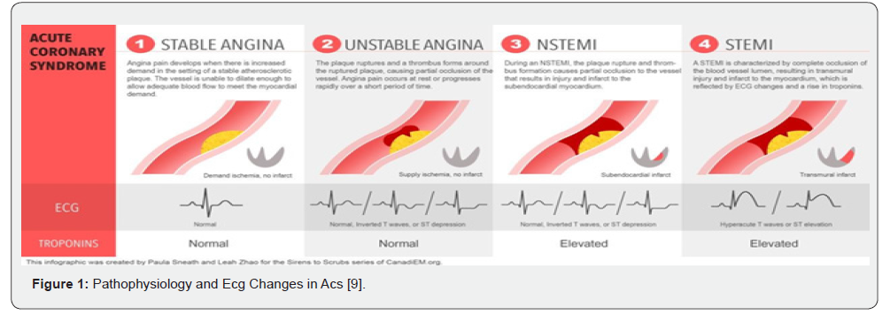
Pathophysiology And Diagnosis of Acs
ACS is caused by the rupture of an atheromatous plaque in the wall of a coronary artery. This provokes a thrombotic response causing variable obstruction to luminal flow. This obstruction cause myocardial ischemia downstream of the obstruction [7]. If this leads to necrosis of myocardial myocytes, this causes the release of cardiac troponins, whose elevation is an essential requirement for the diagnosis of myocardial infarction. The diagnosis is confirmed by the presence of one of the following criteria which were agreed upon in the third international definition of acute myocardial infarction [7];
Increase or decrease in troponin levels with at least one value
>99th centile of upper reference limit, plus at least one of the
following:
• Symptoms of ischemia.
• New ST segment or T wave changes or new left bundle
branch block LBBB on electrocardiography.
• Development of pathological Q waves on electrocardiograph.
• Imaging evidence on new loss of viable myocardium or new
regional motion abnormality.
• Identification of an intracoronary thrombus by angiography
or at post-mortem examination.
In STEMI there is total blockage of the blood flow to the cardiac muscle cells, which leads to cell death and new STsegment changes will be seen on the ECG. In NSTEMI there is partial obstruction to flow but with evidence of myocyte necrosis, while in unstable angina the blood flow is reduced but does not necessarily lead to muscle cell damage. This means the required blood flow to the cardiac muscle cells will be diminished and does not fulfil the cardiac muscle metabolic requirements. In NSTEMI there is no ST-segment elevation, but the diagnosis will be confirmed based on raised cardiac biomarkers [5,8].
Risk Factors for Acs
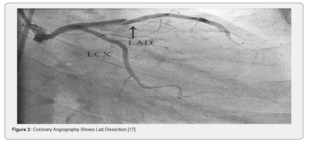
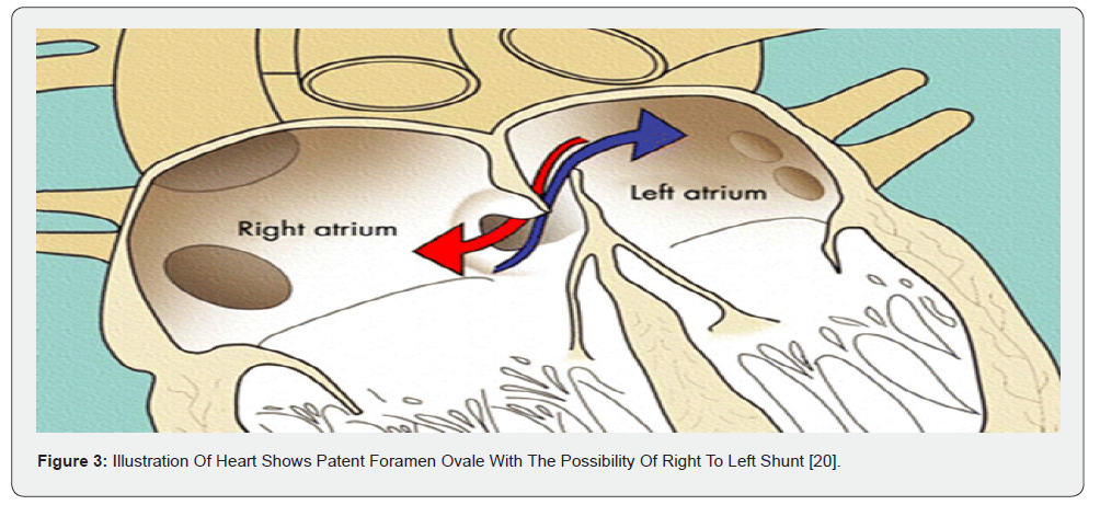
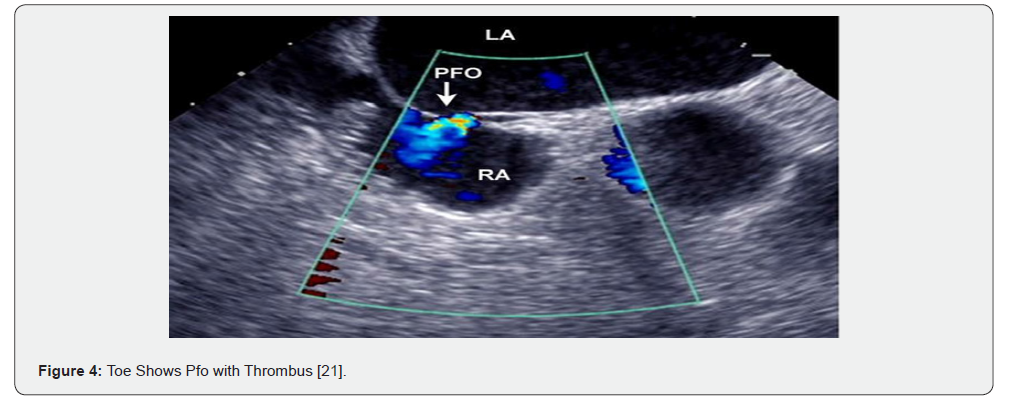
The traditional risk factors for ACS such as family history, smoking, obesity, diabetes mellitus, hypertension and dyslipidaemia were recognised many decades ago. Surprisingly many young patients who present to the emergency departments have few if any of these risk factors [9]. Recent studies find smoking and diabetes mellitus the most important risk factors in the majority of cases, whereas according to Hubacek et al. [9] most patients from Kazakhstan were non-smokers and interestingly the ACS patients had a lower level of total plasma cholesterol than the control group [9]. Studies published by Tamosiunas et al. (2014), Kim et al. (2013), and Petursson et al. (2012) had argued that there might no relation between cardiovascular diseases and total plasma cholesterol level [9]. These finds suggest that other risk factors should be sought among young adults. Diabetes mellitus is one of the known risk factors and often diabetic patients present with vague signs and symptoms and do not necessarily have typical ECG changes. According to Dotevall et al. [10] diabetic patients with ACS are less likely to present with STEMI and this presentation has been seen with a higher percentage among men than women. Moreover, the mortality risk is increased in diabetic patients both during the in-patient admission and the period following discharge [10]. Even though ACS is considered rare among pregnant women, when present it is associated with a very high mortality for both mother and fetus. Pregnancy has been shown to increase the risk of Myocardial Infarction and is reported to occur in 3-10 cases for every 100,000 deliveries. Both maternal age and multiple pregnancies are linked to an increased risk of AMI. One reason, as well as the traditional risk factors which might be behind AMI in pregnant women, is a hypercoagulable state in pregnancy that raises platelet adhesion and decreases fibrinolysis. These haemostatic changes in pregnancy will increase the risk of thrombotic events. Other conditions which increase the risk of ACS in pregnancy are pre-eclampsia, eclampsia, severe postpartum haemorrhage and blood dyscrasias [11]. Illicit drug use is another causal factor for ACS among young adults. Cocaine abuse for example, is a widely recognised cause of ACS among teenagers. Cocaine causes coronary artery vasospasm through the stimulation of both alpha- and beta-adrenergic receptors. It also causes the release of catecholamines which increase the heart rate raising myocardial oxygen demand leading to ischemia and infarction if left untreated. In addition, particularly when associated with severe hypertension it may lead to coronary artery dissection [12]. ACS may be due to the presence of an underlying coronary artery aneurysm or ectasia. In oriental populations, these coronary aneurysms may be a late complication of Kawasaki disease, but it is very rare in a Western population with only single sporadic cases being reported [13]. Spontaneous Coronary Artery Dissection is another uncommon cause for ACS, which can be seen among young adults. It involves intimal tearing in the arterial wall and haematoma formation (Figure 2). This form of ACS which is non-atheromatous in origin is more prevalent in young pregnant women or women in the postpartum period. While the aetiology remains unclear, most of the cases that have been recorded so far, had predisposing arteriopathy such as, fibromuscular dysplasia or other connective tissue disorder. Other precipitating factors were intense exercise, emotional stress and hormonal therapy [14,15]. Another documented possible causal factor of ACS in young patients and pregnant women is a patent foramen ovale (PFO). PFO is a congenital opening between the right and left atrium which normally closes after birth but may remain patent in up to 20-25% of adults (Figure 3). Thrombus from the deep leg veins may enter the right atrium and pass across the PFO to the left atrium and subsequently pass into one of the coronary arteries and cause myocardial infarction. Though unusual, these cases are usually diagnosed through transoesophageal or transthoracic echocardiography after other possible causes have been excluded [16,17].
Presenting Signs and Symptoms
Regardless of novel imaging techniques and advanced blood tests, both patient history and physical examinations remain fundamental in diagnosing ACS. The most common symptoms of ACS are [18];
• Chest pain or discomfort, in form of tightness and
pressure on chest.
• Pain or discomfort in both arms, jaw, neck, back or
stomach.
• Shortness of breath
• Feeling dizzy or lightheaded
• Nausea with or without vomiting
• Sweating
Physiological Signs:
• Signs of sympathetic activation: pallor, sweating,
tachycardia.
• Signs of vagal activation: vomiting, bradycardia
• Signs of impaired myocardial function: hypotension,
oliguria, cold peripheries, narrow pulse pressure, raised JVP, third
heart sound, quiet first heart sound, diffuse apical impulse and
lung crepitation.
• Signs of tissue damage: fever
• Signs of complications: mitral regurgitation, pericarditis.
Biomarkers
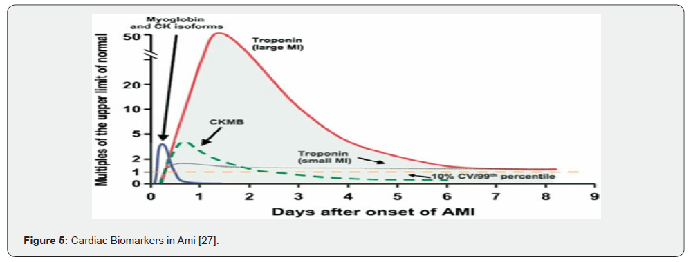
Myocardial injury leads to the release of cardiac biomarkers and their elevation. One of the most essential and classical ways to diagnose myocardial injury is to assess the cardiac biomarkers and is a basic universal requirement to diagnose MI. Biomarkers are proteins which rise during myocardial injury. Several biomarkers can be detected in MI but the most valuable ones are those with very high sensitivity and specificity (Figure 4) [19]. Today’s standard biomarkers used in most of the cardiac and emergency units are the following:
T and I (cTnI and cTnT)
Troponins have a unique cardiac insomnia that only originates from cardiac tissue. They are the gold standard for diagnosing myocardial injury. Several studies have shown them to be the most sensitive and specifically available biomarkers. The high sensitivity comes from the ability to detect a very low level of damage due to myocardial injury. They will be detectable 4-12 hours after the onset of ACS, peak at 12-48 hours and return to normal approximately after 7 days. A high level of Troponin is a powerful indicator of a high short- term risk of death in a patient with NSTEMI. It also helps to determine the need of early invasive management [19,20].
Creatine Kinase (CK-MB)
Creatine Kinase is another marker that may be used if Troponin assays are not available. It rises within 4-6 hours of an AMI and remains high for 24-48 hours. CK-MB is a CK isoenzyme, which is mainly found in the cardiac myocyte. The CK-MB test is highly sensitive and has a very long history in ACS diagnosis. However, this marker is not highly specific as CK-MB can be released from other tissues such as skeletal muscle, intestine, diaphragm, uterus, and prostate. The reliability of CK-MB as a marker for AMI is low as an elevation in CK-MB levels may result from chronic muscle injury or in those who have had a surgical procedure [20,21].
Myoglobin
Myoglobin is a hemeprotein found in skeletal and cardiac muscle. It is a low molecular weight molecule whose levels rise within 2-4 hours after the myocardial infarction returns to normal within 24-36 hours. Repetitive assays are required to improve sensitivity, but overall, it has 90% sensitivity in diagnosing AMI [19,21].
C-reactive protein (CRP)
Even though it is not specific, CRP is one of the inflammatory markers that may rise in ACS due to widespread inflammation in the vessels. Also, it has been shown that in STEMI, CRP levels reflect the extent of myocardial injury [19]. Liuzzo et al. [22] shown that in patients with ACS higher levels of CRP were associated with an adverse prognosis [22].
Imaging Modalities in Acs
Invasive Modalities
Coronary Angiography
The evolution in medical imaging has made various invasive and non-invasive modalities available for ACS diagnosis. The best way to detect ACS in a patient with acute chest pain and high-risk ACS is with imaging of the coronary arteries with catheter-based angiography. Its use is indicated in known high-risk patients or in those who have typical symptoms. Also, in patients with ECG changes and atypical symptoms catheter-based angiography is the most accurate test to look for the vessel with suspected lesions [23]. On the other hand, in intermediate and low-risk patients presenting with chest pain but are subsequently seen to be lesion free, angiography may delay their discharge because of the bed stay required after the procedure. Therefore, in these groups non-invasive procedures such as CT angiography may be more appropriate [23].
Intravascular Ultrasound (Ivus)
In IVUS a tiny ultrasound transducer is placed at the tip of a catheter, which can be introduced into a coronary vessel. During coronary intervention, it gives real time images of the coronary vessel and the degree of the stenosis [24].
Optical Coherence Tomography (Oct)
OCT gives high-resolution images and can detect coronary plaques and intra- luminal thrombus. Also, it has been shown in that OCT can differentiate between Unstable Angina and Myocardial Infarction. This is achieved through differentiating between thrombus types, whether it is red or white. The disadvantages of OCT are the limited penetration in non- transparent tissue and the requirement to displace blood to give adequate visualization [24].
Non-Invasive Modalities
Echocardiography
Echocardiography can be used to detect rapidly wall motion abnormalities. In ACS, when there is a defect in myocardial contractility, echocardiography can show hypokinesis. This technique is highly recommended in low-intermediate risk patients and has a sensitivity of 92-93% in diagnosing AMI [25]. Additionally, echocardiography is widely used in diagnosing PFO cases (Figure 5) [26,27].
Computer Tomography (Ct) And Cardiac Magnetic Resonance (Cmr)
CT and CMR are more applicable in patients who either present with stable angina to risk stratify or post-angioplasty to assess and tailor their future long-term management. The CMR procedure is longer when compared to CT and is susceptible to electromitsnetic interference that limits it use in those with implanted magnetic devices in their body. CT Angiography is contraindicated in patients with poor renal function and the visualization is limited by motion artefacts and heart rate [24,25].
Invasive Management Strategies
The usual management plan depends on several factors such
as,
• Premorbid condition of the patient.
• How many vessels are involved and the severity of the
lesion(s).
Revascularization
Revascularization, which is the mainstay of treatment for most patients with ACS, has been in use for over 50 years. Over time the process has evolved with better outcomes and less complications. Two different strategies are available Coronary Artery Bypass Graft (CABG) and percutaneous revascularization. In CABG the patient’s own vessels from the legs or less frequently the arms are harvested and anastomosed to the epicardial coronary arteries beyond the site of the stenosis, bringing additional blood supply to the area. On the other hand, PCI involves inflating a balloon at the site of the lesion followed by stent implantation to ensure long-term patency of the vessel. This form of management is usually chosen when the critical lesion is in one or two vessels in emergency settings. In cases of more severe and multivessel disease CABG is chosen. Early revascularization is crucial to minimise myocardial damage [28].
The following is NICE guidance on primary PCI and fibrinolysis
for STEMI, “Offer coronary angiography, with follow-on primary
PCI if indicated, as the preferred coronary reperfusion strategy for
people with acute STEMI if:
• presentation is within 12 hours of onset of symptoms
and
• Primary PCI can be delivered within 120 minutes of the
time when fibrinolysis could have been given.
Offer coronary angiography, with follow-on primary PCI if indicated, to people with acute STEMI and cardiogenic shock who present within 12 hours of the onset of symptoms of STEMI’ [29].
Drug Treatment
Whether the patient has undergone primary PCI or not, cardio-protective drugs are given as a part of the treatment plan to minimize recurrent ischemia, protect left ventricular (LV) function and improve mortality. NICE guideline state that all patients who have suffered an acute myocardial should receive the following drug treatment unless there is a contraindication.
• Angiotensin Converting Enzyme Inhibitor (ACEI): ACEI prevents the conversion of angiotensin I to angiotensin II. Studies showed that ACEI lowers blood pressure through dilatation of the blood vessel, reduces recurrence of angina and prevent left ventricular dysfunction and remodelling. NICE recommends using the highest tolerated dose or targeted dose in all post-ACS patients indefinitely unless haemodynamically unstable or contraindicated where Angiotensin II Receptor Blocker ARB can be given [30,31].
• Dual antiplatelet therapy: Aspirin is a thromboxane A2 inhibitor which reduces platelet aggregation and is given indefinitely with another antiplatelet agent (Clopidogrel, Ticagrelor or Prasugrel) for at least 12 months. Clopidogrel which inhibits P2Y12 Adenosine Diphosphate ADP receptors on platelet membranes thereby decreasing their aggregation and clot formation is the most widely used [31-33].
• Beta-blocker (BB): bind to beta-adreno receptors which in turn block the binding of epinephrine and non-epinephrine to those receptors. Through several mechanism BB help protecting the myocardium; decreasing oxygen demand through reduction in heart rate, blood pressure and contractility, decrease the risk of ventricular fibrillation, improvement in LV diastolic function and decrease the microvascular damage. NICE recommends BB should be offered as soon as possible after MI unless contraindicated,
Non dihydropyridine Calcium Channel Blocker (CCB) can be used if BB are not tolerated/contraindicated provided LV function is good [31,34].
• Stations: Atorvastatin is recommended by NICE as a secondary prevention for all patients post MI unless contraindicated or the patient has chronic kidney disease. Statins works by inhibiting HMG-CoA reductase enzyme which is responsible for lipid production within the liver [35,36].
Mi in Young Adult and the Reason Behind my Study
In the last few years the incidence of ACS among young adults has shown a steady increase. The reasons are varied; unhealthy lifestyle, diabetes, smoking and illicit drug use are among the most common causes of this disease. Although there have been a few studies of this topic, the characteristics and clinical courses of ACS in older age group have been reported in depth. Current studies do not give a comprehensive overview of ACS in young adults in the UK. In addition, most ACS guidelines are tailored for older people. In the available data on young adults with ACS, men represented over 70% of cases. One study of adults under 40 years of age who presented to a cardiology centre in Poland over the course of 7 years found that of the ACS 242 cases identified, 84.2% were male [37]. Another study of cases admitted to a Malaysian tertiary centre between January 2005 and December 2013, showed that 71.9% of the total of 28 cases were male [38]. Whereas the incidence of ACS among females of reproductive age was reported to be very low. In Denmark, available data shows 25 cases a year per 100,000 women, while in America the Figure 6 is slightly higher at 57 per 100,000 a year. The reason behind this, is thought to be protective effect of a high level of oestrogen in young females [39].
The aims of this study are to audit the diagnosis, management
and treatment of young patients (age ≤45 years) presenting with
ACS with emphasis on the following questions.
• To identify the risk factors, present in MI in those ≤45
years and compare them to those ≥46 years to assess whether
these risk factors have been adequately addressed.
• To assess whether appropriate pharmacological
secondary prevention such as antiplatelet agents, beta blockers,
CCB and statins have been instituted in appropriate dosage.
• To assess whether coronary angiography has been
undertaken and appropriate revascularisation achieved.
• In those cases where it is unclear whether the myocardial
infarction was due to simple premature coronary atherosclerosis,
has a systematic search been undertaken for the less common
conditions which may disproportionately contribute to ACS in this
younger cohort of patients?
Literature Review
Aims of the Literature Review
This literature review aims to outline published material to date on MI in young adults.
Literature Search
There are very few studies with large numbers of young patients with ACS. The majority of the published papers regarding ACS in young adults are single patient reports. The main reason for this is that the occurrence of ACS in the age group (18 to 45 years of age), which are the focus of this study, is uncommon and has gained little attention from health authorities and researchers. Until now, all studies published on this topic, were carried out outside the UK. Literature research on this topic were performed using three different databases up to and including March 2018: Pubmed, Embase and Google Scholar.
The search terms used are
• ((Acute coronary syndrome [Title]) AND young adult
[Title]) OR young patient [Title].
• ((Acute myocardial infarction [Title]) AND young adult
[Title]) OR young patient [Title].
On Pubmed, the first broad search profiles yielded 517 articles. Refining and changing the search terms resulted in 35 articles, 23 of which were relevant for full article review. When the same searches were performed for Google Scholar and Embase, 3 additional papers were relevant for full literature review. The others were the same as Pubmed or irrelevant. Each paper and their abstract have been read for possible review. Out of the 26 papers highlighted in the above exercise, only 11 papers were chosen for literature review, as most of the papers were single case studies.
General Overview of Well-Structured Study
Among the few studies carried out into ACS in young adults, the characteristics of the disease have been reported to be quite like ACS in older patients. In 2007, Tungsubutra et al. [40] performed a retrospective study on 9,373 patients admitted to hospital in Thailand between August 1, 2002, and October 31, 2005 [40]. This was a retrospective study enrolling patients from several tertiary care centres [7]. In this study the patients were divided into three age categories, <45 years old, 45-54 years old and >54 years old. The researchers not only compared their results with previously published data but also between age groups that were included within the same study. Out of the 9,373 patients, 544 patients comprising 5.8% of the total patients were under the age of 45 years old. While the other two groups, 45-54 years old and >54 years old comprise 16.2% and 78% of the total patients respectively. Male gender predominates in all groups ≤45 (85.3%) compared to the older age groups, the percentage of a family history of coronary artery disease (CAD) was more frequent, the percentage occurrence was 23.6% in the younger age group. Also, tobacco smoking was highest among the younger age group at 65.9%, while diabetes mellitus and hypertension were less frequent. The most common presenting symptom in the young patient group was typical angina, but the time onset of taken from symptoms to hospital arrival was the longest in this age category which took 13.9 hours. This makes the significance of the awareness of symptoms of ACS among the general population crucial in decreasing adverse outcomes. Another significant finding in the study was patients in the younger age group were more likely to have STEMI at 67.3%, while NSTEMI and UA came next at 19.3% and 13.4% respectively. The incidence of congestive heart failure was higher at 25.6%, while the mortality rate was 7.4% [40]. Overall, the study is well structured and should be taken into consideration to gain insight into this subject. The study did not document the site of the lesion(s) in coronary arteries in the over 60% of total patients investigated with coronary angiography. Moreover, most of the patients in this study were from tertiary care centres and therefore largely excluded those patients in hospitals lacking facilities for invasive angiography. Also, other causes for ACS were not studied such as anomalies of coronary arteries and coronary ostia, spontaneous coronary artery dissection and drug taking as a cause of coronary artery spasm [17,41].
Methods Used in Previous Studies
In most previous studies the retrospective method was used to gather the available patient data analysis. Retrospective method means available data which has been recorded previously by the health care professionals up to the date of the study. Sometimes, the data has been collected routinely for another purpose and not necessarily for research purposes. This method helps the researcher investigate the factors which may have triggered the disease, the mode of presentation and all clinical and radiological features of the disease. It also allows analysis of the management plan which has been used for different patients and their ultimate outcome [42,43].
Presentation And Characteristics of Mi Among Young Adults
The typical presentation of myocardial infarction in young adults might not always mirror those that are seen in older patients. This makes the diagnosis much more difficult and challenging for health care professionals. This has been in the interests of Pellaton et al. [44] who studied the features of myocardial infarction and myocarditis among young adults who were admitted into the emergency department of University Hospital of Lausanne in Switzerland with chest pain. The study used retrospective secondary data collection to answer its research question [44]. The recruitment method used in sampling included all the patients admitted between January 2009 and June 2011, aged between 18 and 40 years of age presenting with chest pain. In the study, the patients were divided into two groups: those with a final diagnosis of myocardial infarction and those with myocarditis. The risk factors of the patients in both groups were compared and clearly those with a definite diagnosis of myocardial infarction had more cardiovascular risk factors. Most of the risk factors and clinical investigations were overlapping and not exclusive for either diagnosis [44]. Again, the standard diagnosis of ACS was based on ECG changes and cardiac biomarkers abnormality [7,19]. Coronary angiograms were performed in the patients to identify the culprit lesion precipitating the active presentation. The investigation methodology of choice to diagnose or exclude myocarditis was CMR. There are some potential problems with this study. Firstly, when it comes to the patient inclusion methodology, chest pain as a criterion by itself is too general. In myocarditis, chest pain may not be the main feature in all patients, they may have myocarditis for many weeks before they have been diagnosed. It might therefore mean that some other younger patients with myocarditis might have been admitted to that hospital in the same period but were unfortunately missed because they were not present with chest pains. A second potential problem is the absence of a highly sensitive troponin essay. Although this is a relatively new assay and perhaps was not in routine use at the time of the study, however, this may still cause bias as those with chest pain and conventional low troponin levels would have been ruled out of the study. A patient could have had high troponin levels at the time of the test, but this would have been missed because of the use of the conventional troponin essay test [19,20,44].
In another study the characteristics of ACS among young patients in Oman was investigated by Panduranga et al. [45]. The study included 1,579 patients, of whom 121 patients were under the age of 40 years and were predominantly male. The prevalence of ischemic chest pain was more frequent among the younger patients (85% vs 72%; P = 0.002), resulting in the under 45 years old patients presenting earlier to the hospital compared to their more senior counterparts. This nature of presentation was explained by the presence of other co- morbidities among older patients, making the chest pain vaguer in character or less significant until an advanced stage. Surprisingly, in this study the lipid profile was normal in most of the patients and only 16% had hyperlipidemia. STEMI was the most common presentation of ACS among the younger age group, and they had better outcome in hospital than NSTEMI and UA. Also, patients with a positive family history had ACS one decade earlier in comparison with those that had a negative family history [45]. This also has been observed by Hoo et al. [38] in a study performed in a Malaysian tertiary care centre. The study was a cross sectional, single center, retrospective study including 628 patients who were admitted into the centre and diagnosed with ACS between 2005 and 2013. Patients with a positive family history were presented more than a decade earlier than those with negative family history, however 70.3% had no family history [38]. Again, there was a male predominance among the 628 patients with a ratio of 68.4% to 31.6% male to female. Hypertension was present in nearly half of the young patients. Other modifiable risk factors such as diabetes mellitus 37.8% but dyslipidaemia (16.2%) were not as common as might be expected [38].
Unusual Causes of Acs
Occasionally young patients present with ACS without any precipitating factors such as illicit drug abuse or family history of premature coronary disease [46]. In this group of patients there may be other causes or contributing factors: anatomical abnormalities of the coronary arteries; genetic blood clotting abnormalities such as antiphospholipid syndrome which make them more vulnerable to forming a clot in their circulation; or persistent patent foramen ovale (PFO) which on straining or Valsalva may allow right to left passage of a clot from the venous circulation to the left atrium and cause a systemic or coronary embolism. In the absence of a PFO such small clots would lodge and be cleared in the pulmonary circulation [46-48]. The presence of a PFO may be combined with an additional risk factor such as a hypercoagulable state induced by the oral contraceptive pill as described in a case study by Mohammad et al. Clotting abnormalities in those who may have hypercoagulable states are another cause for ACS among young adults. Usually, they are present early in life, and they may present with recurrent episodes of thrombosis. In one study, which included all age groups, 37% of the patients who had ACS were positive for one or more antiphospholipid autoantibodies such anti‐β2 glycoprotein1, anti-phosphatidylserine, anticardiolipin, anti-prothrombin and anti‐ oxidized LDL/β2GP1. Most patients with these autoantibodies were women 57% [49]. In another study, the prevalence of the autoantibody anti-Apo A-1 was higher among ACS patients at 21% compared to 1% in the normal population. Only 13% of patients diagnosed with Systemic Lupus Erythematous (SLE) and Anti- phospholipid Syndrome (APS) had positive results for anti-Apo A-1 antibody [50]. The same study concluded that a normal lipid profile should not necessarily exclude the risk for developing coronary artery diseases, because most of the patients in the study, who had significant high levels of autoantibodies such anti-Apo A-1 antibodies, had normal or low lipid profiles. This hypothesis suggests that autoantibodies may cause both plaque formation and plaque rupture without interfering with lipid metabolism [50].
Anatomical Pattern of the Culprit Lesions in Young Adults
ACS in young adults may not follow the same anatomical pattern as in older people. This has been studied in 2009 by Teixeira et al. [51]. As with the other published papers on this topic a retrospective method was used which analysed the records of 128 patients who were diagnosed with ACS in a non-tertiary cardiac centre in Portugal between 1999 and 2007. One aspect that made this study different from previously published work was that the site of the culprit lesion of the ACS in the young adults enrolled in the study was recorded. Most patients had lesions within the right coronary artery (RCA) which caused decreased blood flow in the inferior wall. Single-vessel disease was the most prevalent at 45.6%. Two-vessel and three-vessel involvement occurred in 23.9% and 10.9% respectively. Left main coronary artery disease was seen in only 4.5% of the patients. In 15.2% no significant CAD could be identified [51]. Even though reperfusion therapy might not be the best management plan for a young patient with ACS, and primary PCI may be more effective according to published data, most of the patients (76.1%) in this study were offered thrombolysis [51]. Ventricular arrhythmia was noticed to be more frequent than in previous studies, which might be due to the management plan that mainly depended on pharmacological reperfusion therapy. Tungsubutra et al. [40] found the younger patients more frequently had normal or minimal coronary lesion on angiography [40]. In the study of Ewa et al. (2015) 49.4% of the total patients included in the study had more than 50% luminal stenosis of a vessel, of which 13.4% had 50-90% stenosis, and 36% of patients had more than 90% stenosis. Significantly, more than 50% had no obstructive coronary artery stenosis and normal coronary arteries were found in 37.2% of the patients diagnosed with ACS. Again, like other studies, single vessel disease was the most common pattern of stenosis at 61.9% and the most common lesion was in left anterior descending artery (LAD) (61.6%) followed by the RCA (27.4%) [37].
Management Strategies of Acs in Young Adults
In most published studies an aggressive management plan for ACS in young patients was employed. In Panduranga et al. [45] both older and younger age groups received aspirin, statins, thrombolytic agents and anticoagulant equally. However, the younger patients received B-blockers, glycoprotein IIb/IIIa inhibtors, clopidogrel and coronary angiogram more frequently [45]. This might be due to fewer contraindications than are usually present in older patients. This aggressive plan is also reported in the study of Tungsubutra et al. [40] where PCI was performed in a higher percentage of younger patients than in older ones [40]. While in the Teixeira et al. [51] study thrombolysis was the main myocardial reperfusion therapy in most of the patients (76.1%) who presented with STEMI and only 10.2% and 2.3% of the patient underwent primary PCI and rescue PCI respectively [51]. In the Tungsubutra et al. [40] study a high percentage of the patients received aspirin, beta-blockers and statins, while the use of ACEI was relatively low at 58.5% which can help a lot of patients, particularly those with heart failure. Again, the use of GPIIb/IIIa inhibitors was low but higher in the younger age group. The percentage of GPIIb/IIIa inhibitors among <45 years and 45-54 years old patients were 17.8% and 14.6% respectively compared to 9.3% among patients who were >54 years old. Coronary angiography, PCI and fibrinolytic were more frequently employed in younger patients. While the other studies have not mentioned any form of Coronary Artery Bypass Graft (CABG) in younger patients, the Tungsubutra et al. [40] study indicated that emergency CABG was performed in 2.5% of the young patients [40]. An aggressive interventional management plan may not be the right way to deal with all cases. For example, in those with antiphospholipid antibody syndrome, PCI and standard risk factor management alone may not be sufficient, and other preventive measures be required [40,46].
Outcome of Acs Among Young Adults
As expected, in most studies a better outcome has been seen in the younger age group compared to those who were over 45 years old. For example, in Tungsubutra et al. [40], heart failure and cardiogenic shock were both lower in the under 45-year-old patients. The percentage of heart failure among under 45-year-old patients was 25.6%, while among those who were over 54 years old was 41.05%. Similarly for cardiogenic shock the incidence was 9.2% versus 14.4% for both <45 years and >45 years old. Also, other non-cardiac complications such as major bleeding and stroke had a lower incidence rate among the young patients. Regarding the short-term and long-term prognosis in young patients with ACS [40] Panduranga et al. [45] found that in the short-term prognosis was favourable for the younger age group. However, the long-term prognosis would only improve if the patient changed their social habits in the form of cessation of smoking which is regarded as one of the strongest predictors for long-term prognosis. Also, the study found that young patients who had diabetes mellitus with multi-vessel disease would probably have the same prognosis as older patients [45]. After discharge mortality was 7.4% and 8.6% in both Tungsubutra and Teixeira studies [40,51] Almost the same figure has been recorded in Ewa et al. (2015) study and the mortality rate was 7.75% in long-term follow-up in all patients, whereas those who had abnormal coronary arteries had slightly higher mortality rate at 8.5% over five years of follow-up [37].
Methodology
Introduction
This chapter will outline and explain research questions, methodology and the collection of data from the hospital including the ethical considerations for this study.
Choice of Research Method
This study represents an audit on patients admitted with acute MI into the Royal County Hospital in Brighton. A secondary data analysis method has been used for this single centre study [52]. This data is held in the MINAP database and further data was obtained from searching through computer held patient data and collecting information from patients’ notes. Patients were divided into two age groups namely 45 years and under (Group-1) versus over 45 years old (Group-2). Group-1 data was analysed and compared using two separate criteria. The first was an overview of their age and gender in comparison to Group 2. The data was also compared to previously published data of patients aged 45 and over. Secondary data refers to that data originally collected for another purpose. For instance, the data that has been obtained routinely by hospitals and medical centres on inpatients and outpatients including morbidity, births, deaths or data collated proactively by the Department of Health for disease prevention. This data can then be analysed independently for scientific research. Using the secondary data analysis method, an individual or individual who has some familiarity with the data, i.e., a healthcare professional, harvests the data. The selected data is then analysed with respect to a proposed study and researchers analyse data according to the study questions and research hypothesis. The most important advantages of secondary data analysis are the economy, time saving and it is easier for smaller projects that cannot get primary data for any reason. The major drawbacks of using secondary data analysis are having no control over the date and time of data collection, exactly how the data was collected and the exact nature and depth of information gathered. This would result in data that might not exactly fit or fulfil all research criteria. Also, the reliability of the data should be taken seriously as the veracity of the data cannot be checked retrospectively. For these reasons, questions should be asked when considering using any secondary data, such as, where and when the data has been collected and by whom, what was the methodology of collection and the reason the data was collected [42,43,52,53].
Research Approach & Ethical Consideration
Once the goals and aims of this study are established under supervisory guidance, the research proposal is submitted to the BSMS Research Proposal Committee. The Committee, upon review, recommended the change of research methodology from a cross-sectional survey to secondary data analysis on the grounds that this study is based on secondary data gathered. The amended study was resubmitted to the Committee and approved. (Appendix A and B, Research Governance and Ethics Committee (RGEC) Application Form and John Anderson’s confirmation of approval). This study uses patients’ data that is a potential breach of patient privacy and confidentiality; potential ethical conflicts should be addressed through the Research Ethics Committee (REC) via the Integrated Research Application System (IRAS) (Appendix C, IRAS use of service). However, as this study is an audit of existing data ethical approval was not considered necessary (Appendix D, Letter from Dr O’Nunain indicating no need for IRAS approval). As this was a clinical audit, approval for this project was sought and obtained from Dr Nalyaka Sambu, Consultant Audit Lead in Cardiology. Data was then collated from a database of patients’ notes at BSUH. Dr O’Nunain, a consultant cardiologist at BSUH and acting in a supervisory role, harvested data from the PATS system containing the records of all patients admitted into the cardiac unit. Group 1 data, comprised of patients aged between 18 and 45 years in accordance with the research criteria, was compiled from the database. Group-1 data was collated and managed in Microsoft Excel spread sheets. To ensure the anonymised and ethical value of the study, case study numbers were assigned to each patient and all identifiable data within the notes was removed before analysis. Both Bamboo and Panda software systems were used to extract information on discharge summaries, procedural reports, outpatient letters and an echocardiographic report. A nonanonymised data set was stored on the secure hospital computer system. Only anonymised data was held by the author on a secure password protected laptop.
Case/Patient Selection
The cases used in this study were patients who presented with ACS or AMI and admitted into cardiac unit at Royal Sussex County Hospital RSCH between May 2013 and May 2018.
Inclusion Criteria
The patients were divided over two groups:
Group 1: Younger age group inclusion criteria were:
• 45-years-old and younger.
• Admitted to hospital between May 2003 and May 2018.
• Diagnosed with MI.
Group 2: Older age group inclusion criteria were:
• 46-year-old and older.
• Admitted to hospital between May 2003 and May 2018.
• Diagnosed with MI.
Exclusion Criteria
• In Group-1, all patients over 45 years were excluded; in
Group 2 all patients under 45 years were excluded.
• Any patient admitted to the cardiac unit due to anything
other than ACS were excluded.
• Group-2 patients with a previous history of ACS prior to
admission were excluded.
Data Collection
The data has been derived using the data that has been collected for Myocardial Ischaemia National Audit Project MINAP database, together with other datasets for audit including of all surgical, interventional and electrophysiological that is stored on PATS database in RSCH in Brighton.
MINAP Database
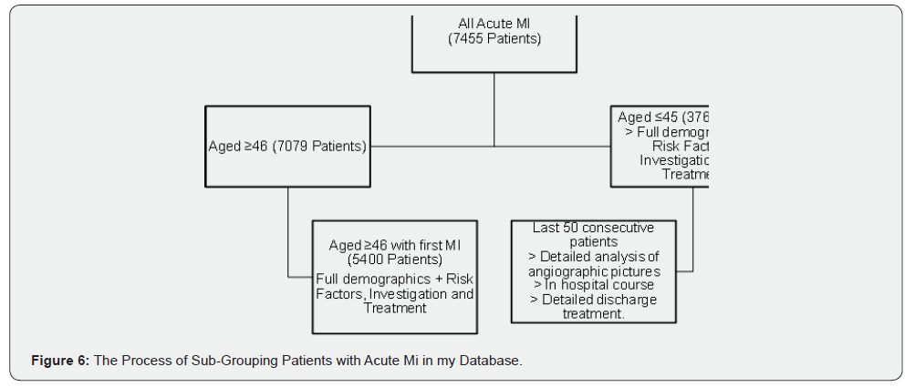
The Myocardial Ischaemia National Audit Project (MINAP) is a compulsory national clinical audit of management of heart attack. It stores the data and supplies participating hospitals and ambulance services in England, Wales and Northern Ireland with records of their management and compares with the standard management that is agreed nationally and internationally. MINAP collects data for National Institutes for Cardiovascular Outcome Research (NICOR) which produces analysis to help hospitals, healthcare providers and improvement bodies to monitor and improve the quality care and outcomes of cardiovascular patients. The MINAP dataset contains many parameters, many of which were irrelevant to our study. The relevant parameters have been selected and stored on an Excel spreadsheet. Data collection also involved analysing databases of the patients attending RSCH. Group-1 data was collected using a two-tiered system. Primarily the data available on the Bamboo and Panda systems yielded 376 patients fitting Group-1 inclusion criteria, which were extracted onto a Microsoft Excel spread sheet. Secondarily, the notes of the last 50 patients to be assessed chronologically by the Group-1 cohort were assessed in depth with regards to angiographic and echocardiographic findings with the details of their medication dosage post discharge. Again, in this process the anonymity of the data from the Group-1 sub- group was maintained. The same primary and secondary processes were performed to identify Group-2 patients, which yielded 7079 cases from the PATS database. A filter applied to extract the first presentation only, which left 5400 cases only.
Additional Data Fields
In addition to previously mentioned databases, discharge summaries of the patients have been examined in detail for pharmacological data. In example, administration of drugs such as ACEI, ARBs and BBs with their dosage.
Angiographic and Echocardiographic Data Collection
In group-1, the angiographic data of 50 patients have been reviewed with Dr O’Nunain. All angiographic films were stored on the mcKesson Horizon system. The pattern and extent of coronary disease of each case was assessed, and the culprit lesion identified. Flow through the culprit vessel was graded using the Thrombolysis in Myocardial Infarction TIMI grading system both before and after angioplasty. Also, Left Ventricular Ejection Fraction LVEF was obtained preferably from the reports of detailed echocardiographic studies that were performed in the echocardiography department. When this was not available, LVEF assessments were obtained on bedside echocardiography were used.
TIMI Flow (TIMI Coronary Grade Flow)
TIMI flow is a grading system used to assess the flow across the specific site of the coronary vessels. Four grading scores are given to the lesion from 0 to 3 according to their patency [54].
Grade 0: no perfusion.
Grade 1: penetration without perfusion.
Grade 2: partial perfusion.
Grade 3: complete perfusion.
Demographic and clinical data collected from Group-1 patients
• Age and gender
• Initial diagnosis
• Systolic BP and Heart rate.
• ECG changes
• Blood tests (S. Cholesterol, S. Glucose, Haemoglobin, S.
Creatinine)
• BMI
• Past medical history (Hypercholesterolemia, PVD, CVS,
Asthma or COPD, CRF, HF and diabetes mellitus)
• Smoking Status
• Family history
• Drug discharge
• Outcome
Demographic and clinical data collected from the 50 patients
in details from Group-1
• Age and gender
• Past medical history (Hypercholesterolemia, PVD, CVS,
Asthma or COPD, CRF, HF and diabetes mellitus)
• Angiographic findings
• Echo-graphic findings
• Smoking, alcohol and illicit drug status
• BMI
• Blood screening for auto antibodies, thrombophilia and
basic clotting.
• Drug on discharge with their dosage.
Demographic and clinical data collected from Group-2 patients
• Age and gender
• Blood tests
• Peak troponin level
• BMI
• Past medical history (Hypercholesterolemia, PVD, CVS,
Asthma or COPD, CRF, HF and diabetes mellitus)
• Smoking Status
• Family history
Data Analysis
All data were analysed in September-December 2018 using Microsoft Excel version 15 and IBM SPSS Statistics version 24.0. For simple demographic data descriptive statistics only was used. For comparison of blood result values unpaired t-tests were used with a cut off value of p< 0.05 being considered statically significant.
Research Limitation
The statistical significance of study would have been improved if it were done on a larger scale including more patients’ data, especially the numbers in Group 1. Assessing the detailed case notes of more than 50 patients across both study groups, over 100 patients would have helped to collect much more information on the presentation and management strategies. However, time constraints and issues of data confidentiality rendered a larger cohort to study unviable.
Data and Results
Chapter 4 will provide the results and analysis of the data which has been collected for the study. In total between (May 2003 to May 2018, 5776 patients were identified as suffered first MI, of these 376 (6.5%) were aged ≤45 years.
Results Of Patients in Group-1 (Patients Aged ≤45 Years)
Age And Gender Distribution
The majority (62%) of the younger patients with first MI were 41-45 years old. The second largest group (27%) were 36-40 years old. Those who were 35 years-old and younger accounted for 11% of the total patients. The mean age of those patients ≤45 years was 39.68 ± 4.07 years with a range (18-45) years (Figure 7). Most of the patients were male (86.2%) (Table 1). The following blood tests were performed as a routine for all patients that were admitted to the hospital. The mean value and standard deviation are presented in Table 2.
Smoking Status
Smoking status on admission was determined in almost all patients. Over 80% of the patients had a history of smoking, being either current smokers at the time of the heart attack or were ex-smokers. Only 50 patients (13.3%) of the young patients had never smoked. In a small percentage (5.6%) smoking history was not available.


Body Mass Index Bmi
The BMI of 51% of patients was over 25 when they experienced their first heart attack. The mean BMI was 29.27 ± 13.07 (Table 3).

Positive Family History of Mi
Nearly half of the patients (47.1%) had a positive family of MI history, 27.1% had no family history of MI and family history was not documented in 25.8% (Table 4).

Previous Medical Conditions
Approximately one fifth (21.3%) of patients had hypertension and 17% had hypercholesterolemia before having MI. The least frequent medical condition among group- 1 was heart failure and chronic renal failure 0.8% and 1.1% respectively (Table 5).

Peak Troponin Level
Cardiac biomarkers were tested on all patients who were admitted with sign and symptoms of ACS and later diagnosed with MI. Among 376 patients, 354 had a raised troponin, while 15 patients (4.07%) had a normal level for troponin. The mean peak troponin was 3316 ± 3432 ng/ml with a range of 1.016 to 10,000 ng/ml (Figure 8).
Ecg At Presentation
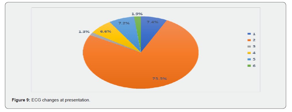
Many of the patients (75.5%) had ST segment elevation at presentation. A much smaller percentage showed T wave changes only (7.2%), ST segment depression (6.6%) and no acute changes (7.4%). Almost over 3% of the patient had either left bundle branch block LBBB or non-specified other acute abnormality (Table 6 and Figure 9).

Initial Diagnosis at the Hospital
The vast majority (97%) of the patients were correctly diagnosed as MI or ACS at the initial assessment. Only 2.9% of the patients were initially diagnosed as suffering from different conditions but were ultimately diagnosed as suffering an MI (Table 7).

Drugs on Discharge
The drugs on discharge are shown in Table 8.

Optimal Medication on Discharge
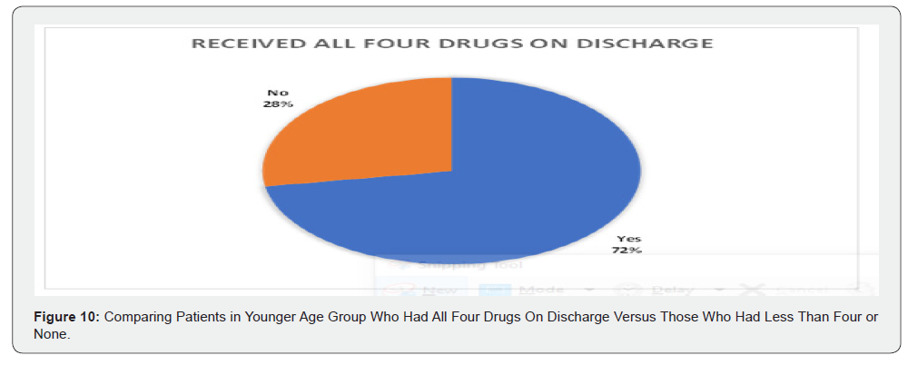
Out of 376 patients in this group, only 72% of patients had been discharged on all four drugs (Aspirin, Beta blockers, ACEI or ARB and Statin) (Figure 10).
Results of Group-2 Patients (Patients Aged ≥46 Years)
Data regarding older age groups has been analysed using the same system that is used for the younger age group. In this group 7079 patient’s data has been retrieved from the system who were admitted and diagnosed with MI in RSCH from May 2003 up to September 2018. This was the first MI of 5400 of these patients.
Age and Gender Discrepancy
Almost two third of the cases were male and 33.9% were females (Table 9). Mean age among this group was 69.7 ± 12.8 with first MI. The oldest patient recorded with the first episode of MI was 103-year-old, whereas the youngest one was 46-year-old.

Blood Tests on Admission
Average blood test results among older age groups revealed the following results (Table 10).

Smoking Status
Around one third of patients of the older age group had never smoked cigarettes at 35.9%. Ex- smokers and current smokers came next at 32.8% and 27.6%. Only 3.7% had an unknown smoking history (Table 11).
Body Mass Index Bmi
The mean BMI among patients in this group was 26.78 ± 5.
Family History
The largest percentage of the older patients’ family history of MI was not available 35.9%. Both positive and negative family history of MI were almost the same 31.1% and 33% respectively (Table 12).


Previous Medical Conditions
Many patients (45%) in group-2 had hypertension, a quarter of the group had hypercholesterolemia and 15.8% had diabetes (Table 13).

Peak Troponin Level
The mean troponin level was 2829 ± 3619 ng/ml with a range of 1.65-10,000 ng/ml.
Risk Factors Scoring Among Group-1 And Group-2
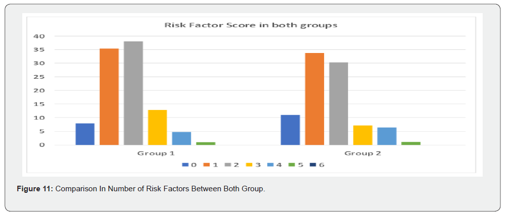
Six denominators have been used to calculate the number of risk factors that were available in each patient of both groups. The risk factors were being overweight, hypertension, diabetes, hypercholesterolemia, smoking history and family history. One point is given to each risk factor, and the results have been compared (Table 14 with Figure 11).

Detailed ≤45 Sub-Group Analysis In 50 Patients
Among the younger age group, 50 consecutive patients who were admitted between March 2017 and May 2018 were studied in greater detail. The full angiographic studies were reviewed and drug therapy analysed in detail.
Age And Gender
In this group, the mean age among the patients were 39.68 ± 4.08 years with 96% being male.
Smoking Status, Alcohol and Illicit Drug Use
Seventy percent were positive for smoking history and only 26% never smoked cigarettes (Table 15). The data regarding alcohol history among younger age groups has not been recorded. The data revealed that 4% of this age group had a positive history of using illicit drugs.

Angiographic Findings
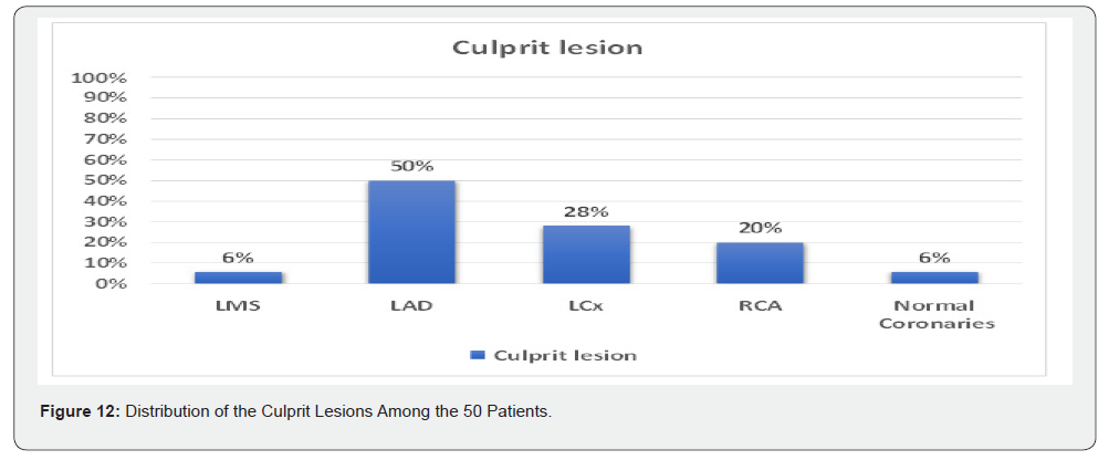
Anatomical distribution of the culprit lesion In the left anterior descending (LAD) 50%, left circumflex artery (LCx) 28% and right coronary artery (RCA) 20% (Table 16 and Figure 12).

Type of the Culprit Lesion
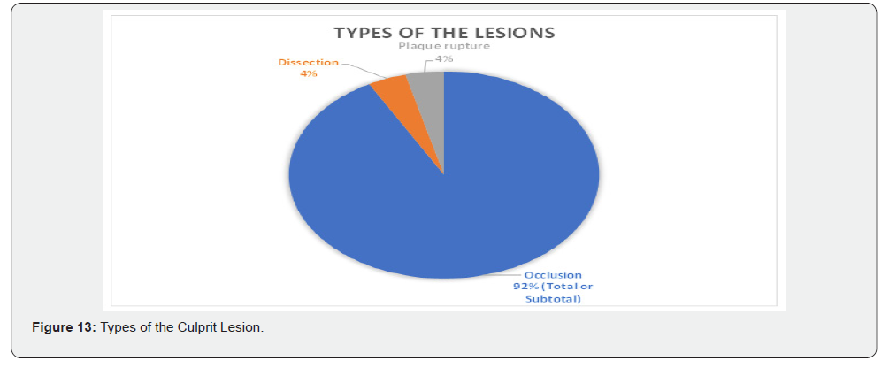
Ninety two percent had MI due to total coronary or subtotal occlusion, equal number of the patients (4%) had either coronary dissection or plaque rupture with minimal luminal stenosis in coronary arteries. This does not include patients with normal coronaries (Figure 13).
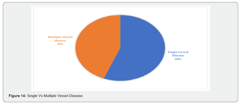
Single Vessel Disease Vs Multiple Vessel Disease
Fifty-six percent had single vessel disease, with 44% of the patients with multiple vessel disease (Figure 14).
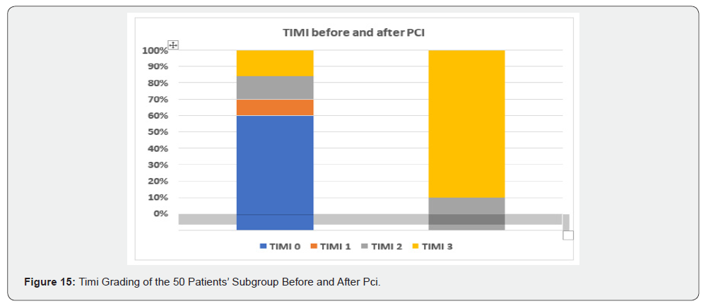
Timi Grading Before and After Pci
Sixty percent of the patients had TIMI grade 0 at the time of angiography, while after PCI 92% of the coronaries with culprit lesion had TIMI grade 3 and the remaining were TIMI grade 2 (Figure 15).
Echocardiographic Findings
Left Ventricular Ejection Fraction LVEF
LFEV has been assessed in 84% of the patients in this group. The mean LVEF on admission was 46.9% ± 9.5% and where repeated post-revascularization on average LVEF fraction improved by 10%.
Bubble Study
Out of 50 patients only one patient had been screened for PFO through using bubble study which was negative.
Clotting and Auto-Antibody Screening
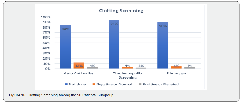
Blood tests for autoantibodies and coagulation abnormalities which promote clotting were not performed on a routine basis. The majority (84%) of patients were not screened for these potential abnormalities. Out of 50 patients, auto-antibody screening has been performed on 8 patients, which was positive in 2 patients and negative in 6 patients. Fibrinogen level was normal among 2 out of 5 patients who were tested. A thrombophilia screen was performed in 3 patients, one of which was positive (Figure 16).
Medication
The main four drug classes which are recommended for patients post MI are: the antiplatelet drug usually Aspirin with or without Clopidogrel, Ticagrelol or Prasugrel; the Angiotensin Converting Enzyme Inhibitor (ACEI) or Angiotensin Receptor Blocker (ARB) usually Ramipril; the Beta-blocker usually Bisoprolol and the lipid lowering agent usually Atorvastatin. Among the 50 young patients, 6 patients which equal 12% of the total subgroup have not been given any type of antiplatelet on discharge. The largest percentage of patients, 68%, have been given a combination of 75mg of Aspirin with 75mg of Clopidogrel.
In 8 patients, 180mg of Ticagrelol substituted Clopidogrel and in only one patient Prasugrel 10mg has been given with Aspirin (Table 17). ACEI and ARB are given to protect and improve LV function. Sixty percent of the patients received either 1.25mg or 2.5mg Ramipril and nearly one third of the patients did not receive ACEI (Table 18). Beta Blockers when given during an acute MI reduce mortality and in the long term reduce reinfarction rates. Same as ACEI, over 50% of the patients received either 1.25mg or 2.5mg Bisoprolol and 32% of the patients did not receive any Beta-Blocker (Table 19). Eighty four percent of patients were administered Atorvastatin, 70% of the patients received 80mg, with 12% and 16% respectively receiving 40mg or none (Table 20).


Presentation And Outcomes in the 50 Patients’ Subgroup
Out of the 50 patients, 10 patients presented with cardiac arrest, while the other 40 presented with typical MI. Regarding the outcome two patients died in hospital, two patients suffered residual severe neurological disability and were discharged on minimal medication. The other 46 patients were discharged on standard medication (Table 21).

Discussion
Introduction
Myocardial infarction was previously considered a disease of older patients but more recently the incidence has been rising in younger patients. In the future, this trend may be further exacerbated by the increase in obesity, lack of exercise and diabetes. Our study compared several factors present in the records in a small group of young MI patients compared to an older cohort. We investigated clinical presentation, initial diagnostic and blood tests, invasive and non-invasive radiological investigation, lesion location, risk factors and the use of secondary preventive medication. Chapter 5 will provide discussion of the main findings in our study and link those findings with previously published literature.
Age And Gender in the Younger Versus Older Age Groups
Our data indicates that within the younger age group the incidence of MI increased with increasing age, with 62% of all patients within the ≤45 group aged between 41 and 45 years old. The mean age for MI within this group was 39.68 ± 4.07 years. In addition, within the younger age group men represented 86.2% of this group compared to just 17.8% women. This data agrees with previous studies indicating that females are less prone to heart attack especially at a young age due to hormonal protection [55- 57]. Among group-2, the mean age was 69.7 ± 12.8 years. Again, as expected males outnumbered of their female counterparts (66.01% versus 33.09%). Male gender predominance and mean age distribution in our study are close to previous studies [37,38,40,51,58] In recently published study in young adults with MI, Yandrapali et. al., found men represented 72.5% of the study population and the mean age was 39 ± 5 years [58].
Blood Tests in Both Groups
Mean serum cholesterol was nearly the same in both groups and above normal level, [(5.4 ± 1.44 mmol/L) in ≤45 group versus (5.1 ± 9.8 mmol/L)] in ≥46 group (t(4466) = 0.609, p = 0.543) [59].These values are consistent with previous studies that most of the patients with MI have high levels of cholesterol level and it is a major risk factor for MI [51,60]. The mean serum glucose level was high in both groups. This is a well-recognized biological feature of MI in any age group regardless of whether it has diabetes or not. Hyperglycaemia is a body reaction due to stress which leads to sympathetic nervous system activation and excess production of catecholamines [61,62] In ≤45 age group, the serum glucose was 8.3 ± 4.03 mmol/L compared to 8.5 ± 3.9 mmol/L in ≥46 age group (t(4621) = - 1.084, p = 0.278). The higher the level of hyperglycaemia the higher risk of adverse outcome. The patients in our study had mild hyperglycaemia, and this gives a more favourable outcome compared to moderate (glucose 7-11 mmol/L) or severe hyperglycaemia (glucose ≥11mmol/L) [61- 63]. Anaemia is recognised as a non-major risk factor for MI. Since red blood cells carry oxygen and delivery it to the cells, the lower level of haemoglobin the higher risk of MI due to inadequate of cell oxygenation [64-66]. Haemoglobin level was normal in both groups, the mean haemoglobin level in ≤45 age group was 15.2 ± 6.2 g/L, while, in ≥46 aged patients the mean value was 13.6 ± 4.8 g/L (t(5700) = 5.715, p = 0.0008). This is consistent with previous studies that anaemia is more common in older patients at the time of MI, most likely due to other chronic comorbidities or gastrointestinal diseases [67]. The serum creatinine level on admission is a predictor of mortality. Creatinine level should be assessed before performing PCI, as the contrast administration may result in further creatinine rise. The higher level of creatinine the higher level of mortality and the more adverse outcome [68,69]. In our study creatinine level was normal in both groups, with slightly lower level in younger age group (84.3 ± 31.12 micromole/L in group-1 vs 99.8 ± 66 micromole/L in group-2) (t(5112) = - 4.145, p = 0.00034). This correlates with the prevalence of chronic renal failure which was very low in both groups (1.1% in group-1 versus 7.1% in group-2).
Smoking Status, Bmi and Family History in Both Groups
Most of the younger patients (70.7%) were smokers at the time of MI, compared to ex-smokers (10.4%). Whereas, in the older age group only 27.6% were current smokers and 32.8% were ex-smoker. In the younger age group 13.3% had never smoked compared to 35.9% in the older age group. This correlates with previous studies where tobacco use is more common in young patients with MI than older age group and it is a major risk factor for MI regardless of age [40,58,70]. An elevated BMI values is another major risk factor for MI. [58,71] Mean BMI in both groups was in the overweight range (BMI ≥25-30), in ≤45 age group was not significantly higher (29.27 ± 13.07) compared to ≥46 patients’ group (26.78 ± 5.0) (t(3949) = 0.606, p = 0.544). Non-modifiable risk factor such as family history of MI among ≤45 patients was significantly high (47.1%), while only 31.1% of the ≥46 patients had a positive family history of MI. Having family history of MI is an independent risk factor for MI and having a first degree relative with MI doubles the risk [60]. Our data could not reveal whether the patients with positive family history had first or second degree relative with MI and the numbers of family members with MI, which may have a greater impact on the patient and determining the nature of the MI.
Previous Medical Conditions in Both Groups
Hypertension (21.3%) and hypercholesterolemia (17%) were the most common conditions concurrently diagnosed with MI within the younger age group, whereas in the ≥46 age group these ratios were higher at 45% and 26% for both conditions respectively. The third most common condition in ≤45 years group was respiratory diseases (11.4%), while in older age group diabetes comes third (15.8%). Other medical conditions were uncommon in the younger age group, whereas with advancing age they become more prominent, such as CVD (8.2%) and chronic renal failure (7.1%). This data confirms previous studies that both hypertension and dyslipidaemia are the most common medical conditions in patients with MI, but surprisingly, the percentage of patients with hypercholesterolemia was not as high as expected and in other studies the percentage was much higher than our study. For instance, a recently published study by Yandrapalli et al. [58] on young patients with first MI, 57% of the male patients and 61% of the female patients had dyslipidaemia [38,40,45,58].
Comparison Of Risk Factors Between Both Groups
Most of the patients in both groups had either one or two risk factors. In group-1, respectively 35.4% and 38% had one and two risk factors at the time of MI. Whereas in group-2, there is a slight decrease to 33.7% and 30.3% for the same numbers of risk factors. The percentage of patients for three or more risk factors was lower in group-1 compared to group-2. Significantly, no patients in either group had six risk factors. Having no risk factors at all for MI was 8% in group-1 compared to 11% in group-2. These values are same as previously published studies that majority of young patients have either one or two risk factors at the time of MI [40,58].
Peak Troponin Level, Ecg Changes and Initial Diagnosis Among Young Adults
Almost 96% of the ≤45 group had raised troponin levels on admission with the mean troponin level in this group being 3316 ± 3432 ng/ml. In the ≥46 aged patients troponin levels were slightly lower (2829 ± 3619 ng/ml) (t(3890) = 2.007, p = 0.045). Four percent of ≤45 patients had normal troponin levels at the time of presentation to hospital. The earlier rise of troponin helps earlier diagnosis and better survival rate [19,72]. Whilst, in our study not all patients with MI had raised troponin, this brings the question whether the troponin level which has been done on our patients was highly sensitive and tested in timely fashion which is after 3-6 hours when symptoms begin [19,72,73]. Previously normal level of troponin in patients with MI has been noticed but not frequently, and this demonstrates that patients with normal level of troponin assays may have lower risk of MI but does not completely exclude it [74-76]. The early identification of ECG changes and elevated troponin levels improves the overall outcome for the patient due to early diagnosis and intervention [51,72,74]. An abnormal ECG was a very good indicator of MI in the younger patients, 92.6% of the patient group had an abnormal ECG. The most common type of ECG change observed was ST segment elevation (75.5%), with LBBB being the least common (1.3%). Not all patients had an abnormal ECG and cardiac biomarkers on admission, with a normal ECG observed in 7.4% of patients. In addition, our data indicates that ACS was the initial suspected diagnosis in 97.1% of admissions. This data indicates that in a small percentage of patients’ ECG abnormalities and elevation of cardiac biomarkers such as troponin were not present and could not be completely relied upon at presentation, therefore clinical judgement remains crucial to diagnose MI in clinical settings. These findings agree with Samuel et. al., who concurred that chest pain without ECG changes did not necessarily exclude ACS, but when ACS was confirmed, mortality amongst those patients was lower compared with patients with early ECG changes [77].
Coronary Involvement and Angiographic Findings
All patients in the younger group underwent invasive investigation and were revascularized if appropriate. The most common site of the culprit lesion was in LAD (50%), while 28% had a culprit lesion in LCx. RCA had the lowest among the three major vessels supplying the heart, which was 20%. In the study of Ewa et. al. study the most common culprit lesion was in the LAD, but RCA came second with 27.4% and LCx with only 11% [37]. LMS also contributed to 6% of the culprit lesions among young patients. Surprisingly, 6% of the patients had normal coronaries. While, in Ewa et. al. study this ratio was much higher (50.6%). Regarding the type of the culprit lesion, 92% was due to thrombus occlusion of one or more of the coronary arteries, both dissection and plaque rupture had similar share of the culprit lesions as 4% each. The number of coronaries involved have always been one of the criteria to tailor the management strategy. In our study 56% of the patients had single vessel disease, while 44% had multiple vessel disease with two or more of the coronary arteries involved. These data are almost the same compared to previously published studies [37,78]. TIMI grading of the coronary arteries shows that 54% of the culprit lesions had TIMI grade 0 before angioplasty, 24% were either TIMI grade 1 or 2 and 22% of the lesions were TIMI grade 3. Whereas, post angioplasty majority of the patients (92%) had TIMI 3 flow rates and only 8% left with TIMI grade 2.
Pfo And Clotting Abnormalities
Not all patients have been assessed by echocardiography. Only 84% had an echocardiogram to assess their ejection fraction and to exclude any other structural abnormality. This may have been due in part to early successful revascularisation with primary PCI with little residual ECG abnormality and early discharge from hospital. An echo bubble study which assesses probable PFO as cause of MI and stroke has been done on only one patient which was negative. Again, a very small number (6%) of the young patients had been fully screened for autoantibodies and clotting abnormalities. 16% of the patients were checked, of which 4% were positive. Thrombophilia screening was performed in only 6%, and 2% of the cases were positive for thrombophilia. Fibrinogen screening has been performed in 10%, and 4% had a raised fibrinogen level. These data shows that both PFO and clotting abnormalities were overlooked and have not been taken in consideration, which increases the possibility of missing patients with PFO or clotting abnormalities. In cases with diffuse or multiple coronary involvement, it might be reasonable to attribute MI to coronary atherosclerosis, while in single vessel disease with no other disease or in normal coronaries the mechanism could be embolic and other causes should be sought [16,17,48,49]. Due to the low incidence of MI in patients with PFO, the pick-up rate could not be found in too many studies and majority of the cases were single case reports. In only one study by Franz et. al. the incidence of PFO related MI was estimated at <1% [79].
Drugs on Discharge and their Dosages
As a secondary prevention protocol patients with MI are prescribed four drug groups: Anti- platelet agents, ß-blocker, ACEI or ARB and Statin. This helps reduce the risk of future MI and stabilize the condition of the heart. In our study, 72% of the young patients with MI have been prescribed all four drugs, whereas in the other 28%, one or more of the four drugs was not prescribed. Looking at the details, the highest percentage of patients 20.2%, had not been prescribed ACEI or ARB, 18.4% were not given ß-blocker. The third least prescribed drug was statin (11.4%). Whereas only 8.8% of the post-MI patients were not given Aspirin or its equivalent class of antiplatelet. This is at variance NICE guidelines that recommend all four drugs post MI [80]. In a systemic review by Victor et. al. of 9 randomised control trials on routine therapy post MI, showed that all four drugs are crucial post MI. The study revealed giving Aspirin reduces mortality and reinfarction in 1 month to 12 months, ß-blocker notably decreases late mortality. ACEI prevents ventricular dilatation and remodelling and improves both long term morbidity and mortality [81]. Regarding the dosages and regimen of the prescribed drugs, the most common given antiplatelet regimen in young adults was dual antiplatelet which is recommended by NICE, combination of Aspirin 75mg and Clopidogrel 75mg was given in 68%, followed by Aspirin 75mg and Ticagrelol 180mg (16%) and Aspirin 75mg with Prasugrel 10mg (2%). Aspirin 75mg as a single antiplatelet has been given to 2% of the patients [80]. No antiplatelet agent was used in 12% of the patients. The reason behind this could not be identified, though in 2 patients (4%) all cardioactive drugs were withdrawn due to severe irreversible hypoxic brain injury. Looking at the data of ACEI class, 30% has not been given any drug from this group. Sixty percent either prescribed Ramipril 1.25mg or 2.5mg. The higher dosage Ramipril (Ramipril 3.75mg to 10mg) was given to only 10% of the group. This tendency to prescribe low dosage has been noticed in ß-blocker’s class as well. Forty percent of the young patients were either given Bisoprolol 1.25mg or 2.5mg, while the higher dosage (Bisoprolol 3.75mg to 10mg) was only given to 14% of the patients. However, 32% were not prescribed any ß-blocker. This illustrate that a very large number of young patients are not given optimal dosage of drugs as a secondary prevention, leaving them at higher risk of further MI [34,80,82,83]. Some patients may have had genuine absolute/relative contraindications to ACEI or Beta Blocker such as hypotension, bradycardia or asthma.
The lower use of these agents may, however, in part reflect the perception that patients immediately revascularized with primary PCI with no residual coronary stenosis and normal LV function may have less to gain with ACEI and Beta blocker therapy than those patients in historical post MI clinical trials in the preprimary angioplasty era. Prescribing lower dosages of medication than required to avoid further MI incidents has been raised previously in many studies. Pedersen et. al. (2016) concluded that the target dose of ß-blocker was infrequently achieved at discharge following MI, with 80% were given ≤25 % of target dose. Barron et. al. (1998) showed that only 11% of patients in their study had been prescribed >50% of the effective dosage of ß-blocker. They observed that the mortality rate and revascularization within three months following MI was higher in those prescribed lower doses of ß-blocker [84,85]. Also, in another study by Herlitz et. al., more clinical benefits were seen in patients prescribed higher dosages of metoprolol than lower dosages [86]. The AIRE study in 1993 revealed the advantages of early prescription of Ramipril following MI in patients showing signs on heart failure, resulting in the reduction of premature death from all causes [87]. Statins were the only drug group given in the proper dosage in most of the patients. Seventy percent were prescribed Atorvastatin 80mg, 12% were given Atorvastatin 40mg and 2% were given Atorvastatin 20mg. While only 16% of the patients were not given any sort of statin on leaving the hospital.
Clinical Outcome Among Young Adults
Almost every patient in the study had a completely open culprit vessel at the end of the procedure and therefore should benefit from an open artery in terms of long-term prognosis. Previous literature suggests that survival may improve even when revascularization performed late, even when irreversible necrosis has occurred [88]. A study by Nakagawa et. al. claims that reperfusion after 24 hours still has benefits and may prevent expansion of the infarct area. Also, they found that late reperfusion may help enhance cellularity of the infarct area [89]. Most patients (92%) were discharged on medication and offered a cardiac rehabilitation program [90-95]. The hospital mortality rate was 4% in young patients and the same percentage suffered of hypoxic brain injury and were not given any further medication as they were severely disabled and not deemed to be suitable for cardiac rehabilitation. Compared to Tungsubutra et al. [40] study, the mortality rate in the hospital was 7.4% and the incidence of stroke was 1.3%, whereas in Ewa et. al. study the mortality rate was 1.25% [37,40].
Conclusion
Young adults diagnosed with MI is a more frequent phenomenon compared to a few decades ago. The presumption and treatment for MI in young adults presenting at A&E is not given the same diagnostic priority as in older patients. In our study on a small group of younger patients, we looked at several key factors. These include mode of presentation, initial blood work and diagnostic tests performed, invasive and non-invasive radiological investigations, lesion location, risk factors and drugs prescribed on discharge.
From the data we concluded the following:
1. Most younger patients post MI diagnosis received all
appropriate secondary prevention drugs.
2. Contrary to NICE guidelines, a significant minority of
younger patients did not receive all four types of preventative
drugs.
3. Where prescribed statins and antiplatelet agents were
administered at clinically effective doses, ß-blockers and ACEI
were routinely prescribed at sub-optimal doses.
4. All younger patients underwent appropriate invasive
investigation and treatment, and the vast majority left the cardiac
catherization laboratory with TIMI 3 flow in the culprit artery.
5. Clinical investigation into less common causes of MI,
such as clotting abnormalities and PFO, were performed in only a
very small number of younger patients.
Acknowledgment
I would like to thank my main supervisor, Dr O’Nunain, for his continuous effort through my journey and Dr Welland, John Anderson, for their support regarding research methods and approval process. Also, special thanks to my family and Dr Nicola Kurdo, who revised all chapters of my dissertation and provided me with valuable notes.
References
- Henderson RA (2013) Acute coronary syndrome: optimising management through risk assessment. Clin Med (Lond) 13(6): 602-606.
- Sanchis-Gomar F, Perez-Quilis C, Leischik R, Lucia A (2016) Epidemiology of coronary heart disease and acute coronary syndrome. Ann Transl Med 4(13): 256.
- WHO (2017) The top 10 causes of death. World Health Organization.
- Foundation Bh (2015) Cvd Statistics-Bhf Uk Factsheet.
- NICE (2014) Quality Improvement in Acs. National Institute for Health and Care Excellence.
- Wilsdon T (2011) The burden of Acute Coronary Syndrome in the United Kingdom. Charles River Associates’ Life Sciences Practice.
- Timmis A (2015) Acute coronary syndromes. BMJ 351: h5153.
- David D McManus, Joel Gore, Jorge Yarzebski, Frederick Spencer, Darleen Lessard, et al. (2011) Recent Trends in the Incidence, Treatment, and Outcomes of Patients with STEMI and NSTEMI. The American Journal of Medicine 124(1): 40-47.
- Hubacek JA, Stanek V, Gebauerova M, Adamkova V, Lesauskaite V, et al. (2017) Traditional Risk Factors of Acute Coronary Syndrome in Four Different Male Populations – Total Cholesterol Value Does Not Seem To Be Relevant Risk Factor. Physiol Res 66(Suppl 1): S121-S128.
- A Dotevall, D Hasdai, L Wallentin, A Battler, A Rosengren (2005) Diabetes mellitus: clinical presentation and outcome in men and women with acute coronary syndromes. Data from the Euro Heart Survey ACS. Diabetic Medicine 22(11): 1542-1550.
- Yilmaz S, Sahinkus S, Kilic H, Gunduz H, Akdemir R (2014) Acute Coronary Syndrome During Pregnancy: A Case Report and Literature Review. Turk J Emerg Med 14(3): 135-138.
- Wood DM, Hill D, Gunasekera A, Greene SL, Jones AL, et al. (2007) Is cocaine use recognised as a risk factor for acute coronary syndrome by doctors in the UK? Postgrad Med Journal 83(979): 325-328.
- Yang EH, Kapoor N, Gheissari A, Burstein S (2012) Coronary and Intracerebral Arterial Aneurysms in a Young Adult with Acute Coronary Syndrome. Tex Heart Inst J 39(3): 380-383.
- Jacqueline Saw, Eve Aymong, Tara Sedlak, Christopher E Buller, Andrew Starovoytov, et al. (2014) Spontaneous Coronary Artery Dissection Association With Predisposing Arteriopathies and Precipitating Stressors and Cardiovascular Outcomes Circulation 7(5): 645-655.
- Meng PN, Xu C, You W, Wu ZM, Xie DJ, et al. (2017) Spontaneous Coronary Artery Dissection as a Cause of Acute Myocardial Infarction in Young Female Population: A Single- center Study. Chin Med J (Engl) 130(13): 1534-1539.
- Moreno-Torres Concha V, Montero Hernández E (2017) Paradoxical coronary embolism through a patent foramen ovale. Medicina Clínica (English Edition) 149(4): 183-184.
- Aleksandr Rovner, Ali A Valika, Attila Kovacs, Andrew M Kates (2018) Possible Paradoxical Embolism as a Rare Cause for an Acute Myocardial Infarction. Echocardiography 23(5): 407-409.
- Davidson S (2006) Davidson's Principles and Practice of Medicine. 21st Edition ed. Colledge NR, Walker BR, Ralston SH, editors p:
- Del Val Martin D, Sanmartín Fernández M, Zamorano Gómez JL (2015) Biomarkers in acute coronary syndrome. IJC Metabolic & Endocrine 8: 20-23.
- Valentina Loria, Milena Leo, Gina Biasillo, Ilaria Dato, Biasucci LM (2008) Biomarkers in Acute Coronary Syndrome. Biomarkers Insights 3: 453-468.
- Kyaw Thu Moe, Wong P (2010) Current Trends in Diagnostic Biomarkers of Acute Coronary Syndrome Annals Academy of Medicine 39(3): 210-215.
- Liuzzo G, Biasucci LM, Gallimore JR, Grillo RL, Rebuzzi AG, et al. (1994) The Prognostic Value of C-Reactive Protein and Serum Amyloid A Protein in Severe Unstable Angina N Engl J Med 331(7): 417-424.
- Roobottom C, Mitchell G, Iyengar S (2011) The role of non-invasive imaging in patients with suspected acute coronary syndrome. Br J Radiol 84(Spec Iss 3): S269-S279.
- Angela S Koh, Chia S (2010) Update on Clinical Imaging of Coronary Plaque in Acute Coronary Syndrome. Ann Acad Med Singapore 39(3): 203-209.
- Zimmerman SK, Vacek JL (2011) Imaging Techniques in Acute Coronary Syndromes: A Review. ISRN Cardiol 2011: 359127.
- Gardin JM (2014) PFO or UFO, How Good Is Transesophageal Echocardiography in Identifying Patent Foramen Ovale as the Cause of a Cryptogenic Stroke? Circulation 7(1): 5-7.
- Mauro Pepi, Arturo Evangelista, Petros Nihoyannopoulos, Frank A Flachskampf, George Athanassopoulos, et al. (2010) Recommendations for echocardiography use in the diagnosis and management of cardiac sources of embolism. European Journal of Echocardiography 11(6): 461-476.
- Kolh P, Windecker S, Alfonso F, Collet JP, Cremer J, et al. (2014) ESC/EACTS Guidelines on myocardial revascularizationThe Task Force on Myocardial Revascularization of the European Society of Cardiology (ESC) and the European Association for Cardio-Thoracic Surgery (EACTS) Developed with the special contribution of the European Association of Percutaneous Cardiovascular Interventions (EAPCI). European Journal of Cardio-Thoracic Surgery 35(37): 2541-2619.
- NICE (2018) Myocardial infarction with ST-segment elevation overview - NICE Pathways. NICE.
- Davies MK (1994) Effects of ACE inhibitors on haemodynamics and angina pectoris. British Heart Journal 72(3 Suppl): 52-56.
- NICE (2018) Drug treatment after myocardial infarction - NICE Pathways. NICE guidance that was used to create this part of the interactive flowchart. Myocardial infarction: cardiac rehabilitation and prevention of further cardiovascular disease (2013) NICE guideline CG172 Ticagrelor for preventing atherothrombotic events after myocardial infarction (2016) NICE technology appraisal guidance 420 Rivaroxaban for preventing adverse outcomes after acute management of acute coronary syndrome (2015) NICE technology appraisal guidance 335 Prasugrel with percutaneous coronary intervention for treating acute coronary syndromes (2014) NICE technology appraisal guidance 317 Ticagrelor for the treatment of acute coronary syndromes (2011) NICE technology appraisal guidance 236 Clopidogrel and modified-release dipyridamole for the prevention of occlusive vascular events (2010) NICE technology appraisal guidance 210.
- Lee SH, Yang JH, Choi SH, Park TK, Jang WJ, et al. (2017) Duration of dual antiplatelet therapy in patients treated with percutaneous coronary intervention for coronary chronic total occlusion. PLoS One 12(5): e0176737.
- S Yusuf, G Tognoni, SR Mehta, S Chrolavicius, S Anand, et al. (2019) Effects of Clopidogrel in Addition to Aspirin in Patients with Acute Coronary Syndromes without ST- Segment Elevation. The New England Journal of Medicine 345(7): 494-502.
- Boudonas GE (2010) β-Blockers in coronary artery disease management. Hippokratia 14(4): 231-235.
- Ostadal P (2012) Statins as first-line therapy for acute coronary syndrome? Exp Clin Cardiol 17(4): 227-236.
- NICE (2016) Lipid modification therapy for preventing cardiovascular disease - NICE Pathways. The NICE guidance that was used to create this part of the interactive flowchart. Cardiovascular disease: risk assessment and reduction, including lipid modification (2014 updated 2016) NICE guideline CG181 Ezetimibe for treating primary heterozygous-familial and non-familial hypercholesterolaemia (2016) NICE technology appraisal guidance 385.
- Maroszyńska-Dmoch EM, Wożakowska-Kapłon B (2015) Clinical and angiographic characteristics of coronary artery disease in young adults: a single centre study 74(4): 314-321.
- Hoo FK, Foo YL, Lim SMS, Ching SM, Boo YL (2016) Acute coronary syndrome in young adults from a Malaysian tertiary care centre. Pak J Med Sci 32(4): 841-845.
- Idris N, Aznal SS, Chin SP, Ahmad WAW, Rosman A, et al. (2011) Acute coronary syndrome in women of reproductive age. Int J Womens Health 3: 375-380.
- Tungsubutra W, Tresukosol D, Buddhari W, Boonsom W, Sanguanwang S, et al. (2007) Acute coronary syndrome in young adults: the Thai ACS Registry. J Med Assoc Thai 90(Suppl 1): 81-90.
- Devlin RJ, Henry JA (2008) Clinical review: Major consequences of illicit drug consumption. Crit Care 12(1): 202.
- Bowling A (2014) Research Methods in Health. Fourth Edition Ed. Ny, Usa: Mcgrawhill Education.
- Long-Sutehall T, Sque M, Addington-Hall J (2010) Secondary analysis of qualitative data: a valuable method for exploring sensitive issues with an elusive population? Journal of Research in Nursing 16(4): 335-344.
- Pellaton C, Monney P, Ludman AJ, Schwitter J, Eeckhout E, et al. (2012) Clinical features of myocardial infarction and myocarditis in young adults: a retrospective study BMJ Open 2(6): e001571.
- Panduranga P, Sulaiman K, Al-Zakwani I, Abdelrahman S (2010) Acute Coronary Syndrome in Young Adults from Oman: Results from the Gulf Registry of Acute Coronary Events. Heart Views 11(3): 93-98.
- Thapamagar SB, Aung TT, Mascarenhas D (2013) Recurrent acute coronary events in a young adult. J Thromb Thrombolysis 35(2): 290-294.
- Uwe Speiser GE, Silvio Quick, Nadine K Waessnig, Stephan Wiedemann, Ivan Platzek, Ruth H Strasser MG (2014) Acute coronary syndrome and single coronary artery ostium. Kardioloia Polska 72(5).
- Silva GV (2014) Patent Foramen Ovale. Tex Heart Inst J 41(3): 306-308.
- Tom P Greco, Ann Marie Conti-Kelly, Eiji Matsuura, Tom Greco, Ken J Dier, et al. (2007) Antiphospholipid Antibodies in Patients with Coronary Artery Disease - new cardiac risk factors? Annals of The New York Academy of Sciences 1108: 466-474.
- Vuilleumier N, Reber G, James R, Burger D, Moerloose PD, et al. (2004) Presence of autoantibodies to apolipoprotein A-1 in patients with acute coronary syndrome further links autoimmunity to cardiovascular disease. Journal of Autoimmunity 23(4):353-360.
- Teixeira M, Sa I, Mendes JS, Martins L (2010) Acute coronary syndrome in young adults. Rev Port Cardiol 29(6): 947-955.
- Boslaugh S (2007) Secondary Data Sources for Public Health: A Practical Guide Cambridge University Press.
- Johnston MP (2014) Secondary Data Analysis: A Method of which the Time Has Come Qualitative and Quantitative Methods in Libraries 3(3): 619-626.
- Sarkar A, Lee JJ (2018) TIMI Grade Flow. StatePearls.
- Barrett-Connor E (1997) Sex differences in coronary heart disease. Why are women so superior? The 1995 Ancel Keys Lecture. Circulation 95(1): 252-64.
- Hayward CS, Kelly RP, Collins P (2000) The roles of gender, the menopause and hormone replacement on cardiovascular function. Cardiovasc Res 46(1): 28-49.
- Group TECW (2019) Hormones and cardiovascular health in women. Human Reproduction Update 12(5): 483-497.
- Srikanth Yandrapalli, Christopher Nabors, Abhishek Goyal, Wilbert S Aronow, William H Frishman (2019) Modifiable Risk Factors in Young Adults with First Myocardial Infarction. JACC 73(5): 573-584.
- UK (2019) NHS High cholesterol -NHS.
- Ranthe MF, Petersen JA, Bundgaard H, Wohlfahrt J, Melbye M (2015) A Detailed Family History of Myocardial Infarction and Risk of Myocardial Infarction – A Nationwide Cohort Study. PLoS One 10(5): e0125896.
- Ishihara M (2012) Acute Hyperglycemia in Patients with Acute Myocardial Infarction. Circulation Journal 76(3): 563-571.
- Goran Koraćević, Sladjana Petrović, Miloje Tomašević, Svetlana Apostolović (2006) Stress Hyperglycemia In Acute Myocardial Infarction. Facta Universitatis Series: Medicine and Biology 13(3): 152-156.
- Eun Jung Kima MHJ, Myung Ho Jeong, Myung Ho Jeong, Ju Han Kima, Tae Hoon Ahnb, et al. (2017) Clinical impact of admission hyperglycemia on in-hospital mortality in acute myocardial infarction patients - International Journal of Cardiology. International Journal of Cardiology 236: 9-15.
- Farhan S, Baber U, Mehran R (2016) Anemia and Acute Coronary Syndrome: Time for Intervention Studies. JAHA 5(11): e004908.
- Fabio Bellotto, Stefano Fagiuoli, Andrea Pavei, Shawn A Gregory, Arianna Cati, et al. (2005) Anemia and ischemia: Myocardial injury in patients with gastrointestinal bleeding. The American Journal of Medicine 118(5): 548-551.
- Shu DH, Ransom TP, O'Connell CM, Cox JL, Kaiser SM, et al. (2006) Anemia is an independent risk for mortality after acute myocardial infarction in patients with and without diabetes. Cardiovasc Diabetol 5: 8.
- Eugenia Nikolsky, Amir Halkin, Cindy L Grines, David A Cox, Eulogio Garcia, et al. (2004) Stone. Impact of anemia in patients with acute myocardial infarction undergoing percutaneous coronary intervention. JACC 44(3): 547-553.
- Zhao L, Wang L, Zhang Y (2009) Elevated admission serum creatinine predicts poor myocardial blood flow and one-year mortality in ST-segment elevation myocardial infarction patients undergoing primary percutaneous coronary intervention. J Invasive Cardiol 21(10): 493-498.
- Jose P, Skali H, Anavekar N, Tomson C, Krumholz HM, et al. (2006) Increase in Creatinine and Cardiovascular Risk in Patients with Systolic Dysfunction after Myocardial Infarction. JASN 17(10): 2886-2891.
- Alberty R, Studencan M, Kovar F (2017) Prevalence of Conventional Cardiovascular Risk Factors in Patients with Acute Coronary Syndromes in Slovakia. Cent Eur J Public Health 25(1): 77-84.
- Lee CH, Wong P (2010) Acute coronary syndrome: from epidemiology to treatment. Ann Acad Med Singapore 39(3): 161-162.
- Maznyczka A, Kaier T, Marber M (2015) Troponins and other biomarkers in the early diagnosis of acute myocardial infarction. BMJ Postgraduate Medical Journal 91(1076): 322-330.
- NICE (2014) Myocardial infarction (acute): Early rule out using high-sensitivity troponin tests (Elecsys Troponin T high-sensitive, ARCHITECT STAT High Sensitive Troponin-I and AccuTnI+3 assays) | Guidance and guidelines | NICE.
- Hammarsten O, Bjurman C, Holzmann M, Lindahl B (2017) [Assessment of troponin levels on the emergency ward]. Lakartidningen 114: ERAH.
- Nitin Mahajan, Yatin Mehta, Malcolm Rose, Jacob Shani, Edgar Lichstein (2006) Elevated troponin level is not synonymous with myocardial infarction. International Journal of Cardiology 111(3): 442-449.
- Bouzas-Mosquera A, Peteiro J, Broullón FJ, Constanso IP, Rodríguez-Garrido JL, et al. (2016) Troponin levels within the normal range and probability of inducible myocardial ischemia and coronary events in patients with acute chest pain. European Journal of Internal Medicine 28(3): 59-64.
- Samuel D Turnipseed, William S Trythall, Deborah B Diercks, Erik G Laurin, J Douglas Kirk, et al. (2009) Frequency of Acute Coronary Syndrome in Patients with Normal Electrocardiogram Performed during Presence or Absence of Chest Pain - Turnipseed - 2009- Academic Emergency Medicine - Wiley Online Library. Journal of the Society for Academic Emergency Medicine 16(6): 495-459.
- Shah SS, Noor L, Shah SH, Shahsawar, Din SU, et al. (2010) Myocardial infarction in young versus older adults: clinical characteristics and angiographic features. J Ayub Med Coll Abbottabad 22(2): 187-190.
- Franz X, Kleber TH, Antonia Schulz, Anne Winkelmann, Leonhard Bruch (2017) Epidemiology of Myocardial Infarction Caused by Presumed Paradoxical Embolism via a Patent Foramen Ovale. Circulation Journal 81(10): 1484-1489.
- NICE (2013) Myocardial infarction: cardiac rehabilitation and prevention of further cardiovascular disease | Guidance | NICE. UK: NICE.
- Victor Lin, Barbara Jamieson (2010) Which drugs should post-MI patients routinely receive? The Journal of Family Practice 59(9): 527-529.
- Freemantle N, Cleland J, Young P, Mason J, Harrison J (1999) β Blockade after myocardial infarction: systematic review and meta regression analysis. BMJ 318(7200): 1730-1737.
- De Matos Soeiro A, De Barros e Silva PGM, De Castro Roque EA, Bossa AS, Zullino CN, et al. (2016) Mortality reduction with use of oral beta-blockers in patients with acute coronary syndrome. Clinics (Sao Paulo) 71(11): 635-638.
- Barron HV, Viskin S, Lundstrom RJ, Swain BE, Truman AF, et al. (1998) β-Blocker Dosages and Mortality After Myocardial Infarction: Data from a Large Health Maintenance Organization. Archives of Internal Medicine 158(5): 449-453.
- Pedersen SB, Nielsen JC, Bøtker HE, Goldberger JJ (2016) Beta-Blocker Therapy Early After Myocardial Infarction: A Comparison Between Medication at Hospital Discharge and Subsequent Pharmacy-Dispensed Medication. Drugs Real World Outcomes 3(3): 279- 288.
- Herlitz J, Dellborg M, Karlson BW, Lindqvist J, Wedel H (2000) Long-term mortality after acute myocardial infarction in relation to prescribed dosages of a beta-blocker at hospital discharge. Cardiovasc Drugs Ther 14(6): 589-595.
- Investigators As (1993) Effect of ramipril on mortality and morbidity of survivors of acute myocardial infarction with clinical evidence of heart failure. The Acute Infarction Ramipril Efficacy (AIRE) Study Investigators. Lancet 342(8875): 821-828.
- Alireza Zarrabi, Hossein Eftekhari, S Ward Casscells, Mohammad Madjid (2006) The Open- Artery Hypothesis Revisited. Texas Heart Inst Journal 33(3): 345-352.
- Kloner RA, Hwang H (2008) New Insights into the Open Artery Hypothesis. Circulation Research 103(1): 1-3.
- Armour R (2018) Sirens to Scrubs: Acute Coronary Syndromes, Part One - Beyond Door-to- Balloon - CanadiEM: CanadiEM.
- Masoud Tarbiat, Safarpoor G (2015) Emergency Coronary Artery Bypass Graft Surgery for Iatrogenic Left Main Coronary Artery Dissection. Journal of Tehran University Heart Center 10(4): 215-218.
- Kenny D, Turner M, Martin R (2008) When to close a patent foramen ovale. Arch Dis Child 93(3): 255-259.
- Bouteina Bentaarit, Anne Marie Duval, Anne Maraval, Djamal Dahmane, et al. (2010) Paradoxical embolism following thromboaspiration of an arteriovenous fistula thrombosis: a case report. Journal of Medical Case Reports 4: 345.
- R Scott Wright, Jeffrey L Anderson, Cynthia D Adams, Charles R Bridges, Donald E Casey, et al. (2011) 2011 ACCF/AHA Focused Update Incorporated Into the ACC/AHA 2007 Guidelines for the Management of Patients With Unstable Angina/Non–ST-Elevation Myocardial Infarction: a report of the American College of Cardiology Foundation/American Heart Association Task Force on Practice Guidelines developed in collaboration with the American Academy of Family Physicians, Society for Cardiovascular Angiography and Interventions, and the Society of Thoracic Surgeons J Am Coll Cardiol 57(19): e215-367.
- Namazi MH, Biglari M, Khani M, Eslami V, Movahed MR (2011) Acute Myocardial Infarction Secondary to Suspected Paradoxical Emboli Through Patent Foramen Ovale in a Young Woman. The American Heart Hospital Journal 9(2): 122-124.

























