A Giant Caseous Calcification of the Mitral Annulus with Uncommon Presentation
Valentina Boasi*
Medical Doctor, Sanremo Hospital, ASL1 Imperiese, Italy
Submission: March 24, 2021; Published: April 20, 2021
*Corresponding author: Valentina Boasi, Medical Doctor, Sanremo Hospital, ASL1 Imperiese, Via Giovanni Borea 56, Sanremo, Imperia, Italy
How to cite this article:Valentina B. A Giant Caseous Calcification of the Mitral Annulus with Uncommon Presentation. J Cardiol & Cardiovasc Ther. 2021; 16(5): 555950. DOI: 10.19080/JOCCT.2021.16.555950
Case Report
We described the case of a 73 years old woman who underwent an abdomen CT for a renal problem and a giant cardiac calcification was detected. The patient bad no cardiac symptoms except for occasional palpitations. The ECG showed sinusal rhythm with right bundle block.
The echocardiography showed a mild mitral regurgitation without stenosis and a not well defined “enlargement of the mitral anulus and of myocardium in the posterior inter-ventricular sept” with an increased hyperechogenicity but not a clear calcification.

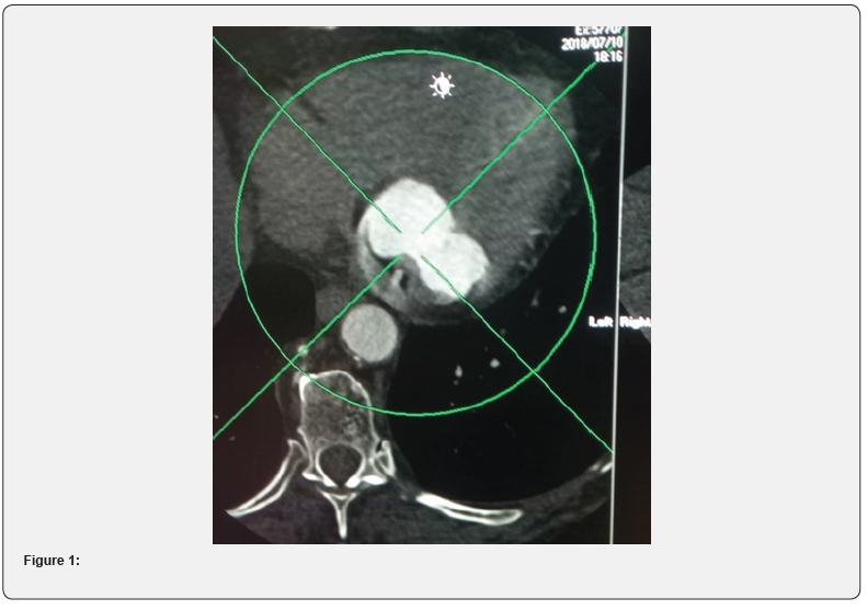
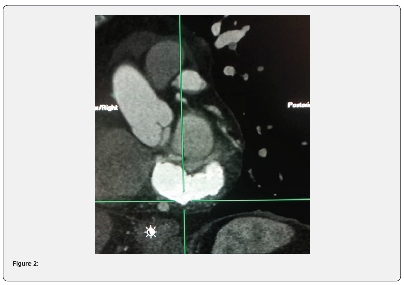
Patient underwent a cardiac CT with and without contrast medium that revealed a hyperdense mass located at the anterior and posterior mitral ring, highly suggestive of caseous calcification of the mitral annulus (see Figures 1 and 2). The mass extended circumferentially for about 75 mm with a maximum thickness of 19 mm. In contrast with other cases of mitral calcification there was not an important involvement of valve apparatus. It showed also coronary calcifications with a stenosis around 50% in the second segment of the anterior descendant artery. The patient was asymptomatic for angor or dyspnea, so she was prescribed an ECG-holter and treated conservatively and followed with echocardiography
Despite its benign prognosis this uncommon finding generated a lot of anxiety for the patient until the correct diagnosis was formulated because it could mimics different diseases and cardiologist and radiologist should become used to recognize it.


