Supra-Cardiac Total Anomalous Pulmonary Venous Connection via Rare Right-Sided Vertical Vein
Atiq Mehnaz¹* and Rizvi Mehwish²
1Department of Pediatrics, Liaquat National hospital and Medical Center, Pakistan
2Department of Pediatrics, Aga Khan University hospital, Karachi, Pakistan
Submission: September 09, 2019; Published: September 20, 2019
*Corresponding author: Mehnaz Atiq, Department of Pediatrics, Liaquat National Hospital and Medical Center, Karachi, Pakistan
How to cite this article:Atiq Mehnaz, Rizvi Mehwish. Supra-Cardiac Total Anomalous Pulmonary Venous Connection via Rare Right-Sided Vertical Vein. J Cardiol & Cardiovasc Ther. 2019; 15(1): 555904. DOI: 10.19080/JOCCT.2019.15.555904
Abstract
A Total Anomalous Pulmonary Venous Connection (TAPVC) is an uncommon congenital anomaly which occurs as result of defective development or early atresia of common pulmonary vein. TAPVC is categorized into four with supracardiac variety occurring most frequently. Common pulmonary vein anomaly results in the retention of cardinal or umbilico-vitelline drainage systems. If cardinal venous system persists, it provides channels to the innominate or azygous vein, Superior Vena Cava (SVC). This case series reports two cases of supracardiac TAPVC draining into SVC via right-sided vertical vein. Careful evaluation before surgical repair is vital for such complex cardiac anomalies.
Keywords: Anomalous pulmonary venous connection; Right vertical vein; Complex heart disease; Cyanotic congenital heart disease
Abbrevations: TAPVC: Total Anomalous Pulmonary Venous Connection; SVC: Superior Vena Cava; CHD: Congenital Heart Disease; RVV: Right Sided Vertical Vein
Introduction
Total anomalous pulmonary venous connection (TAPVC)is a rare congenital anomaly which occurs as result of defective development or early atresia of common pulmonary vein. The incidence of TAPVC ranges from 0.6 to 1.2 per 10,000 live births and it is the fifth most common cause of cyanotic congenital heart disease (CHD) [1]. TAPVC is categorized as supra cardiac, cardiac, infra cardiac, or mixed forms [2]. The most common supra cardiac drainage into innominate vein and then to superior vena cava (SVC) occurs via a left sided vertical vein [3]. We present two cases of a rare variety of total supra cardiac TAPVC which opened directly into superior vena cava via Right Sided Vertical Vein (RVV).
Case 1
A 1-month-3-days old child was referred with the suspicion of congenital heart disease. The child was previously admitted twice for chest infection. He was cyanosed with moderate respiratory distress. There was no murmur. Transthoracic echocardiography revealed a situs solitus, levocardia, small to moderate atrial septal defect of 5mm in size with right to left shunt and a hugely dilated right atrium and right ventricle. All pulmonary veins opened into a small chamber behind left atrium which formed a common pulmonary vein that opened into the base of superior vena cava. No abnormal flow to inferior vena cava was seen.

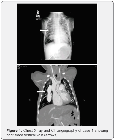
Chest X-ray showed cardiomegaly with multiple rounded shadows in the right perihilar region. The suspicion was that of a right sided vertical vein. The findings were confirmed by a CT angiography (Figure 1) which showed a common pulmonary vein connected to the base of SVC via right sided vertical vein.
Complete repair of supra cardiac TAPVC with ligation of common vein was performed and atrial septal defect was reduced to a PFO. Postoperative recovery was uneventful with laminar flow in pulmonary veins on echocardiography
Case 2
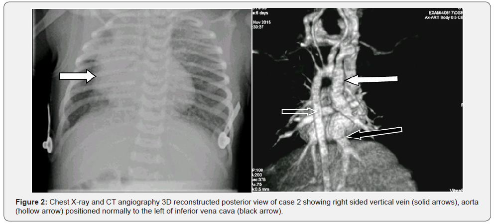
A neonate, with antenatal diagnosis of complex congenital heart disease with complete atrioventricular septal defect, total anomalous pulmonary venous connection, and d-transposition of great vessels, was born after 39 weeks of gestation. Transthoracic echocardiography confirmed the diagnosis along with heterotaxy syndrome. The TAPVC was to a chamber behind LA which was connected to SVC via a right-sided-vertical-vein (Figure 2).

A CT angiography was done which confirmed the diagnosis and showed a right sided vertical vein. Neonate was followed at 17-days of life, had mild tachypnea and moderate cyanosis (transcutaneous oxygen saturation of 72–83%). He was referred for surgery which was planned in stages. The first was to perform an arterial switch operation, correction of anomalous veins and band the pulmonary artery. Later in infancy the plan was to repair the complete atrioventricular septal defect. The child had significant hemodynamic complications after the first surgery, was put on extracorporeal membrane oxygenator, but succumbed 4 after the surgery.
Discussion
Pulmonary vein develops by canalization of endothelial strand, which connects the developing left atrium to the mesenchyme of primary lung bud through dorsal mesocardium [4]. Total anomalous pulmonary venous connection arises due to the failure of the left atrial connection to the pulmonary venous channel. This results in the retention of connections to the primitive cardinal or umbilico-vitelline drainage systems, thus providing collateral channels for anomalous venous drainage [5].
The cardinal venous system provides channels to the innominate or azygous vein, superior vena cava, or right atrium and umbilico-vitelline system provides channels to the portal or hepatic vein, or inferior vena cava. Any of these venous tributaries can persist and enlarge if pulmonary venous connection does not develop, thus resulting in broad variety of TAPVC [2]. Rarely, the right cardinal venous system persists resulting in RVV connection. RVV usually follows oblique course and drains to SVC or superior vena cava-right atrium junction [5]. Rubino et al. [3] described extracardiac connections as the largest group (42 of 72) with supra cardiac connections occurring most frequently (30 of 72 cases).
TAPVC is a frequent component in heterotaxy syndrome [5]. Postmortem study of visceral heterotaxy reports a great variety of anomalous pulmonary venous connections but only one single case out of 72 cases had a right vertical vein [6].
In the reported case 1, it is evident that isolated cases of TAPVC, even though are difficult to diagnose, have better prognosis with straightforward surgical repair. Surgical repair of TAPVC associated with other complex anomalies and syndromes, as reported in case 2 maybe associated with unfavorable prognosis [7].
Conclusion
Total anomalous pulmonary venous connection has always been a challenging diagnosis. Rare varieties can be suspected on echocardiography but need confirmation by further imaging with conventional angiography or CT angiography. The importance of accurate diagnosis has surgical implications in planning surgical approach and complete repair of the anomaly.
Ethical Standards
The authors assert that all procedures contributing to this work comply with the ethical standards of the relevant guidelines on human experimentation in our institution, and with the Helsinki Declaration of 1975, as revised in 2008. Permission for publication as rare cases was sought from the parents of both infants.
References
- Reller M, Strickland M, Riehle-Colarusso T, Mahle W, Correa A (2008) Prevalence of Congenital Heart Defects in Metropolitan Atlanta, 1998-2005. J Pediatr 153(6): 807-813.
- Geva T, Van Praagh S (2001) Anomalies of the pulmonary veins. In: Allen H, Gutgesell H, Clark E, Driscoll D (eds.), Moss and Adams’ Heart Disease in Infants, Children, and Adolescents. 6th ed. Philadelphia, Pa: Lippincott Williams & Wilkins 736-
- Rubino M, Van Praagh S, Kadoba K, Pessotto R, Van Praagh R (1995) Systemic and pulmonary venous connections in visceral heterotaxy with asplenia: Diagnostic and surgical considerations based on seventy-two autopsied cases. J Thorac Cardiovasc Surg 110(3): 641-650.
- Anderson RH, Brown NA, Moorman AF (2006) Development and structures of the venous pole of the heart. Dev Dyn 235(1): 2-9.
- Lehner A, Kozlik-Feldmann R, Herrmann F, Dalla-Pozza R, Netz H, et al. (2012) An unusual form of supracardiac total anomalous pulmonary venous return via a right-sided vertical vein in a heterotaxy syndrome case. Pediatr Cardiol 33(7): 1200-1202.
- Juneja R, Gupta S, Gulati G, Devagourou V (2012) Total anomalous pulmonary venous connection with descending vertical vein: Unusual drainage to azygous vein. Ann Pediatr Cardiol 5(2): 188-190.
- Delisle G, Ando M, Calder A, Zuberbuhler J, Rochenmacher S, et al. (1976) Total anomalous pulmonary venous connection: Report of 93 autopsied cases with emphasis on diagnostic and surgical considerations. Am Heart J 91(1): 99-122.


