Spontaneous Coronary Artery Dissection and Left Ventricular Diverticulum. A Rare Associationy
Jorge Antonio Cervantes-Nieto1, Luis Mario Gonzalez-Galvan1 and Nilda Espinola-Zavaleta2,3*
1 Department of Medical Teaching, National Institute of Cardiology Ignacio Chavez, Mexico
2 Department of Nuclear Cardiology, National Institute of Cardiology Ignacio Chavez, Mexico
3 Department of Echocardiography, ABC Medical Center, Mexico
Submission: January 10, 2019; Published: February 21, 2019
*Corresponding author: Nilda Espinola-Zavaleta, National Institute of Cardiology Ignacio Chavez, Juan Badiano Nº 1, Colonia Sección XVI, Tlalpan, C. P. 14030, Mexico City, Mexico
How to cite this article: Jorge A C-N, Luis M G-G, Nilda E-Z. Spontaneous Coronary Artery Dissection and Left Ventricular Diverticulum. A Rare Association. J Cardiol & Cardiovasc Ther. 2019; 13(1): 555853. DOI: 10.19080/JOCCT.2019.13.555853
Keywords: Coronary Artery; Left ventricular diverticulum; Drug addiction; Diaphoresis; Dyspnea; Rhythmic heart; Pericardial rub; Cardiothoracic index
Clinical Case
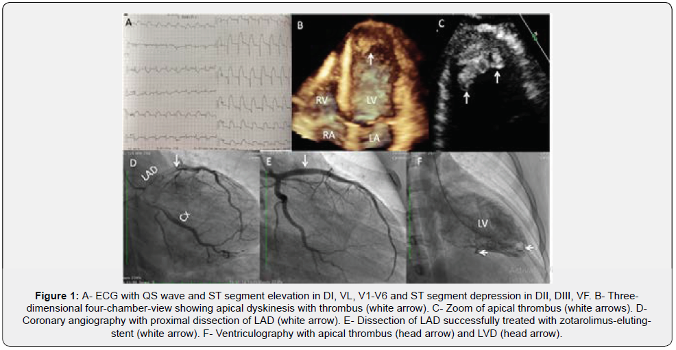
Male, 20 years-old, without background of drug addiction. He came to emergency department, for chest pain with intensity 8/10, profuse diaphoresis and dyspnea. Physical examination revealed rhythmic heart sounds, physiological splitting of the second noise and pericardial rub. Laboratory analysis with reactive protein C of 24nmol / L, troponin I 12.6μg / L, CKMB 727mg / L and pro BNP 1200pg / ml. ECG in sinus rhythm, with QS in ID, VL, V1-V6 and ST-segment elevation in these leads and ST segment depression in DII, DIII, VF (Figure 1A). Chest x-ray with cardiothoracic index of 0.58 and pulmonary venocapillary hypertension. Transthoracic echocardiogram showed left ventricular dilation, apical dyskinesia with thrombus (Figures 1B-C, Clips 1 & 2), septal akinesia and hypokinesia of middle segments and ejection fraction of 35%.
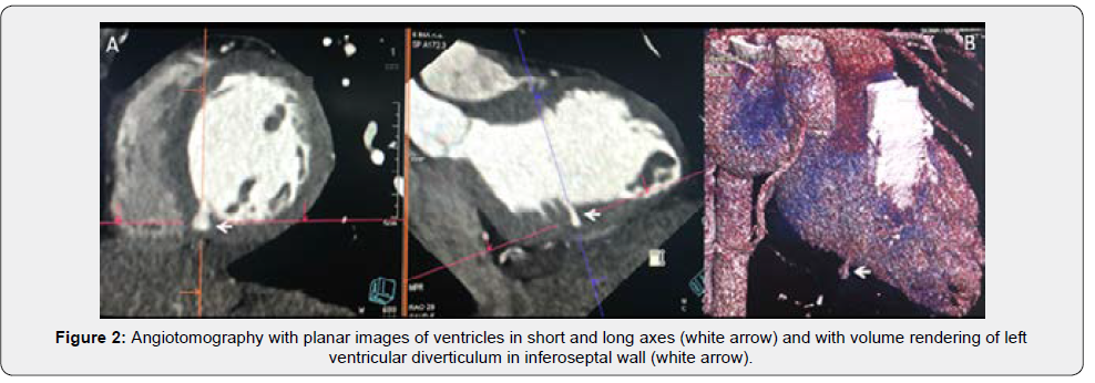
Coronary angiography demonstrated proximal dissection of Left Anterior Descending Artery (LDA) (Figure 1D), treated with zotarolimus-eluting stent (Figure 1E) and ventriculography with apical thrombus and ventricular diverticulum (Figure 1F). Double platelet antiaggregation and anticoagulation were started. Coronary angiotomography demonstrated permeable stent in proximal LDA, presence of Left Ventricular Diverticulum (LVD) in inferoseptal wall (Figures 2A & B), anteroseptal and apical infarction, apical and mural thrombus in apical third of anterior wall. The rheumatologist sought to rule out primary antiphospholipid syndrome. Spontaneous Dissection of Coronary Arteries (SDCA) is an unusual cause of acute coronary syndrome. The incidence of SDCA varies from 0.1% to 1.1% by angiography. Systematic analysis of all published cases concluded:


A. Approximately 20% of cases were diagnosed postmortem and the rest by coronary angiography
B. Isolated coronary involvement was the most frequent lesion
C. Early intervention was superior to conservative treatment
D. Administration of thrombolytics (before diagnosis of SCAD) worse this condition in 100% of patients [1].
Ventricular diverticula are sacculations of myocardial wall that are presented by an embryological failure of the ventricular muscle. Its prevalence is 0.02 to 0.04% of all cardiac malformations. They can evolve asymptomatically or present systemic embolization, heart failure, valvular insufficiency, ventricular arrhythmia and sudden death when there is spontaneous rupture of diverticulum [2]. Coexistence of spontaneous dissection of coronary artery with LVD hasn´t been reported previously.
References
- Shamloo BK, Chintala RS, Nasur A, Ghazvini M, Shariat P, et al. (2010) Spontaneous coronary artery dissection: Aggressive vs conservative therapy. J Invasive Cardiol 22(5): 222‑228.
- Treistman B, Cooley DA, Lufschanowski R, Leachman RD (1973) Diverticulum or aneurysm of left ventricle. Am J Cardiol 32(1): 119- 123.


