A Novel Approach to Extending DF-4 AICD Leads using A DF-4 Y Splitter/Adaptor as Preparation for Stereotactic Radiosurgery to The Chest - A Case Report
Roberto Cerrud-Rodriguez1*, Roman Castillo1, Adonis Castillo1, Brian B Chiong2 and Salim Baghdadi3
1 Department of Internal Medicine, SBH Health System, USA
2 Department of Radiology, SBH Health System, USA
3 Department of Cardiac Electrophysiology, SBH Health System, USA
Submission:July 13, 2018Published: August 14, 2018
*Corresponding author: Roberto Cerrud-Rodriguez, Department of Internal Medicine, SBH Health System 4422 Third Avenue Bronx, New York, 10457, Tel no: +1 929-351-5781; Email: robertocerrud@gmail.com
How to cite this article: Roberto C R, Roman C, Adonis C, Brian B C, Salim B. A Novel Approach to Extending DF-4 AICD Leads using A DF-4 Y Splitter/Adaptor as Preparation for Stereotactic Radiosurgery to The Chest - A Case Report. J Cardiol & Cardiovasc Ther. 2018; 11(4): 555820. DOI: 10.19080/JOCCT.2018.11.555820
Abstract
This is an 80-year-old Hispanic male, former smoker with 30+ pack-years, COPD, ischemic cardiomyopathy with reduced Left Ventricular Ejection Fraction (LVEF) of 15-20% refractory to an appropriate trial of optimal medical therapy, requiring upgrade to Biventricular Implantable Cardioverter-Defibrillator (BIV-ICD) implantation. Five months after BIV-ICD implantation, the LVEF had improved to 55%. During routine lung cancer screening a left upper lobe mass measuring 1.5x1.1 cm was found. A biopsy was done, and further histology showed a spiculated squamous cell carcinoma. Given his frail clinical condition it was decided that he was to receive radiotherapy only, for which the AICD generator had to be relocated away from the radiation target. To extend the DF-4 ICD lead, we used a specialized 27-cm-long DF-4 Y splitter/adaptor (Medtronic Model 5019 HV Splitter/Adaptor) which allowed enough length to extend the DF-4 ICD lead from upper chest to Left Upper Quadrant (LUQ) of the abdomen. The procedure was tolerated well by the patient, after which he made a satisfactory recovery with no postoperative complications. The patient then, in due course, underwent radiotherapy for his lung cancer.
Our technique of using a DF-4 Y Splitter/Adaptor as a DF4 lead extender could be used in any patient needing an extender, such as those in which the leads had to be tunneled to the contralateral side, and not only for patients requiring radiotherapy.
Keywords:AICD; HFrEF; Lung cancer; Radiotherapy; VT prevention; DF-4 lead; DF-4 Y Splitter/Adaptor
Abbrevations:CIED: Cardiac Implantable Electronic Devices; RT: Radiotherapy; PPM: Permanent Pacemaker; LVEF: Left Ventricular Ejection Fraction; LUL: Left Upper Lobe; SRS: Stereotactic Radiosurgery; EP: Electrophysiology; LUQ: Left Upper Quadrant; LV: Left Ventricular
Introduction
With the progressive increase in the age of the general population, clinicians will be faced more and more with challenges that in previous years would have been seen only every so often. Such is the case of patients with Cardiac Implantable Electronic Devices (CIED) who, for a multitude of reasons, go on to develop lung malignancies requiring focal Radiotherapy (RT). To decrease the chance of device malfunction due to RT, different strategies have been devised, including complete explantation of the existing CIED. In a selected population of patients, such an approach would be too invasive and risky given their frail clinical condition, so alternative approaches have to be sought. In the present article, we present the case of a patient with lung cancer requiring focal RT in whom we used a DF-4 Y Splitter/Adaptor as a lead extender for an ICD with DF-4 leads, for which there are no dedicated lead extenders currently in the market. This way, we achieved a shorter intervention time with less operative risk, while obtaining the same benefits of reimplanting the ICD pulse generator out of the way of the RT field.
Case Report
This is the case of an 80-year-old male, former smoker with 30+ pack-years, Chronic Obstructive Pulmonary Disease, chronic atrial fibrillation s/p Permanent Pacemaker (PPM) on oral anticoagulation, significant coronary artery disease s/p percutaneous intervention in June, 2016. He developed subsequent ischemic cardiomyopathy with reduced Left Ventricular Ejection Fraction (LVEF) of 15-20% which did not improve after an appropriate trial of optimal medical therapy, thus requiring upgrade to biventricular implantable cardioverter-defibrillator (BIV-ICD) implantation in December, 2016. Five months after BIVICD implantation, the patient’s clinical status had improved, with a concomitant improvement of his LVEF to 55%.
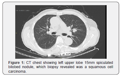
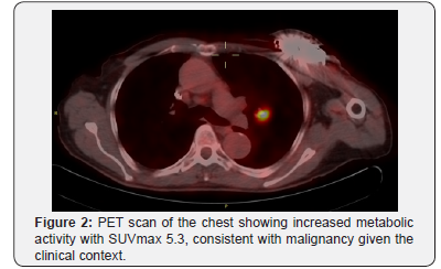
Given his history of smoking, he was referred by his primary care doctor for routine lung cancer screening. It was after one of these screenings that the patient was found to have Left Upper Lobe (LUL) mass measuring 1.5x1.1 cm (Figure 1 & 2). A biopsy was done, and further histology showed a spiculated squamous cell carcinoma. Given his advanced age, his comorbidities, frail condition and following a shared decision-making approach with the patient and his family, it was decided that he would receive Stereotactic Radiosurgery (SRS) to the chest as the only treatment modality for his lung cancer.


In order for the patient to receive SRS, which would exceed the radiation limit of 2 Grays for an AICD, (1) the AICD pulse generator had to be repositioned away from the upper chest. We, in the cardiac Electrophysiology (EP) department determined that the best option was to reposition the pulse generator to the Left Upper Quadrant of the abdomen (LUQ) by extending the leads of the existing BIV-ICD system. The patient’s oral anticoagulation therapy was suspended in preparation for the procedure.
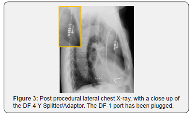
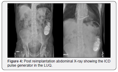
The patient was taken in the fasting state to the EP suite and he was placed under general anesthesia. To extend the DF-4 ICD lead, we used a specialized 27-cm-long DF-4 Y splitter/adaptor (Medtronic Model 5019 HV Splitter/Adaptor) which allowed enough length to extend the DF-4 ICD lead from upper chest to left upper quadrant (LUQ) of the abdomen (Figure 3). The Left Ventricular (LV) lead had enough length to reach the LUQ of the abdomen without the need for an extender. The DF-4 Y splitter/ adaptor and the LV lead were tunneled subcutaneously from the left upper chest to the LUQ of the abdomen and subsequently were connected to the BIV-ICD pulse generator (Medtronic Model VIVA QUAD XT CRT-D DTBA1QQ) which was implanted in the LUQ of the abdomen (Figure 4). The DF-1 port in the splitter/adaptor was plugged.


The procedure was tolerated well by the patient, after which he made a satisfactory recovery with no postoperative complications. Oral anticoagulation was restarted after his recovery. And eventually, the patient underwent SRS treatment for his lung cancer.
We consider the previously described technique to be a novel approach to DF-4 ICD lead extension when an AICD pulse generator needs to be re-positioned, as the device that we used (the DF-4 Y Splitter/Adaptor) is routinely used to connect a new DF-1 coil to the system, not as a DF-4 lead extender.
Discussion
As the age of the general population increases, health care professionals will have to take care of an increasing number of patients with Cardiac Implantable Electronic Devices (CIED) who require Radiotherapy (RT). A single center cohort study of patient’s undergoing radiotherapy revealed that the prevalence of CIEDs is nearly 1% [1].
Current CIEDs use complementary metal-oxide semiconductors (CMOS), which makes them more susceptible to ionizing radiation from RT. This happens due to stochastic effects related to interactions with high energy neutrons. These negative effects range from mild programming corruption, to power-on-reset (where the programming for tachycardia and bradycardia is reset to factory default settings), to total device failure, and tend to increase with cumulative radiation exposure [1-3].
The specific mechanism of how RT affects CMOS technology is still under study. It is hypothesized that exposure of the device to ionizing radiation causes excess positive charge within the circuitry, which in turn produces aberrant electrical pathways, alters the current-voltage characteristics and voltage threshold of the device, as well as cause leakage currents inside the CMOS [1-3].
RT also produces Electromagnetic Interference (EMI) or scatter radiation of neutrons; in the case of Implantable Cardioverters (ICDs), EMI waves can disrupt appropriate internal functioning of the device and be confused for myocardial potentials [1,3]. Several developments, including titanium casings and EMI-recognition software have been designed to counteract this, but it is still difficult to predict or detect the moment of an ICD breakdown [4].
There are no up-to-date guidelines regarding the optimal management of patients with CIEDs who require RT. The repositioning of CIEDs in these cases hasn’t been well studied yet, so there is no standard of care. There are a variety of different approaches to manage patients in situations similar to ours [1].
• One of them is to remove the device and use a LifeVest for the duration of the RT; in the case of our patient, this was undesirable, as we wanted to preserve the improvement in LVEF seen after the BIV-ICD implantation.
• A second possibility was to explant the entire BIV-ICD system and re-implant a new one with a pulse generator and leads originating in the right side of the thorax – in the case of our patient, this would have led to an overly invasive procedure considering our patient’s advanced age, comorbidities, frailty and life expectancy.
• A third option was to attempt to use lead extenders to allow repositioning of the pulse generator to the right hemithorax or to the lower abdomen; this was not feasible as the patient had an ICD with DF-4 leads, for which no extender has been developed yet.
For our patient, who required RT to the left thorax, we chose to re-implant the pulse generator in the abdominal LUQ to decrease the amount of radiation it would receive. This way we aimed to decrease the chance of device failure, even though no clear guidance is available to the clinician in the existing literature.
Regarding the technical aspects, the newer DF-4 connector is less bulky, designed to facilitate lead-to-device connection, minimize the risk of incorrect device connection, and allows for easier implantation with shorter procedure time [5]. It was initially introduced in 2010 as a standard four-pole inline connector system which allows interchangeability from all manufacturers and ensures compatibility with future implanted devices [6].
The specialized DF-4 Y splitter/adaptor (Medtronic Model 5019 HV Splitter/Adaptor) has a DF-4 connector at one end (which connects to the device header) and 2 separate connections (one for the DF-4 ICD lead and other for the additional coil with DF-1 connector) at the other end.
The DF-4 Y splitter/adaptor has been routinely used to connect an additional DF-1 coil to the ICD system when the energy requirements for defibrillation are too high [5].
In the case of our patient, the DF-4 Y splitter/adaptor was used as an extension to the existing leads in order to re-implant a new pulse generator in the abdominal LUQ. This allowed the procedure to be less invasive, as it was not necessary to explant the existing leads. It also required a much shorter operative time and thus, less risk to the patient. To the best of our knowledge, this is the first time a DF-4 Y splitter/adaptor has been used for this purpose. Our MEDLINE search for previous similar reports yielded no results.
Conclusion
The abovementioned procedure can potentially benefit patients with CIEDs using DF-4 leads who require re-implantation of the device by using the DF-4 Y Splitter/Adaptor as an ad hoc DF-4 lead extender, to help reposition the pulse generator in the abdomen or in the contralateral chest wall.
IRB approval:
This case report was approved by the Institutional Review Board of our institution (IRB Study Number 2017.105).
Funding:
This research did not receive any specific grant from funding agencies in the public, commercial, or not-for-profit sectors.
References
- Karnik A, Helm R, Chatterjee S, Monahan K (2015) Abdominal implantable cardioverter-defibrillator placement in a patient requiring bilateral chest radiation therapy. Heart Rhythm Case Reports 2(5): 395-398.
- Brambatti M, Mathew R, Strang B, Dean J, Goyal A, et al. (2015) Management of patients with implantable cardioverter-defibrillators and pacemakers who require radiation therapy. Heart Rhythm 12(10): 2148-2154.
- Frizell B (2009) Radiation therapy in oncology patients who have a pacemaker or implantable cardioverter-defribillator. Community Oncology 6: 469-471.
- Hurkmans C, Scheepers E, Springorum B, Uiterwaal H (2005) Influence of radiotherapy on the latest generation of implantable cardioverterdefibrillators. Int J Radiat Oncol Biol Phys 63(1): 282-289.
- Lim H (2013) Overcoming the Limitations of the DF-4 Defibrillator Connector. The Journal of Innovations in Cardiac Rhythm Management 4: 1205-07.
- Bhargava K (2014) DF-4 Lead Connector: Innovative Technology, Unexpected Problems and Novel Solution. Indian Pacing Electrophysiol J 14(3): 108-111.


