A Cavernous Sinus of Valsalva Aneurysm
Roland Fasol1, Berit BODE1, Xue LI2, Noell FASOL2 and Anneli SEPPÄLÄ-LINDROOS2
1Department of Cardiac Surgery, Tree Top Hospital Maldives, Maldives
2Department of Cardiology, The Jilin Heart Hospital, China
Submission: November 30, 2016; Published: December 09, 2016
*Corresponding author: Roland Fasol, Department of Cardiac Surgery, Tree Top Hospital Maldives Filigas dhoshuge, Ameer Ahmed Magu, Male’ 20066, Maldives, China
How to cite this article: Roland F, Berit B, Xue L, Noell F, Anneli S. A Cavernous Sinus of Valsalva Aneurysm. 2016; 2(3): 555587 DOI: 10.19080/JOCCT.2016.02.555587
Abstract
A 47 year old female healthy, asymptomatic farmer underwent routine medical screening in rural China. A early diastolic murmur was detected. Subsequent color doppler echocardiogram showed a competent aortic valve but a fistulous communication of a perforated Valsalva aneurysm into the left ventricle. Computed tomography revealed a multi cavernous cauliflower-shaped Valsalva aneurysm involving the whole aortic basisor bulbous aortae and all sinuses.
Keywords: Aneurysm; Valsalva; Aorta
Introduction
A sinus of Valsalva aneurysm (SVA) was first described by Thurman in 1840 [1]. A rare abnormality of the heart, a sinus of Valsalva aneurysm, may be caused by a separation or localized connective tissue dystrophy of the aortic tunica media and the annulus fibrosus of the aortic valve [2]. Such a SVA may be congenital or acquired [3] with an incidence as low as <1.5% among congenital heart disease repairs [4]. It is the least common of all aortic aneurysms. If not congenital but acquired causes include atherosclerosis, cystic media necrosis, infective or post-traumatic injuries [5]. Sinus of Valsalva aneurysmmore prevalent originates from the right coronary sinus (70-90%), have been seen less commonly from the non-coronary sinus (10-20%), and rarely from the left sinus (<5%) [6]. The incidences of SVA are reported to be higher in Asian than Western populations, and the male: female ratio was found to be 4:1 [7]. Un-ruptured sinus of Valsalva aneurysms normally do not cause any symptoms unless they rupture, causing fistulous communications, heart failure and death [3,4,8,9].
Case Report
A 47 year old female patient was admitted because of an early diastolic murmur which was detected during a routine medical screening of the rural population in north-east China in Jilin Province, close to the North Korean border. The female patient, working as a farmer and doing heavy work in the fields, was completely asymptomatic and had no shortness of breath and no dyspnea during her work. On admission she was in New York
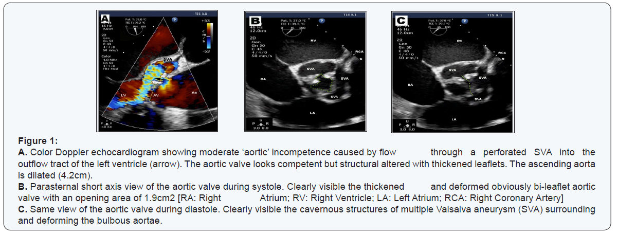
Heart Association class I, regular sinus rhythm and normal blood pressure, no diabetes, normal body mass index, no prevalence of metabolic syndrome, but a heavy smoker. The early diastolic murmur was confirmed but color doppler echocardiogram showed a somewhat ‘difficult’ result. Investigated by different cardiologists we received different interpretations. The first reported signs of a congenital heart disease and a moderate ‘aortic’ incompetence but a competent aortic bicuspid valve, however some signs of aortic valve (AV) perforation.
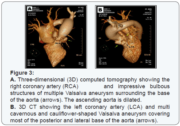
The AV opening area was calculated 1.9cm2, the AV max peak gradient (PG) 56mmHg, Ejection Fracture (Simpson) 74%, the ascending aorta with a post-stenotic dilatation with a diameter of ø4.2cm, LVIDd 4.2cm and AV V max 376 cm/sec. The other reported a combined aortic valve lesion with stenosis and incompeence, some AV vegetations, Vmax 471cm/sec and AV max PG 89mmHg. Repeated color doppler echocardiograms showed a moderate flow through a perforated SVA into the outflow tract of the left ventricle and thickened and deformed obviously bi-leaflet aortic valve leaflets (Figure 1). Computed tomography confirmed cavernous sinus of Valsalva aneurysmatic structures right below the level of the sinutubular junction and fistulous communications of a perforated SVA (Figure 2).Three-dimensional computed tomography confirmed a dilated ascending aorta and multiple cavernous, cauliflower-like Sinus of Valsalva structures surrounding the deformed base of the aorta (Figure 3).


Results and Discussion
There was a clear indication for surgery due to a significant aortic stenosis and a perforated SVA. However, the patient, a farmer from the countryside in rural China, refused surgery because the family could not afford the surgical treatment. There are significant socio-economic disparities in China and a massive gap between urban and rural population groups [10]. While the wealthier share of the Chinese population has benefited from advanced health technologies and spending on health care, the poor have lost access to even the most essential services. In terms of rural-urban disparity across provinces, life expectancy drops parallel to a rising share of rural population [11].
References
- Thurman J (1840) On aneurisms, and especially spontaneous varicose aneurisms of the ascending aorta, and sinuses of Valsalva: with cases. Med Chir Tr 23: 323-384.
- Aliye Ozsoyoglu Bricker, Bindu Avutu, Tan-Lucien H Mohammed, Eric E Williamson, Imran S Syed, et al. (2010) Valsalva sinus aneurysms: findings at CT and MR imaging. Radio graphics 30(1): 99-110.
- Post MC, Braam RL, Groenemeijer BE, Nicastia D, Rensing BJ, et al. (2010) Rupture of right coronary sinus of Valsalva aneurysm into right ventricle. Neth Heart J 18(4): 209-211.
- Abu Saleh WK, Lin CH, Reardon MJ, Ramlawi B (2016) Right ventricular outflow tract obstruction caused by isolated sinus of Valsalva aneurysm. Tex Heart Inst J 43(4): 357-359.
- Pujos C, Morgant MC, Malapert G, Bouchot O (2015) Aortic root remodeling and external aortic annuloplastyto treat sinus of Valsalva aneurysm in a patient with complete situsinversus. J Cardiothorac Surg 10: 182-185.
- Ring WS (2000) Congenital heart surgery nomenclature and database project: aortic aneurysm, sinus of Valsalva aneurysm, and aortic dissection. Ann Thorac Surg 69(4): S147-163.
- Wang ZJ, Zou CW, Li DC, Li HX, Wang AB, et al. (2007) Surgical repair of sinus of Valsalva aneurysm in Asian patients. Ann Thorac Surg 84(1): 156-160.
- Cronin EM, Walsh KP, Blake GJ (2011) Ruptured noncoronary sinus of Valsalva aneurysm. An unusual cause of chronic right-sided heart failure. Circ Heart Fail 4(1): e3-e4.
- Liu FQ, Zhu ZF, Ren J, Mu J (2014) A rare cause of sudden dyspnea and unexpected death in adolescence: fistula from aortic sinus of Valsalva to right atrium. Int J ClinExp Med 7(9): 2945-2947.
- Meng Q, Zhang J, Yan F, Hoekstra EJ, Zhuo J (2012) One country, two worlds - the health disparity in China. Glob Public Health 7(2): 124- 136.
- WHO´s macroeconomics and health initiative (2016) China: Health, poverty and economic development.


