An Audit of Clinical Records and Radiographs in the Assessment of Patients Referred for Third Molar - Compliance to Third Molar Guidelines
Nishma Hindocha N 1*, Bell C2 and Revington P3
1Department of Oral and Maxillofacial Surgery Zealand University Hospital, Europe
2Christopher Bell, Department of Oral and Maxillofacial Surgery University Hospitals, England
3Peter Revington, Department of Oral and Maxillofacial Surgery University Hospitals, England
Submission: June 04, 2019; Published: July 31, 2019
*Corresponding author: Nishma Hindocha, Department of Oral and Maxillofacial Surgery, Zealand University Hospital, Europe
How to cite this article: Nishma Hindocha N, Bell C, Revington P. An Audit of Clinical Records and Radiographs in the Assessment 0011 of Patients Referred for Third Molar - Compliance to Third Molar Guidelines. J Child Adolesc Dent. 2019; 1(1): 555553.
Abstract
Aim: Increasingly many patients are referred for one or two symptomatic third molars. They may have other third molars present, which have been asymptomatic at the time of referral. It is important that all patients undergo a thorough clinical examination together with appropriate radiographs that should include assessment of other third molars if present. The aim of this audit was to investigate if guidance regarding third molar assessment was being met and if the level of staff carrying out the assessment was of any significance.
Material and Methods: A retrospective case note review was carried out. The 50 first patients from September 2015 who had at least one wisdom tooth removed at UH Bristol were included in the study.
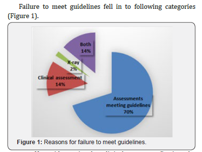
Results:< The data showed that the recorded patient assessment did not meet guidance in 30% (15/50) of the cases. This was due to lack of clinical entry in notes and/or inappropriate radiographs. This was irrespective of level of staff carrying out the assessment.
Conclusion: The audit revealed that the clinical entries in the patient notes and radiographs taken at the patient assessment failed to meet this part of the guidelines that state that in all cases third molars other than those being the subject of the referral should also be assessed.
Clinical Relevance: It is important that patients who are referred for third molar assess, and management undergo a clinically thorough clinical assessment. The possibility of pathology associated with other as yet asymptomatic third molars should also be assessed. It is important that the entries in the clinical notes together with radiographs taken should be of such a standard as to demonstrate that this has been undertaken
Keywords: Third Molar Referral RCS Guidance Wisdom Tooth Third Molar Guidance Orthopantomogram
Introduction
Since the introduction of guidelines for the management of third molars within the UK, three guidance documents have been available. These have been published by Royal College of Surgeons (RCS), NICE and Scottish Intercollegiate Guidance Network (SIGN). From the three guidance documents available the importance of assessing all third molars was best described by the RCS paragraphs [1-3]. “If there are indications for removal of one third molar it is in the patient’s best interests to determine whether the other three are present and if so, whether their excision is required on the grounds of other clinical indications. This was seen to be particularly important for patients who may be listed for treatment under Day case general an aesthetics (GA) where patients may declare on the day of treatment. concernabout other third molars since the date of listing. Furthermore, the guidance suggests the following:
i. It is suggested that removal of other teeth should only be carried out when treatment under general an aesthetic is planned or selected by the patient and there is no evidence of increased risk of post-operative complications such as sensory nerve impairment ….”
ii. Two main points of concern were listed for the audit. Firstly, that although at consultation an appropriate clinical assessment of the patient may have been undertaken, the clinical notes did not show confirmation that other third molars had been recorded or assessed. Secondly, thatconcern about radiographic exposure levels may result in some patients only having half orthopantomograms (OPGs) taken, mainly for single tooth symptoms, and possible pathology on the contralateral side not being assessed.
iii. The aim of this audit was to investigate if clinical notes and radiographs met these criteria as best described in the RCS guidance and if the level of staff carrying out the assessment was of any significance.
Materials and Methods
The audit examined the clinical entries in the notes and radiographs for evidence that the assessment of all third molars potentially present had been undertaken and recorded. A retrospective case note review was carried out. The 50 first patients from September 2015 who had at least one wisdom tooth removed at UH Bristol were included in the study? There were no criteria for exclusion? The notes of these patients were reviewed to complete the data collection sheet (appendix I). All data was then summarized in an excel sheet (appendix II).
Results
The data showed that the recorded patient assessment did not meet guidance in 30% (15/50) of the cases.

a. No evidence in the clinical notes confirming the assessment of third molars other than the tooth specified in the referral
b. Failure to take appropriate radiographs to assess other third molars potentially present, confirming asymptomatic or non-impacted status
c. Patients progressing to GA, where there was no recorded evidence in the notes clarifying if a patient may have expressed the possibility of removal of additional teeth.
d. That all levels of staff failed to meet the standard of the audit.
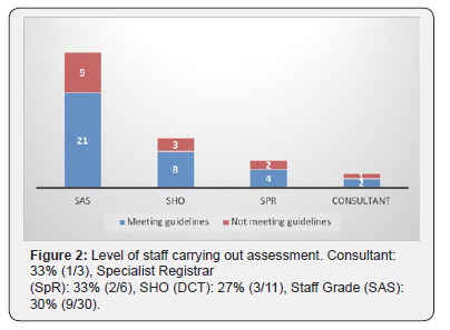
Out of the cases not meeting guidelines, the clinical level of staff carrying out the assessment was as follows: Consultant: 33% (1/3), Specialist Registrar (SpR): 33% (2/6), SHO (DCT): 27% (3/11), Staff Grade (SAS): 30% (9/30) (Figure 2).

Discussion
Clinical records of patients who are referred for third molar opinion, must be able to demonstrate that an assessment and a discussion has taken place. Where patients have pathology that is limited to one or two teeth only, it is important that the records account for another third molars present together with evidence of appropriate assessment. The finding that so many of the assessments failed to meet guidance, shows an area where there is need for improvement. With respect to radiographic exposure careful use of newer radiographic techniques now demonstrate that a sectional bilateral OPG for third molars may carry as little exposure as equivalent to two intra-oral periapical radiographs [4] and that extending the radiographic examination to include asymptomatic teeth may be justified. This is further supported by Section 4.2 of the sign Guidance 3.
There are several reasons for assessing asymptomatic wisdom teeth, e.g. increasing incidence of distal caries in lower second molars when associated with asymptomatic, partially erupted wisdom teeth [5]. Furthermore, distal-cervical caries in second molars is not specifically related to overall caries risk. This implies that patients, if not risk-assessed properly, may require further third molar surgery and incur a higher risk of delayed healing and complications caused by surgical intervention in later years [6]. If the patient requires a GA for the wisdom tooth removal, there is also a risk for an unnecessary second GA, and subsequently all associated risks. To improve our standards, the actions outlined below was implemented:
i. Irrespective of whether a patient is referred for one or more third molars the clinical notes should carry comments about the absence, normal eruption or possible potential symptomatic position of all four third molars.
ii. Where appropriate, bilateral OPG even for asymptomatic third molars should be obtained to support the above assessment
iii. Those patients listed for GA should show evidence of comment on all four third molars and discussion with patients regarding treatment options of removal of additional third molars.
Conclusion
When a patient is referred for third molar surgery, whether for single or multiple teeth, evidence must be present in the notes to account for thorough assessment and discussion with the patient of all other third molars either by clinical or radiographic assessment.
References
- Royal College of Surgeons (2004) The Management of Patients with Third Molar Teeth.
- The National Institute for Health and Care Excellence (2000) Guidance on Extraction of Wisdom Teeth.
- Scottish Intercollegiate Guidelines Network (2000) Management of Unerupted and Impacted Third Molar teeth.
- Ngan DC, Kharbanda OP, Geenty JP, Darendeliler MA (2003) Comparison of radiation levels from computed tomography and conventional dental radiographs. Aust Orthod J 19(2): 67-75.
- Toedtling V, Yates JM (2015) Revolution vs status quo? Non-intervention strategy of asymptomatic third molars causes harm. Br Dent J 219(1): 11-12.
- McArdle LW, McDonald F, Jones J (2014) Distal cervical caries in the mandibular second molar: an indication for the prophylactic removal of third molar teeth? Updassste. Br J Oral Maxillofac Surg 52(2): 185-189.


